
Drug Discovery
&
Development
Neal G. Simon, Ph.D. Professor
Department of Biological Sciences
ebook
+91-9884350006www.pubrica.com [email protected]
Disclaimer:

DRUG DISCOVERY
AND DEVELOPMENT
Volume 1: Drug Discovery
Edited by
MUKUND S. CHORGHADE
“Epothilone” cover art by Doug Scard
www.sputniknewmedia.com
Copyright © 2006 by John Wiley & Sons, Inc. All rights reserved.
Published by John Wiley & Sons, Inc., Hoboken, New Jersey.
Published simultaneously in Canada.
No part of this publication may be reproduced, stored in a retrival system, or transmitted in any form or by any
means, electronic, mechanical, photocopying, recording, scanning, or otherwise, except as permitted under
Section 107 or 108 of the 1976 United States Copyright Act, without either the prior written permission of the
Publisher, or authorization through payment of the appropriate per-copy fee to the Copyright Clearance Centre,
Inc., 222 Rosewood Drive, Danvers, MA 01923, (978) 750-8400, fax (978) 750-4470, or on the web at
www.copyright.com. Requests to the Publisher for permission should be addressed to the Permissions
Department, John Wiley & Sons, Inc., 111 River Street, Hoboken, NJ 07030, (201) 748-6011,
fax (201) 748-6008, or online at http://www.wiley.com/go/permission.
Limit of Liability/Disclaimer of Warranty: While the publisher and author have used their best efforts in
preparing this book, they make no representations or warranties with respect to the accuracy or completeness
of the contents of this book and specifi cally disclaim any implied warranties of merchantability or fi tness for a
particular purpose. No warranty may be created or extended by sales representatives or written sales materials.
The advice and strategies contained herein may not be suitable for you situation. You should consult with a
professional where appropriate. Neither the publisher nor author shall be liable for any loss of profi t or any other
commercial damages, including but not limited to special, incidental, consequential, or other damages.
For general information on our other products and services or for technical support, please contact our Customer
Care Department within the United States at (800) 762-2974, outside the United States at (317) 572-3993 or
fax (317) 572-4002.
Wiley also publishes its books in a variety of electronic formats. Some content that appears in print may not be
available in electronic formats. For more information about Wiley products, visit our web site at
www.wiley.com.
Library of Congress Cataloging-in-Publication Data:
Drug discovery and development/edited by Mukund S. Chorghade.
p. cm.
Includes bibliographical references and index.
ISBN-13: 978-0-471-39848-6
ISBN-10: 0-471-39848-9 (cloth : v. 1)
1. Drug development. I. Chorghade, Mukund S. (Mukund Shankar)
[DNLM: 1. Drug Design. 2. Chemistry, Pharmaceutical–methods.
3. Drug Evaluation, Preclinical–methods. QV 744 D79334 2006]
RM301.25C488 2006
615'.19–dc22
2005021297
Printed in the United States of America
10 9 8 7 6 5 4 3 2 1
CONTENTS
Contributors xiii
Preface xv
1 From Patent to Prescription: Paving the Perilous Path to Profi t 1
Richard J. Pariza
1.1 Introduction, 1
1.2 A Simple Solution to a Complex Problem, 3
1.3 An Intriguing Patent Problem, 8
1.4 Another Structural Insight, 10
References, 15
2 Medicinal Chemistry in the New Millennium: A Glance into the Future 17
Paul W. Erhardt
2.1 Introduction, 17
2.2 Practice of Medicinal Chemistry, 19
2.2.1 Emergence as a Formalized Discipline, 19
2.2.2 Early Developments, 23
2.2.3 Present Status, 26
2.2.4 Examples Involving Site-Directed Mutagenesis, 27
2.2.5 Latest Trends, 31
2.3 Evolving Drug Discovery and Development Process, 35
2.3.1 Working Defi nition for Medicinal Chemistry, 35
2.3.2 Immediate- and Long-Term Roles for Medicinal Chemistry, 36
v
2.4 Pursuing Effi cacy, 40
2.4.1 Gathering Positive, Neutral, and Negative SARs During HTS, 41
2.4.2 Example Involving Multidrug Resistance of Anticancer Agents, 42
2.4.3 Compound Libraries: Example of Working with Nature to Enhance
Molecular Diversity, 45
2.5 Assessing and Handling Molecular Conformation, 46
2.5.1 Chemoinformatics, 46
2.5.2 Obtaining Chemically Correct 3D Structures, 49
2.5.3 Infl uence of Biological Environments: Example Involving Drug
Metabolism, 50
2.5.4 Dynamic Energy Relationships: Example Involving a Small Ring
System, 52
2.5.5 Druglike Properties and Privileged Structures, 54
2.5.6 Tiered Structural Information and Searching Paradigms, 55
2.6 ADMET Considerations, 57
2.6.1 Assuring Absorption, 57
2.6.2 Directing Distribution, 58
2.6.3 Herbal Remedies: Example of Working with Nature to Discover
ADMET-Related Synergies, 59
2.6.4 Brute Force HTS to Uncover Multicomponent Synergies, 62
2.6.5 Controlling Metabolism: Example Involving a Soft Drug Strategy, 63
2.6.6 Optimizing Excretion, 65
2.6.7 Avoiding Toxicity, 65
2.6.8 Weighting Decision Criteria from Effi cacy and ADMET SAR, 67
2.7 Process Chemistry Considerations, 70
2.7.1 Cost and Green Chemistry, 70
2.7.2 Defi ning Stereochemistry: Example Involving Benzylamine Chiral
Auxiliary Synthetic Reagents, 71
2.8 Analytical Chemistry/X-ray Diffraction, 74
2.8.1 Latest Trends, 74
2.8.2 Examples Involving Dopamine Receptors, c-AMP Phosphodiesterase
Enzymes, and the Dynamics of Protein Folding, 75
2.9 Summary, 78
2.9.1 General Points, 78
2.9.2 Attributes of Drug Discovery Libraries, Compound Hits, and Lead
Compounds, 81
2.9.3 Formalized Instruction of Medicinal Chemistry, 81
2.9.4 Intellectual Property Considerations, 83
2.9.5 Knowledge Versus Diversity Paradox, 84
Acknowledgments, 85
References and Notes, 85
3 Contemporary Drug Discovery 103
Lester A. Mitscher and Apurba Dutta
3.1 Introduction, 103
3.1.1 Getting Started, 103
3.2 Characteristics of a Suitable Lead Substance, 104
3.2.1 Potency and Selectivity, 105
vi CONTENTS
3.2.2 Structure–Activity Relationships, 107
3.2.3 Toxicity, 107
3.2.4 Changing Appellation of the Best in Series: Analog Attrition, 108
3.3 Some Criteria That a Hit Must Satisfy to Become a Drug, 108
3.3.1 Level of Potency, 109
3.3.2 Comparison of Potency and Effi cacy, 110
3.3.3 Druglike Character, 110
3.3.4 Effi cacy Following Oral Administration, 110
3.3.5 Lipinski Rules for Oral Absorption, 112
3.3.6 Injectable Medications, 113
3.3.7 Distribution, 113
3.3.8 Serum Protein Binding, 114
3.3.9 Metabolism, 114
3.3.10 Distribution, 114
3.3.11 Excretion, 115
3.3.12 Patenting, 115
3.3.13 Pharmaceutical Properties, 115
3.3.14 Idiosyncratic Problems, 115
3.3.15 Summary, 115
3.4 Example of Drug Development That Illustrates Many of the Aforementioned
Considerations, 116
3.4.1 Control of Blood Pressure with Drugs, 116
3.4.2 Historical Background, 116
3.4.3 Finding a Starting Point: A Clue from Nature, 117
3.4.4 Renin–Angiotensin–Aldosterone System, 117
3.4.5 Attempts to Inhibit Renin, 119
3.4.6 Attempts to Inhibit Angiotensin-Converting Enzyme, 119
3.4.7 Peptides Make Poor Orally Active Drugs, 120
3.4.8 Analoging Studies of Pit Viper–Inspired Peptides, 120
3.4.9 Peptidomimetics, 120
3.4.10 Adaptation to Inhibition of ACE, 121
3.4.11 Success Inspires Competition, 123
3.4.12 Taking a Different Approach, 124
3.4.13 Analoging to Enhance Absorption, 124
3.4.14 Clinical SAR, 126
3.4.15 More Recent Work, 128
3.4.16 Résumé, 128
3.5 Conclusions, 128
Additional Reading, 128
4 Combinatorial Chemistry in the Drug Discovery Process 129
Ian Hughes
4.1 Introduction, 129
4.1.1 The Birth of Combinatorial Chemistry, 130
4.1.2 Development of Screening Strategies for Libraries, 131
4.1.3 From Peptides to Small Molecule Synthesis, 132
4.1.4 Beyond Solid-Phase Chemistry, 133
4.2 The Role of Combinatorial Chemistry in Drug Discovery, 135
CONTENTS vii
4.3 Designing Combinatorial Libraries, 137
4.3.1 Describing and Measuring Diversity, 137
4.3.2 A More Focused Approach, 139
4.4 Tools for Synthesis of Combinatorial Libraries, 141
4.4.1 Nonautomated Tools, 141
4.4.2 Mix-and-Sort Systems, 143
4.4.3 Automated Synthesizers, 143
4.4.4 Postsynthesis Processing, 144
4.5 Managing the Combinatorial Process, 146
4.5.1 Specifi cation of Combinatorial Libraries, 146
4.5.2 Controlling the Automated Workfl ow, 146
4.6 From Specialist Discipline to Standard Tool, 148
4.7 Application of Combinatorial Chemistry in Drug Discovery, 149
4.7.1 Case History 1, 150
4.7.2 Case History 2, 150
4.7.3 Case History 3, 151
4.7.4 Case History 4, 152
4.8 The Future of Combinatorial Chemistry, 154
4.8.1 Dynamic Combinatorial Libraries, 154
4.8.2 Miniaturization, 154
4.9 Conclusions, 155
References, 156
5 Parallel Solution-Phase Synthesis 169
Norton P. Peet and Hwa-Ok Kim
5.1 Introduction, 169
5.2 Ahead of Our Time, 169
5.3 Recent Reports of Parallel Solution-Phase Synthesis, 172
5.4 Solid Supported Reagents, Scavengers, and Catalysts, 178
5.5 The Future, 191
References, 191
6 Timing of Analog Research in Medicinal Chemistry 199
János Fischer and Anikó Gere
6.1 Introduction, 199
6.2 Early Phase Analogs, 199
6.2.1 ACE Inhibitors, 199
6.2.2 AT
1
Antagonists, 200
6.2.3 Proton Pump Inhibitors, 200
6.2.4 Insulin Sensitizers: Glitazones, 200
6.2.5 HMG-CoA Reductase Inhibitors, 202
6.2.6 Antimigraine Drugs, 202
6.3 Drug Analogs, 202
6.3.1 Metoclopramide Analogs, 203
6.3.2 Azatadine Analogs, 205
6.3.3 Miconazole Analogs, 205
6.3.4 Nifedipine Analogs, 206
viii CONTENTS
6.3.5 Propranolol Analogs, 207
6.3.6 Clodronate Analogs, 207
6.4 Summary, 208
Acknowledgments, 210
References and Notes, 210
7 Possible Alternatives to High-Throughput Screening 213
Camille G. Wermuth
7.1 Introduction, 213
7.2 Analog Design, 214
7.2.1 Defi nitions, 214
7.2.2 Pharmacophere-Based Analog Design: Scaffold Hopping
or Scaffold Morphing, 215
7.2.3 Natural Compounds as Models, 216
7.2.4 Emergence of New Activities, 216
7.3 Physiopathological Hypotheses, 217
7.3.1 Discovery of Levodopa, 217
7.3.2 H
2
-Receptor Antagonists, 219
7.3.3 Rimonabant and Obesity, 220
7.4 Contributions from Clinical Investigations, 221
7.5 New Leads from Old Drugs: The SOSA Approach, 223
7.5.1 Rationale, 223
7.5.2 Examples, 223
7.5.3 Discussion, 226
7.6 Conclusion, 228
References, 229
8 Proteomics and Drug Discovery 233
Susan Dana Jones and Peter G. Warren
8.1 Introduction, 233
8.2 Drug Discovery Process, 234
8.2.1 Process Overview, 234
8.2.2 Motivation for Improvement, 236
8.3 High-Throughput Screening Approaches to Drug Discovery, 236
8.4 Emerging Technologies and Approaches: Scale and Speed, 237
8.5 Genomics, 237
8.6 Proteomics, 238
8.6.1 Functional Areas of Proteomics, 239
8.6.2 Fractionation and Purifi cation, 239
8.6.3 Identifi cation, 240
8.6.4 Quantitation, 242
8.6.5 Characterization, 243
8.7 Protein Chip Technology, 248
8.7.1 Issues Addressed, 248
8.7.2 Current State of the Technology, 249
8.8 Proteomics Data Analysis: Computational Biology and
Bioinformatics, 253
CONTENTS ix
8.9 Proteomics and Drug Discovery, 256
8.9.1 Target Identifi cation, 256
8.9.2 Target Validation, 258
8.9.3 Screening for Hits, 259
8.9.4 Lead Optimization, 261
8.9.5 Pharmacology and ADME-Tox, 262
8.9.6 Clinical Trials: Biomarkers and Pharmacogenomics, 263
8.9.7 Case Study, 265
8.10 Conclusions, 266
Acknowledgments, 267
References, 267
Appendix: Public-Domain Software Tools and Databases, 269
9 Using Drug Metabolism Databases During Drug Design
and Development 273
Paul W. Erhardt
9.1 Introduction, 273
9.2 Historical Perspective, 275
9.3 Present Status, 276
9.4 Future Prospects, 280
9.5 Summary, 287
References and Notes, 288
10 Discovery of the Antiulcer Drug Tagamet 295
C. Robin Ganellin
10.1 Historical Background, 295
10.1.1 Prologue, 295
10.1.2 Pharmacological Receptors, 296
10.1.3 Peptic Ulcer Disease, 296
10.1.4 Search for New Antiulcer Drugs, 298
10.2 Search for an H
2
-Receptor Histamine Antagonist, 298
10.2.1 Histamine Receptors, 298
10.2.2 Biological Approach to a Histamine Antagonist at Non-H
1
Receptors, 299
10.2.3 Chemical Approach to an Antagonist: Generating a Lead, 300
10.2.4 Lead Optimization, 301
10.2.5 Validating the Research Program, 303
10.3 Development of a Clinical Candidate Drug, 305
10.3.1 Dynamic Structure–Activity Analysis, 305
10.3.2 Imidazole Tautomerism and Sulfur Methylene Isosterism, 306
10.3.3 Isosteres of Thiourea and the Discovery of Cimetidine, 307
10.3.4 Cimetidine: A Breakthrough in the Treatment of Peptic Ulcer
Disease, 308
10.4 Summary and Further Observations, 309
References, 310
x CONTENTS
11 Discovery of Potent Nonpeptide Vasopressin Receptor Antagonists 313
Bruce E. Maryanoff
11.1 Introduction, 313
11.2 Genesis of the Vasopressin Receptor Antagonist Project, 315
11.3 Vasopressin, Its Receptors, and Disease, 315
11.4 The Game Plan, 317
11.5 Novel Chemotypes: Variations on a Theme, 319
11.5.1 Azepinoindoles, 319
11.5.2 Bridged Bicyclic Derivatives, 322
11.5.3 Thiazino-, Oxazino-, and Pyrazinobenzodiazepines, 324
11.6 Epilogue, 332
Acknowledgments, 333
References and Notes, 333
12 Discovery and Development of the Ultrashort-Acting
Analgesic Remifentanil 339
Paul L. Feldman
12.1 Introduction, 339
12.2 Discovery of Remifentanil, 340
12.3 Chemical Development of Remifentanil, 344
12.4 Human Clinical Trials with Remifentanil, 349
Acknowledgments, 350
References, 350
13 Discovery and Development of Nevirapine 353
Karl Grozinger, John Proudfoot, and Karl Hargrave
13.1 Introduction, 353
13.2 Lead Discovery and Optimization, 355
13.3 Chemical Development and Process Research, 357
13.4 Mechanism of Action, 360
13.5 Clinical Studies, 361
Acknowledgments, 362
References, 362
14 Applications of Nuclear Imaging in Drug Discovery and Development 365
John W. Babich and William C. Eckelman
14.1 Introduction, 365
14.1.1 Process and Challenges of Drug Development, 365
14.1.2 Role and Contribution of Position Emission Tomography, 366
14.2 Principles and Evolution of Technology, 366
14.2.1 Introduction to PET Principles, 366
14.2.2 Suitable Targets, 367
14.2.3 Suitable Animal Models, 367
14.3 Role in Drug Discovery, 368
CONTENTS xi
14.3.1 Target Validation and Drug Design, 368
14.3.2 Preclinical Studies, 371
14.3.3 Clinical Studies, 373
14.4 Summary and Outlook, 376
References, 377
15 Polymeric Sequestrants as Nonabsorbed Human Therapeutics 383
Pradeep K. Dhal, Chad C. Huval, and S. Randall Holmes-Farley
15.1 Introduction, 383
15.2 Polymers as Specifi c Molecular Sequestrants, 384
15.3 Sequestration of Inorganic Ions in the GI Tract, 385
15.4 Polymeric Potassium Sequestrants: A Nonabsorbed Therapy for
Hyperkalemia, 385
15.5 Polymeric Drugs for Chronic Renal Failure, 386
15.6 Polymeric Iron Sequestrants for the Treatment of Iron Overload
Disorders, 389
15.7 Sequestration of Bile Acids: Polymers as Cholesterol-Lowering
Agents, 392
15.8 Sequestration of Pathogens: Polymeric Anti-infective Agents, 396
15.9 Sequestration of Toxins, 397
15.10 Polymeric Antimicrobial Agents, 400
15.11 Conclusions and Outlook, 401
References, 402
16 Botanical Immunomodulators and Chemoprotectants in Cancer Therapy 405
Bhushan Patwardhan, Sham Diwanay, and Manish Gautam
16.1 Introduction, 405
16.2 Immunomodulation, 406
16.3 Ethnopharmacology and Botanical Immunomodulators, 406
16.4 Adaptogens or Adjustive Medicine, 407
16.4.1 Botanicals with Adaptogenic Activity, 407
16.4.2 Rasayana Botanicals as Adaptogens, 408
16.5 Chemoprotection, 409
16.5.1 Drug Targets and Current Trends, 409
16.5.2 Chemoprotectants for Antimetabolites, 410
16.5.3 Thiol-Based Chemoprotectants for Cisplatin and
Oxazophosphorine-Based Alkylating Agents, 411
16.5.4 Chemoprotectants for Anthracyclines, 414
16.5.5 Botanical Immunomodulators as Chemoprotectants, 414
16.6 Radioprotection, 417
16.6.1 Radioprotectants from Botanicals, 418
16.6.2 Botanical Immunomodulators as Antitumor Agents, 418
16.7 Conclusions, 419
References, 420
Index 425
xii CONTENTS
CONTRIBUTORS
John W. Babich, Molecular Insight Pharmaceuticals, Inc., 160 Second Street, Cambridge,
MA 02142, USA
Pradeep K. Dhal, Genzyme Corporation, 153 Second Avenue, Waltham, MA 02451, USA
Sham Diwanay, Department of Microbiology, Abasaheb Garware College, Pune 411004,
India
Apurba Dutta, Department of Medicinal Chemistry, Malott Hall, 1251 Wescoe Hall
Drive, Kansas University, Lawrence, KS 66045-7582, USA
William C. Eckelman, Molecular Tracer, LLC, Bethesda, MD 20814, USA
Paul W. Erhardt, Center for Drug Design and Development, The University of Toledo
College of Pharmacy, 2801 West Bancroft Street, Toledo, OH 43606-3390, USA
Paul L. Feldman, GlaxoSmithKline Research and Development, Research Triangle Park,
NC 27709, USA
János Fischer, Gedeon Richter Ltd., H-1475 Budapest 10, Hungary
C. Robin Ganellin, University College London, Department of Chemistry, Christopher
Ingold Laboratories, 20 Gordon Street, London WC1H 0AJ, UK
Manish Gautam, Bioprospecting Laboratory, Interdisciplinary School of Health
Sciences, University of Pune, Pune 411007, India
Anikó Gere, Gedeon Richter Ltd., H-1475 Budapest 10, Hungary
Karl Grozinger, Boehringer-Ingelheim Pharmaceuticals, 900 Ridgebury Road,
Ridgefi eld, CT 06877-0368, USA
Karl Hargrave, Boehringer-Ingelheim Pharmaceuticals, 900 Ridgebury Road,
Ridgefi eld, CT 06877-0368 , USA
xiii
S. Randall Holmes-Farley, Genzyme Corporation, 153 Second Avenue, Waltham, MA
02451, USA
Ian Hughes, GlaxoSmithKline Pharmaceuticals, New Frontiers Science Park (North),
Third Avenue, Harlow, Essex CM19 5AW, UK
Chad C. Huval, Genzyme Corporation, 153 Second Avenue, Waltham, MA 02451, USA
Susan Dana Jones, BioProcess Technology Consultants, Inc., Acton, MA 01720, USA
Hwa-Ok Kim, CreaGen Biosciences, Inc., 25-K Olympia Avenue, Woburn, MA 01801,
USA
Bruce E. Maryanoff, Johnson & Johnson Pharmaceutical Research and Development,
Spring House, PA 19477-0776, USA
Lester A. Mitscher, Department of Medicinal Chemistry, 4010 Malott Hall, 1251 Wescoe
Hall Drive, Kansas University, Lawrence KS 66045-7582, USA
Richard J. Pariza, Cedarburg Pharmaceuticals, 870 Badger Circle, Grafton, WI 53024,
USA
Bhushan Patwardhan, Bioprospecting Laboratory, Interdisciplinary School of Health
Sciences, University of Pune, Pune 411007, India
Norton P. Peet, CreaGen Biosciences, Inc., 25-K Olympia Avenue, Woburn, MA 01801,
USA
John Proudfoot, Boehringer-Ingelheim Pharmaceuticals, 900 Ridgebury Road,
Ridgefi eld, CT 06877-0368, USA
Peter G. Warren, Independent Biotechnology Consultant, Lexington, MA 02421, USA
Camille G. Wermuth, Prestwick Chemical, Inc., Boulevard Gonthier d’Andernach,
67400 Illkirch, France
xiv CONTRIBUTORS
PREFACE
The pharmaceutical sector has traditionally been a vibrant, innovation-driven, and highly
successful component of industry at large. In recent years, a confl uence of spectacular ad-
vances in chemistry, molecular biology, genomics, and chemical technology and the cog-
nate fi elds of spectroscopy, chromatography, and crystallography have led to the discovery
and development of numerous novel therapeutic agents for the treatment of a wide spec-
trum of diseases. To facilitate this process, there has been a signifi cant and noticeable effort
aimed at improving the integration of discovery technologies, chemical outsourcing for
route selection and delivery of active pharmaceutical ingredients, drug product formula-
tions, clinical trials, and refi ned deployment of information technologies. Multidisciplinary
and multifunctional teams focusing on lead generation and optimization have replaced the
traditional, specialized research groups. To develop a drug from conception to commer-
cialization, the biotechnology and biopharmaceutical industries (which have been highly
entrepreneurial) have reached out and established global strategic partnerships with numer-
ous companies.
Currently, there is no single book in the market that provides an overview of strategies,
tactics, milestones, and benchmarks in the entire sequence of operations involved in discov-
ering a drug and delivering it to the armamentarium of clinicians and medical practitioners.
A large number of advanced texts dealing exclusively with medicinal chemistry have been
published; process chemistry has not received the attention it deserves (the journal Organic
Process Research and Development is a useful and overdue step in this direction). Stra-
tegic in licensing, virtual company interactions and related topics have hitherto not been
chronicled in books on drug discovery. There is usually a great gulf between the medicinal
and process chemists in industry; neither has the opportunity to delve into the disparate
literature of the other. This book is designed to bridge this gap and provide greater under-
standing of the target areas.
Conversely, the book is not designed to be a treatise or an encyclopedia. Its scope pre-
cludes complete coverage of any defi ned area. Ideally, it is envisioned to be an advanced-
level monograph with appeal to active researchers and investigators in the entire gamut of
xv
operations comprising the drug discovery and development process. This two-volume text
will be useful to a broad community of academic and industrial chemists. An overview of
several recent developments is presented; this will make it valuable as a reference primer.
The topics and the extent to which they are summarized are based on decisions by the
editor and authors. Each contributor has achieved international distinction in the relevant
fi elds.
The introductory chapter in the fi rst volume, by Dr. Richard Pariza, delineates all the
essential elements that comprise the development process, from the initial conception of
a program to the successful marketing of a new drug. A time line for making critical deci-
sions, conducting pivotal studies, and the approximate duration of different activities is
described. The time line helps to put the entire developmental process into perspective for
the reader and serves as a conceptual index that unifi es all the contributions. Dr. Pariza
elaborates on these concepts by describing some fascinating aspects of the work done on
commercially successful analogs of erythromycin.
Professor Paul Erhardt describes the competition in the pharmaceutical industry to be
“fi rst to the market” in a chosen therapeutic area and the strategies currently being pursued.
These include research in combinatorial chemistry, collaboration with biopharmaceutical
and “virtual companies,” and strategies in the licensing of drug candidates, among others.
Increasingly, the large pharmaceutical corporations have turned to the establishment of stra-
tegic links with small biotechnology and biopharmaceutical companies for in-licensing of
drug candidates and enhancement of drug portfolios. The author takes a futuristic look at
what medicinal chemistry is expected to be in the new millennium. Dr. Erhardt is chairman
of the Division of Chemistry and Human Health of the International Union of Pure and Ap-
plied Chemistry; his insights gleaned from expertise and experience constitute a valuable
lesson.
Professor Lester Mitscher, an internationally renowned academician and expert, and
Professor Apurba Dutta take us through the next critical phase of the drug discovery pro-
cess: detailed studies of the absorption, metabolism, and excretion of potential drug can-
didates. Such studies are of pivotal importance in determining the suitability of a new
compound for further clinical evaluation. His chapter on contemporary drug discovery
presents a broad overview of the successive steps in the progression of a drug from mind
to marketplace.
Combinatorial chemistry has played a highly visible role in the drug discovery effort
in several companies; numerous new companies have been set up to partner established
companies in the discovery of new molecular entities. The strategic focus in this fi eld is
continually shifting; Dr. Ian Hughes reviews the state of the art with selected examples
from his own research at GlaxoSmithKline. This is followed by an excellent exposition by
Drs. Norton Peet and Hwa-Ok Kim regarding effi cient design and development of paral-
lel solution-phase synthesis. Specifi c examples of lead identifi cation and optimization are
presented.
Dr. János Fischer and Dr. Anikó Gere delve into the important area of the timing of
analog research in medicinal chemistry. This work is a remarkable synthesis of knowledge
of drugs and their functional congeners and has formed the basis of a major IUPAC proj-
ect. Professor Camille Wermuth presents fascinating examples of specifi c new drugs being
derived via the functionalization of old drugs. This approach uses the old drugs as new
scaffolds and derives benefi t from new molecules already having a propensity to be “drug-
like.” Professor Wermuth has worked at the academia–industry interface for collaboration
in drug discovery.
xvi PREFACE
Drs. Susan Dana Jones and Peter Warren focus on the impact of proteomics on the
discovery of drugs: newer methods for effi cient, economical, and safer production, and
the development of novel targets and assays for the application of traditional medicinal
chemistry methods. A brief survey of novel therapeutic concepts such as gene therapy,
antisense, transgenic animals, and pharmacogenomics that have opened new vistas in
drug development are surveyed. The authors have familiarized readers with several newer
biology-based technologies. Next, Professor Paul Erhardt introduces the concept of using
drug metabolism databases during the drug discovery and development process.
Professor C. Robin Ganellin exemplifi es the discovery of Tagamet using classical
structure–activity relationships and modeling of pharmacophore receptors. This drug was
the fi rst “billion-dollar drug.” The research work by Sir James Black and Robin Ganellin
has long been considered to be a tour de force in modern medicinal chemistry.
The art and science of medicinal chemistry is exemplifi ed and epitomized clearly in
the next few chapters. The exponents of the art are highly distinguished and prolifi c in-
dustrial researchers whose work spans the gamut of the therapeutic spectrum. Dr. Bruce
Maryanoff brilliantly summarizes research into the discovery of potent nonpeptide vaso-
pressin receptor antagonists. The work is a great tribute to the perseverance and persistence
of researchers. Valuable insights are presented into the discovery process: A key idea is
followed through despite initial adversity. Dr. Paul Feldman presents an informative case
study on the discovery of Ultiva (remifentanil). This is an ultrashort-acting analgesic used
as an adjunct to anesthesia. Dr. Paul Feldman introduces the rationale for its discovery and
discusses how remifentanil fi ts into the anesthesia drug regimen. The desire to discover
an ultrashort-acting analgesic, the group’s medicinal chemistry efforts, and the structure–
activity relationships are discussed. The divergent syntheses of analogs and the fi nal pro-
cess route are described. Finally, the clinical trial data and clinical uses are incorporated in
the chapter to give a complete picture of Ultiva. Drs. Karl Grozinger, John Proudfoot, and
Karl Hargrave discuss the discovery and development of nevirapine. This drug was a key
ingredient in our efforts to combat AIDS, and the success of the researchers is an object
lesson in creativity and how various skills were brought to the forefront of research.
Drs. John Babich and William Eckelman present insights into the applications of
nuclear imaging in drug discovery and development; the work is technologically complex
and involves radiopharmaceuticals. An increasing number of biopharmaceutical compa-
nies are involved in this activity; readers will fi nd this to be a new and exciting domain of
expertise.
Drs. Pradeep Dhal, Chad Huval, and Randal Holmes-Farley take the reader into a new
and somewhat unexplored area of polymer therapeutics. The exciting idea of using a poly-
mer as an active pharmaceutical ingredient was introduced in the 1990s and led to the
discovery of drugs such as Renagel and Welchol. A large-molecular-weight polymer when
used as a drug manifests its action in the gastrointestinal tract by adsorbing and removing
unwanted analytes. The drug is not systemically absorbed in the blood and therefore does
not generate any hazardous metabolites or lead to any toxic effects. It is also unnecessary
to do long-term toxicity tests. This leads to a signifi cant acceleration of the time required
to introduce a drug to meet unmet medical needs.
Professor Bhushan Patwardhan and his collaborators demonstrate the utility of botani-
cal immunomodulators and chemoprotectants in cancer therapy. Much of this work has its
genesis in the Indian medicine systems of ayurveda; this turns pharmacology “on its head.”
It starts with plant extracts that have been used extensively in medicine in Asia and identi-
fi es the active ingredients from a complex mixture of ingredients. There is considerable
PREFACE xvii
scientifi c debate and discussion about whether the active moieties exhibit their pharmaco-
logical action in tandem or singly.
A detailed introduction to the second volume will be presented in its preface; given here
are glimpses of what is to come to whet the reader’s appetite. Drs. G. N. Qazi and S. Taneja
provide a unique perspective on the therapeutic action of bioactive molecules in medicinal
plants. Their group has several years of experience in prospecting natural products in plants
and following up with the isolation, characterization, and structure elucidation of natural
products.
Professor Steven Ley and his collaborators at Cambridge University enlighten read-
ers is to how natural products have served as inspiration for the discovery of new high-
throughput chemical synthesis tools. A salient feature of this masterpiece is the creative
use of polymer-supported reagents.
Drs. Braj and Vidya Lohray elaborate on the role of insulin sensitizers in emerging
therapeutics. A noteworthy feature of this work is that it was done entirely in India and
represents a fast-growing trend: the discovery of new chemical entities in that country.
Drs. Raymond McCague and Ian Lennon at Dowpharma next discuss the criteria for
industrial readiness of chiral catalysis technology for the synthesis of pharmaceuticals.
They exemplify how and why stereoselective reactions are invented for pharmaceutical re-
searchers: The methodology is applicable in both the discovery and development phases of
a drug in making analogs rapidly and by scalable transformations. Dr. Mukund Chorghade
then introduces readers to the fi eld of process chemistry: the quest for the elucidation of
novel, cost-effective, and scalable routes for production of active pharmaceutical ingre-
dients. The medicinal chemistry routes used in the past have often involved the use of
cryogenic reactions, unstable intermediates, and hazardous or expensive reagents. A case
study of the development of a process for an antiepileptic drug is presented; readers will
also see how problems in the isolation, structure elucidation, and synthesis of metabolites
were circumvented.
Drs. Mukund K. Gurjar, J. S. Yadav, G. V. M. Sharma, P. Radha Krishna, C.V. Ramana,
Yatendra Kumar, Braj and Vidya Lohray, and Bipin Pandey have each made seminal con-
tributions to process chemistry. They have invented commercial processes for key phar-
maceuticals that have resulted in signifi cant economies in cost and minimization of waste,
and have engineered “green chemistry” and the development of eco-friendly processes.
These scholars describe their work in the next few chapters with case studies of specifi c
compounds. The work is an eloquent testimony to the collaboration and cooperation inher-
ent in the strategic triad of academics institutions government, and industry. The work is
applicable to the synthesis of both agricultural and fi ne chemicals.
Over the last few years, an increasing number of pharmaceutical and biopharmaceutical
companies have resorted to outsourcing activities in chiral synthesis, process development,
and manufacturing. Dr. Peter Pollack demonstrates this strategy, provides useful pointers
about the do’s and don’ts, and beautifully elaborates the risks and rewards inherent in out-
sourcing in the pharmaceutical industry.
Dr. Shrikant Kulkarni exemplifi es solving regulatory problems via thorough investiga-
tions of processes and processing parameters. Dr. Peter Pollack delineates the fascinating
impact of specialty chemicals on drug discovery and development, providing further illus-
tration of the power and utility of outsourcing in drug manufacture.
Chemical engineering plays a central and pivotal role in scale-up operations. Dr. Andrei
Zlota discusses chemical process scale-up tools, mixing calculations, statistical design of
experiments, and automated laboratory reactors.
xviii PREFACE
Dr. Richard Wife explains how some novel initiatives will lead to rescue of “lost
chemistry and molecules,” how the net will make research results accessible to the entire
chemical world, and how information sharing will lead to better and more effi cient re-
search. Thought-provoking and novel studies aimed at predicting compound stability are
presented.
In the concluding chapter, Dr. Colin Scott describes some general principles and prac-
tices in drug development. A brief review is presented of the history of the requirements
for clinical studies leading to the registration of a drug prior to being marketed. This is
followed by a discussion of ethical issues related to clinical studies, the phases of drug
development, and clinical trial design features. The support operations necessary for the
initiation of clinical trials and optimization of results are described. Finally, a global devel-
opment plan, accelerated development opportunities, international regulatory procedures,
and postmarketing requirements are summarized.
There are few courses in academic chemistry departments that deal with drug discov-
ery and development. Graduating students typically have scant exposure to the fascinating
world of industrial chemistry. I am confi dent that the material will excite students inter-
ested in careers in the pharmaceutical industry. A salient feature of the book is the inclusion
of several case studies that exemplify and epitomize the concepts detailed in each chapter.
An instructor interested in developing a course in pharmaceutical chemistry will fi nd the
book useful as a teaching text for a one-semester course.
Dr. Raghunath A. Mashelkar, Director General of the Council of Scientifi c and Indus-
trial Research, has stated: “Rapid paradigm shifts that are taking place in the world as it
moves from superpower bipolarity to multipolarity, as industrial capitalism gives way to
green capitalism and digital capitalism, as information technology creates netizens out of
citizens, as the nations move from ‘independence’ to ‘interdependence,’ as national bound-
aries become notional, and as the concept of global citizenship gets evolved, will see a
world full of new paradigms and new paradoxes; there is no doubt that the rapid advance of
science and technology will directly fuel many of these. The global pharmaceutical and, in
particular, the contract R&D organizations have seen a dramatic change in their capabilities
and sophistication. International pharmaceutical companies should now be ideally poised
to seek collaborations to bring innovative drugs to the consumers at an affordable price.”
Finally, I wish to thank my wife, Veena, my son, Rajeev, and my parents for
their encouragement, emotional support, understanding, and love. They have helped
immeasurably during this endeavor.
M
UKUND S. CHORGHADE
PREFACE xix

1
Drug Discovery and Development, Volume 1: Drug Discovery, Edited by Mukund S. Chorghade
Copyright © 2006 John Wiley & Sons, Inc.
1
FROM PATENT TO PRESCRIPTION:
PAVING THE PERILOUS PATH TO PROFIT
RICHARD J. PARIZA
Cedarburg Pharmaceuticals
Grafton, Wisconsin
1.1 INTRODUCTION
A research director at a major pharmaceutical fi rm used to tell the new scientists in his
company that there was no nobler career than to discover and develop a drug that would
help alleviate human suffering or cure a deadly disease without causing serious side effects.
Many others have doubtless said the same, and added that the complexity of this adventure
can be compared to landing people on the moon and getting them home safely to Earth.
Notice that safety is paramount in both endeavors. Although we must at fi rst do no harm, our
drugs must also do some good. Ethical drug companies spend millions of dollars studying
new drugs over many years to determine both safety and effi cacy, in order to legitimately
promote new chemical entities and formulations to physicians, and more recently directly
to the public. Even with enormous research expenditures and careful regulatory scrutiny,
safety issues with blockbuster drugs are frequently in the news. Patients do not all respond
adequately to existing drugs or even drug classes, and new agents are regularly needed to
fi ght infections caused by microorganisms that become resistant to available antibiotics. So
how do we get started along this path to better and safer drugs?
First, a target must be identifi ed. This is a medical and marketing exercise, where a
problem is recognized that could be treated with a pharmaceutical drug that fi ts into a
company’s portfolio. It is necessary to assure that adequate fi nancial and human resources
will be available for this daunting task. Once the commitment is established, teams of sci-
entists must determine how a chemical could possibly be used to help patients. After all,
pharmaceuticals are chemicals, and pharmaceutical companies sell chemicals.
2 FROM PATENT TO PRESCRIPTION: PAVING THE PERILOUS PATH TO PROFIT
Biochemists, molecular biologists, physicians, pharmacologists, and others team up
with synthetic chemists to determine a strategy to attack a disease. Often, these scientists
are in what might be considered a virtual team: not in the same company, not on the same
continent, nor even working in the same decade. By following the medicinal literature
carefully over many years, often in fi elds seemingly unrelated to their own, scientists can
gain insight into possible treatments and apply their own unique talents to come up with
a new drug. There is an enormous amount of information available online, on the World
Wide Web and various scientifi c databases, and modern search engines make it easy to fi nd
both obvious and obscure relationships. A small well-equipped startup company with the
right mix of desire and talent can make breathtaking strides only dreamed of a few decades
ago. They need to understand biology and chemistry, law and economics. To do so, they
must seek the wisdom from the past that often made success achievable even without these
modern tools. Wisdom translates knowledge into understanding.
Very sophisticated approaches are often envisaged that involve inhibiting complex en-
zyme pathways, preventing invading microorganisms or invasive cancer cells from multi-
plying, replacing natural hormones that are lacking in the body, or a host of other possible
ways to treat medical conditions. Chemists are involved in every phase, from planning to
execution of the research, from the laboratory to the clinic. The resulting product sold will
be a chemical, a pure chemical, or a well-defi ned mixture, often a single enantiomer. It
must be stable enough to ship to pharmacies and consumers, who will store it, dispense it,
and use it. It must be safe to handle and have unambiguous safety and a predictable side-
effect profi le once administered. These days especially, it must be cost-effective, offering
worthy advantages over cheaper generic drugs, often helping a patient avoid an expensive
hospital stay and getting him or her back to work sooner. There is always competition to
deal with, so the patent literature must be studied carefully, and risks must sometimes be
taken when working in areas where other companies may have also begun research, be-
cause earlier priority dates may already have been secured. As you will see below, you may
be sowing the seeds for a future partnership by doing research in a crowded fi eld.
The chemical that will become the drug substance or API (active pharmaceutical ingre-
dient) will often be chosen by a process of screening thousands of contending structures,
with various attributes evaluated at each stage. Any structural insights that scientists have
in the early stages can help enormously to abbreviate this development. Rules of thumb re-
garding stability, solubility, and toxicity are ubiquitous, and the successful team will know
these well. ADME (adsorption, distribution, metabolism, and excretion) concepts must be
studied and applied to the drug candidates and their biochemical targets.
Modern approaches that can gain real advantages often involve computer-assisted mod-
eling of potential drug molecules and the sites of their activity. If an x-ray structure of a
target enzyme is known, especially with an inhibitor molecule fi rmly docked, computer
modeling can be used to determine what other drug candidates may also bind strongly with
that site. NMR techniques are also used to screen and assess the interactions of hosts and
potential drugs. With this fl ood of new technologies only now becoming available to me-
dicinal chemists, it is amazing indeed that so many powerful wonder drugs were discovered
and developed in the antediluvian days of the recent past.
At fi rst the cost of producing samples for early testing may not be a major factor, but it
will become more and more important as larger quantities are needed for testing and prog-
ress is made toward clinical trials and commercialization. It is also essential that chemists
and engineers use the most cost-effective syntheses and modern approaches as early as
possible along the drug development time line so that when scale-up issues arise, as they
always do, the best options are available to solve problems quickly. In 2001, the top 16
pharmaceutical companies spent $90 billion to manufacture their products.
1
Manufacturing
costs have become more than twice the cost of R&D and nearly as much as marketing and
administrative costs. This is due partially to the enormous regulatory and quality issues,
which can lock ineffi ciencies into a manufacturing process very early in the fi ling strategy.
Detailed process information, equipment specifi cations, testing protocols, and storage and
stability programs must all be put in place long before clinical studies on a new drug are
completed and reviewed by the Food and Drug Administration (FDA). This is caused by
concerns that any process changes may lead to new impurities or higher levels of extant
impurities, or may make a product that will decompose more quickly and lose potency or
develop harmful by-products. Companies must choose between delaying a fi ling, which
could allow competitors to move ahead or could lose precious patent life, or must submit a
fi ling with a less than ideal manufacturing process. Because of the enormous profi t incen-
tives to get a drug onto the market quickly, the latter is often the course chosen. The drugs
discussed in this chapter each sell at least $1 million to $3 million per day, so any delays
requested to investigate new chemical processes, even for a few weeks, may be considered
too costly. This often plays into the hands of the generic companies, which can start refi n-
ing the manufacturing processes years before the innovator’s patents expire. Chemistry is
always on the critical path.
A glance into the past may convince the reader that human ingenuity, recognizing the
essential features of a problem, and applying Occam’s razor
2
can often lead to success.
Such cleverness may sometimes be rewarded with a dash of serendipity as well. Genius
transforms understanding into beauty.
1.2 A SIMPLE SOLUTION TO A COMPLEX PROBLEM
Erythromycin was introduced into the clinic in 1952, and although it was a useful antibiotic
with an excellent safety profi le, allowing its use even in children and pregnant women,
blood levels were erratic and there were often annoying side effects, such as nausea, upset
stomach, and diarrhea. In fact, in 1984, the director of antibiotic sales for Abbott Laborato-
ries announced in a meeting with scientists that if they could come up with “[a compound
identical to] erythromycin, but without the belly ache,” he could triple the sales. Newer
formulations of erythromycin were tried but had only limited success in reducing this rela-
tively benign but market-limiting side effect.
It was recognized very early on that acid instability in the digestive track could be a
major cause of these problems. Although the mechanism of the acid-catalyzed degradation
was explored in a one-page publication by Abbott chemists in 1971,
3
the acid degradation
of erythromycin is not as simple as fi rst envisaged. It was known that erythromycin A (IA)
formed enol ether (IIA), as did erythromycin B (IB). Due to the ⫺OH group on C-12 of
IIA, a further reaction can take place to form anhydroerythromycin A (III), a spiroketal
that is nearly devoid of antimicrobial activity. A paper published in 1986
4
corroborated this
idea, and a more detailed kinetic study in 1989
5
suggested that there is equilibrium between
IA and IIA. This equilibrium was confi rmed by the very simple deuterium labeling study
shown in Scheme 1.1.
6
Work continues on this intriguing system.
7
Erythromycins A and B (IA and IB) were treated with anhydrous CH
3
CO
2
D to form
IIA and IIB, with the ⫺OH’s exchanged for ⫺OD’s. When IIB was treated further with
CH
3
CO
2
D in D
2
O, a more acidic medium, erythromycin B was recovered, with deuterium
A SIMPLE SOLUTION TO A COMPLEX PROBLEM 3

4 FROM PATENT TO PRESCRIPTION: PAVING THE PERILOUS PATH TO PROFIT
incorporation and some epimerization
8
at C-8. It was known that under similar protic con-
ditions, IIA would convert to a single epimer of III.
3
However, when, after exchanging
the ⫺OH’s for ⫺OD’s, IA was treated directly with CH
3
CO
2
D in D
2
O, within a few min-
utes the anhydroerythromycin A (III) that was formed contained about 50% deuterium at
C-8, as analyzed by
13
C-NMR. No deuterium was detected at C-10. Furthermore, when
naturally labeled (III) was treated similarly with CH
3
CO
2
D in D
2
O, deuterium was slowly
incorporated at C-8.
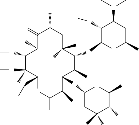
Physiologically active compounds often have emergent properties that are due to the
unique spatial arrangement and interactions of their functional groups. For example, the
macrolactone (macrolide) ring appears to have a hydrophobic and a hydrophilic side in
its low-energy conformations, perhaps accounting for the amphiphilic nature of the mol-
ecule, with the OH at C-6 sticking out on the hydrophilic side. IR spectra of erythromycin
A indicate that there is one OH that is not involved in a hydrogen bond. The x-ray struc-
tures as well as molecular modeling show that the OH on carbon 6 is the only one in the
molecule not involved in an internal hydrogen bond with a neighboring polar functionality
(see Scheme 1.2).
O
OH
OO
R
O
O
O
O
O
OMe
OH
N
O
H
H
O
OO
R
Me
O
O
O
O
O
OMe
OH
N
H
H
O
O
OO
O
O
O
O
O
OMe
OH
N
H
H
O
O
X
DOAc
6
11
12
9
8
10
IA = erythromycin A, R = OH
IB = erythromycin B, R = H
DOAc/D
2
O
HOAc/H
2
O
IIA = ery A enol ether, R = OH
IIB = ery B enol ether, R = H
III = anhydroerythromycin
(spiroketal)
DOAc/D
2
O
100%
D-incorporation
expected
X = 52% H, 48% D
Scheme 1.1

Scheme 1.3 shows that the three secondary hydroxyl groups in erythromycin A (2', 4",
and 11) can readily be differentiated chemically. The most reactive ⫺OH group is on the
desosamine sugar moiety by virtue of the 3'-dimethylamino group acting as an intramo-
lecular catalyst. Thus, erythromycin A can easily be converted to its 2'-acetate (IV) in
dichloromethane by reacting with acetyl chloride and sodium bicarbonate as base, or acetic
anhydride and triethylamine, rendering the amino group nearly two pK units less basic,
due to the neighboring group interaction.
9
Further reaction when DMAP is present leads
to acetylation on the cladinose sugar at the 4"-hydroxyl group, and the hydroxyl group
at C-11 can be acetylated only after heating. The lone pairs of electrons on the oxygen at
C-4" are more readily accessible to the reagents than those at C-11, which is involved in a
hydrogen bond. However, if IV is treated with strongly basic conditions capable of fully
deprotonating an ⫺OH on the molecule, such as sodium hexamethyldisilazide in THF at
⫺78⬚C, and acetic anhydride is added, the ⫺OH at C-11 is preferentially acetylated over
the ⫺OH at C-4". This can be understood by stabilization of the C-11 alkoxide by the
neighboring proton on the C-12 hydroxyl, while an alkoxide at 4" is relatively less stable.
Interestingly, compound V was shown to be a 12,9-hemiacetal by NMR, and in the pres-
ence of water–deuterium oxide mixtures undergoes slow hydrogen–deuterium exchange of
the proton at the hydroxyl at C-6 on the NMR time scale. A similar 12,9-hemiacetal was
reported to be a major contributor to the equilibrium structures of erythromycin A (IA)
itself.
10
Although many scientists were studying these issues, a small group of scientists work-
ing at Taisho Pharmaceuticals in Japan read Abbott’s brief 1971 publication in Experientia
3
and thought of a very simple solution to the acid instability of erythromycin. In a meeting
with Abbott executives in the 1985, the lead Taisho chemist, Dr. Yoshiaki Watanabe, in
somewhat broken English, thanked Abbott for this one-page revelation. The head of the
Abbott delegation then mumbled to his scientists, on his side of the table, something to
the effect that they were never going to publish another (expletive deleted) paper. Thank
O
OO
O
O
O
O
O
O
O
O
N
O
H
H
H
H
O
H
9
11
12
6
2′
4′′
Scheme 1.2 Internal hydrogen bonds of erythromycin A.
A SIMPLE SOLUTION TO A COMPLEX PROBLEM 5

6 FROM PATENT TO PRESCRIPTION: PAVING THE PERILOUS PATH TO PROFIT
goodness he was joking! A partnership was soon born, bridging time and space, and has
fl ourished. Both clarithromycin and azithromycin, discussed below, have achieved annual
sales around $1 billion.
By the amazingly simple idea of blocking the hydroxyl group on carbon 6 with a
methyl group, these Japanese chemists were able to prevent formation of the enol ether
(IIA) or anhydroerythromycin A (III). The compound they fi rst made in 1980,
11
now
sold as clarithromycin VII (Scheme 1.4), not only has superior acid stability, but pro-
duces less stomach irritation, a major drawback to the widespread use of erythromycin
itself. This serendipitous result may be due at least in part to the inability of clarithro-
mycin to form the enol ether (II), which was later shown in animal studies to increase
gastrointestinal motility to a much greater extent than the parent structure. This effect
was seen even after intravenous administration, so it is not simply the result of contact
of the drug with the stomach. Abbott had developed a gastrointestinal motility assay to
screen new drug candidates. Pressure transducers were attached along the outside of
the GI tract in an anesthetized beagle dog, and peristaltic contractions were recorded
after administering an erythromycin analog. Clarithromycin not only demonstrated a
O
OH
OO
O
O
O
O
O
O
OMe
OH
N
O
H
H
O
O
OO
Me
O
O
O
O
O
OMe
OH
N
Ac
Ac
O
O
O
H
H
O
OO
O
O
O
O
O
OMe
OAc
N
H
Ac
O
HO
H
O
6
11
12
9
Erythromycin A 2′-acetate
IV
3′-Dimethylamino group:
2′O-ester: p
K
b
= 7.1;
2′-OH: p
K
b
= 5.2
Ac
2
O, Et
3
N,
DMAP
Na(TMS)
2
,
THF, –78 °C,
Ac
2
O
4′′
2′
Erythromycin 2′,4′′-diacetate (VI)
Erythromycin 2′,11-diacetate-
12,9-hemiacetal (V)
3′
Scheme 1.3
reduction in the recorded contractions relative to erythromycin, but fewer belly aches
were reported in the clinic.
12
Clarithromycin has also improved absorption from the
GI tract and enhanced blood levels, coupled with lower intrinsic minimum inhibitory
concentrations (MICs) against important pathogens. Thus, this second-generation semi-
synthetic macrolide is a better antibiotic overall than the direct fermentation product
from which it is made.
The original synthesis
11
of VII involved protecting both the 2'-OH and the 3'-
dimethylamino functions on the desosamine sugar with benzyloxycarbonyl groups
(Z-groups). This was a method that had been used by many others and results in the loss
of one of the methyl groups on the nitrogen. This step was then followed by simple and
somewhat selective methylation of the 6-OH with methyl iodide. The Z-groups were then
removed by hydrogenolysis, and a methyl group was put back on the nitrogen by reduc-
tive amination with formaldehyde. However, as mentioned above, the mere presence of an
ester functionality on the 2'-OH, such as an acetate, renders the neighboring nitrogen group
much less basic and much less nucleophilic. Therefore, the fi rst process used to prepare
larger quantities of VII was simply to methylate IV, erythromycin 2'-acetate, a compound
that is much more easily prepared and subsequently deprotected. This chemistry is shown
in Scheme 1.5, where each structure is purposely drawn with a different convention taken
from contemporaneous literature, to illustrate how information, even accurate information,
does not always lead to clarity!
13
A highly crystalline product resulted from the meth-
ylation of IV, albeit in low yield, which could be purifi ed suffi ciently for early studies.
In these early studies large supplies of drug were more important than the effi ciency of
the manufacturing process. The only signifi cant impurity was the 6,11-dimethylated com-
pound, similar to what was seen with the more onerous Z-group protection–deprotection
scheme. Dissolving IV in methanol, and allowing the methanolysis to take place at room
temperature overnight, quantitatively removed the acetyl group. It is interesting to realize
O
OO
O
O
O
O
O
O
OMe
OH
N
O
H
H
H
3
CO
H
Scheme 1.4 Clarithromycin (VII).
A SIMPLE SOLUTION TO A COMPLEX PROBLEM 7

8 FROM PATENT TO PRESCRIPTION: PAVING THE PERILOUS PATH TO PROFIT
how close many scientists were to this novel second-generation macrolide when they were
working with the simple 2'-esters of erythromycin many years earlier. This is something
to keep in mind when working with a readily available derivative of an active compound:
What new chemistry can you do with it?
1.3 AN INTRIGUING PATENT PROBLEM
A different solution to the acid instability and erratic blood-level problems of erythromy-
cin was found with another analog. Ironically, this new compound has such low blood
levels that at fi rst it seemed to some researchers that infectious disease physicians would
not trust it. The old paradigm was that an antibiotic had to exhibit blood concentra-
tions above the MIC for the particular strain of bacteria causing the illness. In fact, the
O
OO
O
O
O
O
O
O
OMe
OH
N
O
H
H
O
HO
O
O
H
3
C
OH
OCH
3
CH
3
HO
H
3
C
CH
3
O
O
O
CH
3
CH
3
H
3
C
O
OCH
3
CH
3
OH
CH
3
O
N
CH
3
H
3
CCH
3
HO
O
O
Me
OH
Me
Me
HO
Et
O
O
O
Me
Me
OMe
Me
O
O
H
Me
H
AcO
NMe
2
Me
OH
OMe
Me
room temp.
6
11
12
9
Erythromycin A 2′-acetate
(IV)
3′-Dimethylamino group:
2′O-ester: p
K
b
= 7.1;
2′-OH: p
K
b
= 5.2
NaH in
DMSO–THF,
–5
°C, CH
3
I
4′′
2′
Clarithromycin (VII)
6-
O
-Methyl erythromycin
2′-acetate
3′
methanol,
Scheme 1.5

infectious organism is often compartmentalized in particular tissues such as the tonsils
and the prostate gland. An antibiotic that can penetrate and sustain therapeutic levels in
those diseased tissues would actually be more useful than one that was largely in the blood
serum. This concept is also true of cancer chemotherapy agents, which need to accumulate
in the tumor cells rather than in the bloodstream or healthy tissue. Scientists at Sour Pliva
in Zagreb, in what was then Yugoslavia,
14
and at Pfi zer in Groton, Connecticut,
15
were
able, almost simultaneously and concurrently to solve the tissue penetration problems and
acid instability issues by cleverly adding an additional basic nitrogen atom in a Beckman
rearrangement process followed by reduction. Almost simultaneously—and the resulting
blockbuster drug, azithromycin (VIII), is the subject of two U.S. composition of matter
patents! Although Pliva fi led their patent more than a year earlier, Pfi zer’s patent was issued
seven months sooner. It turns out that the two companies drew their new structures and
named their compounds using quite different conventions. They even numbered the macro-
cyclic ring differently than the classical structures shown in Schemes 1.1, 1.3, and 1.4. Due
to this confusion, the U.S. Patent and Trademark Offi ce thought the groups were claim-
ing two distinctly different compounds. Pliva had pioneered work with the ring-expanded
macrolides,
16
and since they fi led their patent fi rst, Pfi zer had to negotiate the rights to a
compound that it had discovered and patented independently (Scheme 1.6 and Table1.1). In
today’s competitive world, the Patent Cooperation Treaty requires publication of patents 18
months after fi ling, or earlier claimed priority date: for example, from a provisional patent
application. Had this process been in place in the early 1980s, it would have allowed Pfi zer
scientists to see the Pliva application much sooner (the Pliva application would have been
published four months after the Pfi zer fi ling), and the Pfi zer experts would doubtless have
realized that the compounds they both claimed were identical.
AN INTRIGUING PATENT PROBLEM 9
O
N
H
3
C
H
3
C
HO
CH
3
CH
3
HO
H
3
C
C
3
H
5
O
O
OH
O
CH
3
O
O
HO
CH
3
CH
3
CH
3
N(CH
3
)
2
CH
3
O
OH
H
3
C
O
N
O
H
3
C
HO
HO
O
O
HO
O
O
HO
OCH
3
OH
N(CH
3
)
2
11-Methyl-11-aza-4-O-cladinosyl-6-O-desosaminyl-
15-ethyl-7,13,14-trihydroxy-3,5,7,9,12,14-hexamethyl
oxacyclopentadecane-2-one
11
1
1′
2′
1′′
4′′
2
7
6
13
11
2′
4′′
N-Methyl-11-aza-10-deoxo-10-
dihydroerythromycin A
(
a
)(
b
)
Scheme 1.6 (a) U.S. patent 4,517,359, May 14, 1985; (b) U.S. patent 4,474,768, October 2, 1984.

10 FROM PATENT TO PRESCRIPTION: PAVING THE PERILOUS PATH TO PROFIT
The fi rst generic formulations of azithromycin were approved by the FDA on Novem-
ber 14, 2005 for two companies, Teva Pharmaceuticals and Sandoz, to sell this block-
buster drug for a wide variety of indications, permitting Pfi zer and Pliva almost exactly a
20.5-year head start, due to the extension of 3.5 years granted May 20, 1993 for the Pliva
patent. At that time, patents expired 17 years after issuing rather than 20 years from the date
of fi ling, as is now the case (unless extensions are granted).
Note that in Scheme 1.7 azithromycin is drawn in two additional ways, as shown cur-
rently in SciFinder
17
(clockwise numbering) and the Merck Index
18
(counterclockwise
numbering), reversing the order of the sugars. The Physicians’ Desk Reference
19
draws
azithromycin in a fashion related, but not identical, to the Pfi zer patent (Scheme 1.6b),
which depicts the stereochemistry of C-6 as S when in fact it is R. Clarithromycin and
erythromycin are drawn in a format similar to that used in SciFinder, except inverted
(so the numbering runs traditionally counterclockwise); there are some ridiculously long
bonds, so the drawings don’t overlap; and all the necessary centers are reversed to maintain
the correct stereochemistry. It is highly unlikely that anyone has ever looked at structures
presented in that fashion and gained any useful insights. How could anyone be expected to
see the crucial interaction of the C-6 OH group with the carbonyl at C-9 in such a rendi-
tion? [Compare erythromycin (R ⫽ H) in Scheme 1.8b with Scheme 1.1 and 1.2.] The enor-
mous confusion caused by following such diverse conventions when drawing and naming
signifi cant compounds has restricted an understanding of the literature to the few experts
who take the time to become familiar with the structures and conventions. It is fortunate
that modern desktop computer programs can recognize instantly that these structures are
equivalent, calculate empirical formulas, assign stereochemistry, and even predict NMR
spectra.
20
The use of such powerful computer routines, CAS Registry numbers, and other
modern library tools can save time for the expert and be invaluable to the uninitiated. It is
hoped that the confusion over structures such as these will become a relic of the past.
1.4 ANOTHER STRUCTURAL INSIGHT
Recently, it has been widely reported that the new class of wonder drugs called COX-2
inhibitors exhibit serious cardiovascular side effects, and several of these drugs have been
withdrawn from the marketplace. Meanwhile, another class of blockbuster drugs, the statins,
may not only be safe and effective in their intended role of lowering cholesterol, but may
have a plethora of other potentially valuable properties. Cancer, Alzheimer’s disease, dia-
betes, osteoporosis, high blood pressure, multiple sclerosis, and macular degeneration are
Patent Filed 18 months
a
Issued Extended
Sour Pliva,
U.S. 4,517,359
9/22/81 3/22/83 5/14/85 (43.5 months) 5/20/93 (42.2 months)
b
Pfi zer
U.S. 4,474,768
11/15/82 5/15/84 10/2/84 (23 months)
TABLE 1.1 Comparison of Paths to Patents
a
Under current regulations the patents would have been published 18 months after fi ling.
b
1,267 days (3.5 years).

O
OH
OH
CH
3
HO
H
3
C
CH
3
O
O
O
CH
3
H
3
C
O
OCH
3
CH
3
OH
CH
3
O
N
CH
3
H
3
C
CH
3
HO
N
H
3
C
H
3
C
CH
3
O
N
Me
Me
HO
Me
O
Et
HO
Me
Me
O
O
OH
O
O
H
Me
H
OH
NMe
2
OH
Me
Me
OMe
(
R
)
(
R
)
(
R
)
(
R
)(
R
)
(
R
)
(
R
)
(
R
)
(
R
)
(
R
)
(
R
)
(
S
)
(
S
)
(
S
)
(
S
)(
S
)
(
S
)
(
S
)
(
a
)(
b
)
Scheme 1.7 Azithromycin: (a) SciFinder, registry number 83905-01-5; (b) from the Merck Index.
11

O
N
O
H
3
C
H
3
C
HO
CH
3
O
O
HO
H
3
C
CH
3
CH
3
O
O
HO
O
OH
CH
3
H
3
C
CH
3
N
H
3
C
CH
3
CH
3
OH
CH
3
O
O
O
HO
CH
3
OCH
3
CH
3
H
H
CH
3
H
CH
3
HO
H
O
CH
2
CH
3
H
3
C
HO
H
H
3
C
H
3
C
H
O
O
O
OH
N(CH
3
)
2
CH
3
CH
3
OR
H
H
(a)(b)
Scheme 1.8 Structures similar to those in the 2002 Physicians’ Desk References; (a) azithromycin, pp. 2739, 2743,
2748; (b) clarithromycin, R ⫽ CH
3
, pp. 403, and erythromycin, R ⫽ H, pp. 454, 456.
12
among the diseases that the statins may ameliorate.
21
It has often been said that drugs are
discovered in the clinic. In this sense the clinic consists of the patients using these drugs in
the general population. Observational studies on the millions of people taking these drugs
revealed the problems of COX-2 inhibitors and the additional potential indications for the
statins. Another example of this in a much smaller population can serve as an illustration.
In the late 1960s, Pfi zer fi led patents on an α
1
-adrenergic blocker that came to be known
as prazosin.
22
The structure is shown in Scheme 1.9, next to a very similar compound,
terazosin, patented in 1977 by Abbott.
23
Both of these compounds are effective in lowering
blood pressure and have benefi cial effects on the plasma lipid profi le. Dr. Marty Winn, a
chemist at Abbott, looked at a drawing of the structure of prazosin, and realizing that its
failings included problematic intravenous formulation and short duration of action, thought
that a similar molecule with higher water solubility might be more effective. He knew
that furan is only sparingly soluble in water, whereas tetrahydrofuran (THF) is completely
miscible with water. He concluded correctly that simply saturating the furan ring in prazo-
sin might lead to a much more soluble compound. In fact, his fi rst samples were made by
direct hydrogenation of prazosin, leading, of course, to a racemate. This was not a problem
since in those days the FDA did not require compounds to be pure single enantiomers. In
the 1990s, Abbott considered making a new compound as the single enantiomer, a chiral
switch, but did not pursue the issue. It turns out that the base form of terazosin is 25 times
more water soluble than prazosin, and its elimination half-life is about three times greater,
permitting once-daily administration of the new drug. The difference for the correspond-
ing hydrochloride salts is even more dramatic. The terazosin salt is over 500 times more
soluble than the corresponding prazosin salt!
Since terazosin was projected to be a relatively low volume (about a ton per year) high-
potency (10 mg/day) drug, the cost of manufacturing was not deemed a big issue. Note that
one patient would take 3.65 g per year, so 1 metric ton of API is enough to treat 274,000
patients per year. At a price of $1.00 per day, the annual sales would be $100 million. In
the late 1980s a clever process chemist
24
in the pharmaceutical division suggested ways to
streamline the process and save as much as $500,000 per year. Management decided not
to pursue the new chemistry because it was estimated that it would cost over $2 million
to run several successful manufacturing scale batches, place the API on stability studies,
manufacture tablets, put the tablets on stability for a year, fi le all the data with the FDA,
and wait for approval, before being able to switch over to the new process. The FDA would
have to be convinced that the new API made tablets that were identical to those made by the
old process, and the review process could take many months. Little did Abbott realize that
the market for terazosin was about to increase dramatically, so the new process, despite the
costs of implementing it, could have saved them many millions of dollars in manufacturing
the drug over the long term.
Soon after the introduction of terazosin into the marketplace as an antihypertensive, it
was noticed anecdotally that men with symptomatic benign prostatic hyperplasia (BHP)
who were given the drug to treat high blood pressure began reporting relief of their urethral
pressure and bladder outlet problems. Sales of terazosin increased slowly as word got out
of this promising new treatment for BPH, as it was evidently being prescribed for off-label
use. Physicians have the authority to prescribe drugs for conditions other than those pro-
moted by the pharmaceutical companies, so this is quite common: for example, with anti-
cancer drugs. However, Abbott had to conduct costly clinical studies and get FDA approval
to advertise and market the drug for this new use. Once the new indication was approved by
ANOTHER STRUCTURAL INSIGHT 13

N
N
H
3
CO
H
3
CO
NH
2
N
N
O
O
N
N
H
3
CO
H
3
CO
NH
2
N
N
O
O
H
R,S
(a) (b)
Scheme 1.9 (a) Prazosin; solubility of the hydrochloride salt in water (pH ca. 3.5) at ambient temperature (mg/mL): 1.4. (b) terazosin;
solubility in water at 25⬚C (mg/mL): 29.7-hydrochloride salt (mg/mL): 761.2 (544 times more soluble!).
14
the FDA for detailing, an unexciting drug that had been third or fourth tier for hypertension,
selling much less than $100 million per year, became a major seller at about $500 million
per year worldwide. Terazosin was becoming a very profi table drug just as its patents be-
gan to run out. It quickly became a very attractive target for generic drug manufacturers.
Abbott was able to make deals with the generic competitors to keep them off the market
temporarily, extending its very profi table franchise for about four years after the patent ran
out. They paid several companies millions of dollars per month to keep them off the market
with their cheaper generic version of terazosin. However, faced with an antitrust investiga-
tion in 1999, they canceled such arrangements with potential competitors.
25
Abbott’s sales
of terazosin fell 70% in the next year alone, to $141 million.
26
The insight of a chemist who looked at a structural drawing, and spotted the Achilles’
heel of the compound represented, and so elegantly corrected it is astounding. A clearly
drawn chemical structure can reveal the beauty of subtle emergent properties of the func-
tional groups to the astute imagination of a skilled scientist. The fact that this increase in
solubility makes terazosin a superior drug for BPH makes a mark on the positive side of the
ledger of unintended consequences. Serendipity can convert beauty into profi ts!
We should learn never to ignore the clues that any piece of the puzzle is revealing: by
itself or as part of the emerging picture. Each fact that builds toward further understanding
can be exploited, but since our fellow scientists, competitors, or future partners may be far
from us in time and space, our discoveries must be published with clarity. Abbott’s brief
paper in the 1970s paved the way for its future Japanese partner to solve a long-standing
problem, and Pfi zer wisely joined with Pliva when it found success in a research area that
the Yugoslavians had pioneered. Despite the dazzling advances in the tools of modern sci-
ence that you will see in the following chapters, there is no substitute for the wisdom of
good common sense, along with painstaking attention to basic details and the occasional
fl ashes of genius that reveal the true beauty of nature. If we only learn from mistakes, then,
after we have made all the mistakes possible, we will fi nally do things right. Discovering
wisdom from the past is a much more effi cient process!
REFERENCES
1. Leila Abboud and Scott Hensley, Wall Street Journal, September 3, 2003, p. A1.
2. The English philosopher William of Occam (1300–1349) propounded Occam’s razor: “Entities
are not to be multiplied more than necessary.” This is especially appropriate when trying to keep
microorganisms or cancer cells from dividing.
3. Paul Kurath, P. Hal Jones, Richard S. Egan, and Thomas J. Perun, Experientia, 27 (4), 362
(1971).
4. Paul J. Atkins, Tristan O. Herbert, and Norbert B. Jones, International Journal of Pharmaceu-
tics, 30(2–3), 199–207 (1986).
5. T. Cachet, R. Hauchecorne, J. Hoogmartens, G. Van den Mooter, and C. Vinckier, International
Journal of Pharmaceutics, 55(1), 59–65 (1989).
6. Richard J. Pariza, and Leslie A. Freiberg, Pure and Applied Chemistry, 66(10–11), 2365–2358
(1994).
7. Yong-Hak Kim, Thomas M. Heinze, Richard Beger, Jairaj V. Pothuluri, and Carl E. Cerniglia,
International Journal of Pharmaceutics, 271(1–2), 63–76 (2004).
8. Under these conditions, erythromycin B epimerizes to about a 1 : 1 mixture of epimers at C-8 in
a day or two.
REFERENCES 15
16 FROM PATENT TO PRESCRIPTION: PAVING THE PERILOUS PATH TO PROFIT
9. The Merck Index entry for erythromycin (Monograph 03714) gives the pK
a1
value as 8.8; for
erythromycin propionate (Monograph 03719) the pK
a
value is 6.9. Merck & Co., Inc., White-
house Station, NJ, 2001–2005. pK
b
⫽ 14 – pK
a
.
10. J. Barber, J. I. Gyi, G. A. Morris, D. A. Pye, and J. K. Sutherland, Journal of the Chemical Soci-
ety, Chemical Communications, 1040, (1990).
11. Yoshiaki Watanabe, Shigeo Morimoto, and Sadafumi Omura, Novel erythromycin compounds,
U.S. patent 4,331,803, May 25, 1982; fi led May 19, 1981. Merck Index, Monograph 02362,
Merck & Co., Inc., Whitehouse Station, NJ, 2001–2005.
12. Although it is imperative that animal models be validated in the clinic, demonstrating that the
responses in the animal model translate to the human condition, the real proof is often found only
after large populations have taken the drug.
13. During a plenary lecture on his monumental total synthesis of erythromycin at the 1977 ACS
National Organic Symposium in Morgantown, West Virginia, R. B. Woodward had a cartoon
on a slide depicting a tombstone with words to the effect: “Cahn–Ingold–Prelog R.I.P.” He was
annoyed that each time he changed the protecting groups on his intermediates the R, S nomen-
clature changed, so it appeared that stereogenic centers had been inverted when they had not. The
problem of conveniently conveying data by use of various nomenclature conventions has plagued
even Nobel laureates in this fi eld! Thank goodness for computers.
14. Gabrijela Kobrehel and Slobodan Djokic, 11-Methyl-11-aza-4-O-cladinosyl-6-O-desosaminyl-
15-ethyl-7,13,14-trihydroxy-3,5,7,9,12,14-hexamethyloxacyclopentadecane-2-one and deriva-
tives thereof, U.S. patent 4,517,359, May 14, 1985; fi led September 22, 1981.
15. Gene M. Bright, N-Methyl-11-aza-10-deoxo-10-dihydroerythromycin A, intermediates thereof,
U.S. patent 4,474,768, October 2, 1984; fi led November 15, 1982.
16. Gabrijela Kobrehel, Gordana Radobolja, Zrinka Tamburasev, and Slobodan Djokic, 11-Aza-10-
deozo-10-dihydroerythromycin A and derivatives thereof as well as a process for their prepara-
tion, U.S. patent 4,328,334, May 4, 1982, fi led March 28, 1980; cited in ref. 15.
17. SciFinder, registry number 83905-01-5, American Chemical Society, Washington, DC, 2005.
18. Merck Index, Monograph 00917, Merck & Co., Inc., Whitehouse Station, NJ, 2001–2005.
19. 2002 Physicians’ Desk Reference, 56th ed. Medical Economics Company, Inc., Montvale, NJ.
Since the data in the PDR are identical to the package inserts from the drug companies, it is
surprising that Pfi zer would approve an incorrect rendition of its own drug.
20. Programs such as ChemDraw Ultra 9.0 from CambridgeSoft Corporation, Cambridge, MA.
21. Ronald Kotulak, Chicago Tribune, December 25, 2005, p. 1.
22. Hans-Jurgen E. Hess, 2,4,6,7-Tetrasubstituted quinazolines, U.S. patent 3,511,836, May 12,
1970; fi led December 13, 1967.
23. Martin Winn, Jaroslav Kyncl, Daniel Ambrose Dunnigan, and Peter Hadley Jones, Antihyperten-
sive agents, U.S. patent 4,026,894, May 31, 1977; fi lled October 14, 1975.
24. Bruce W. Horrom, who started at Abbott in 1946 and became the longest-serving Abbott em-
ployee ever when he retired with more than 50 years of service. He came up with this new pro-
cess idea after having been at Abbott for over 40 years.
25. New York Times (National Edition), July 23, 2000, p. 1.
26. Pharmaceutical Business News Incorporating Biotechnology Business News, February 7, 2001,
p. 11.

17
2
MEDICINAL CHEMISTRY IN THE NEW
MILLENNIUM: A GLANCE
INTO THE FUTURE
PAUL W. ERHARDT
The University of Toledo College of Pharmacy
Toledo, Ohio
Who of us would not be glad to lift the veil behind which the future lies hidden; to cast a
glance at the next advances of our science and at the secrets of its development during future
centuries?
—David Hilbert in 1900, as quoted recently by R. Breslow
1
2.1 INTRODUCTION
Given the highly interdisciplinary nature of medicinal chemistry and the potential for its de-
ployment across a myriad of future life science research activities, this review seeks to high-
light only those possibilities that stand out upon taking a broad purview of the fi eld’s most
prominent trends. From this vantage point, however, at least a glance will have then been cast
toward some of the more noticeable of the exciting opportunities seemingly in store as me-
dicinal chemistry moves forward into the new millennium.
1,2
An overview of the document’s
several sections is provided below. It lists what topics are covered as well as those that are
not covered and indicates the reasoning behind these choices. The overview also describes
the consistent tone that was sought while attempting to elucidate the numerous technologies
that necessarily become encompassed by the variously highlighted activities.
While initially contemplating how medicinal chemistry might continue to evolve as both
a basic and an applied science, it became apparent that it would fi rst be useful to consider
Drug Discovery and Development, Volume 1: Drug Discovery, Edited by Mukund S. Chorghade
Copyright © 2006 John Wiley & Sons, Inc.
This chapter has been used by permission of the International Union of Pure and Applied Chemistry (IUPAC) with
only minor modifi cation from its original form as published in Pure and Applied Chemistry 74, 703–785 (2002).
18 MEDICINAL CHEMISTRY IN THE NEW MILLENNIUM
where medicinal chemistry has been and how it has come to be what it is today. Thus,
toward quickly establishing a context from which the future might be better appreciated,
and perhaps even seen already to be repeating itself among a new set of players and tech-
nologies, in Section 2.2 a short discourse about medicinal chemistry’s emergence as a for-
malized discipline, its early developments, and its present status is provided by considering
how medicinal chemistry has been practiced across jumps of about 25-year increments.
This section does not include a chronological list of medicinal chemistry’s many contribu-
tions, nor does it highlight the many accomplishments of its noted investigators. Both of the
latter can be found elsewhere as part of more traditional historical treatments.
3
From the backdrop provided in Section 2.2, medicinal chemistry’s near- and longer-
term futures are considered in Section 2.3 relative to several of today’s trends that are
already having a major impact on the drug discovery process. A working defi nition for
medicinal chemistry is recited at the opening of this section so that medicinal chemistry’s
immediate and future roles can be ascertained more clearly. Section 2.3 also sets the stage
for a later consideration of where several drug development topics may be headed in the
near and longer terms.
Gene therapy, vaccines, and biotech-derived therapeutic agents are not discussed in
Section 2.4, which addresses medicinal chemistry’s continued pursuit of effi cacy. The
aforementioned topics reside primarily within the domains of other disciplines. Readers
are, however, encouraged to consult other reviews offered for these areas in order to appre-
ciate how their advances are sure to have a dramatic impact on future life science research
and its interface with medicinal chemistry (e.g., refs. 4 to 6, respectively). Alternatively,
because assessing molecular conformation is such an integral part of practicing medicinal
chemistry along any venue, several aspects of this key topic are considered in Section 2.5.
In particular, the handling of chemical structures in database settings (e.g., chemoinformat-
ics) is discussed in detail.
Several drug development topics are regarded as critical factors that will have a pivotal
infl uence on medicinal chemistry’s continuing evolution in the new millennium. Each of
these topics is addressed briefl y in Section 2.6. These key topics include assuring absorption,
directing distribution, controlling metabolism, assisting excretion, and avoiding toxicity
[i.e., the traditional absorption, distribution, metabolism, excretion, and toxicity (ADMET)
studies that previously have been undertaken by pharmaceutical companies during the
secondary stages of preclinical drug development]. As an important extension of the
ADMET discussions, nutraceuticals considered in parallel with pharmacological synergy
are also addressed in Section 2.6.
Issues pertaining to medicinal chemistry’s future roles in pharmaceutical intellectual
property (IP) and to trends associated with process chemistry are raised in Section 2.7.
With today’s highly publicized emphasis on genomics and proteomics, at least an abbrevi-
ated discourse about process chemistry is included at this juncture so that this fundamental
aspect of medicinal chemistry’s link with synthetic chemistry remains appreciated. Thus,
toward providing just some of such coverage, the unmet need for large-scale stereoselec-
tive synthetic methodologies is discussed briefl y in Section 2.7. As part of this discussion,
an example is cited that concludes by conveying the medicinal chemistry logic that was
encompassed as a critical component of the example’s investigations.
Although it is beyond the scope of this review to discuss the impact that progress in
each of several analytical methods is likely to have on medicinal chemistry,
7
x-ray dif-
fraction has been selected to provide a representative discussion in Section 2.8. As is often
acknowledged by researchers from various disciplines, “science moves forward according
to what it can measure,” and presently, there appear to be numerous promising advances
among various analytical techniques that can be used to study drug–receptor interactions.
For example, readers are encouraged to seek other reviews in order to appreciate the poten-
tial impact that anticipated developments in nuclear magnetic resonance (NMR inclusive of
LC-NMR and high-fl ow-through techniques), mass spectrometry (MS inclusive of LC-MS
and LC-MS/MS), microcalorimetry, and surface plasmon resonance may have on medicinal
chemistry’s future (e.g., refs. 7 to 10, 11 to 13, 14 and 15, and 16 and 17, respectively).
In Section 2.9, practical implications that stem from some of the earlier discussions are re-
visited in what also serves as an overall summary for this document. After restating medicinal
chemistry’s anticipated roles in future life science research, concerns pertaining to the training
of medicinal chemists, inventorship, and the interplay of patent trends and future research within
the context of the IP arena have all been reserved for comment in the concluding summary.
The document’s running dialogue has been developed from future possibilities suggested
by the current medicinal chemistry and drug discovery literature, as well as from general
observations afforded while consulting in both the private and public sectors. Descriptions of
specifi c research projects have been interspersed throughout so that real case examples, along
with their chemical structures, could be conveyed explicitly. A concerted effort has been made
to keep hype to a minimum. Alternatively, jargon has been used whenever it was thought that
such terms portray the mind sets that were important for a given period, or because a par-
ticular term or phrase appears to be taking on an enduring signifi cance. Some of the more
technical of these terms are listed in Table 2.1 along with a short defi nition in each case.
Since several acronyms have been used for repeating phrases, an alphabetical listing of all
acronyms and their defi nitions is provided in Table 2.2 to assist readers as they move deeper
into the document. Numerous references to secondary scientifi c/primary news journals have
been cited because these journals are doing an excellent job of both reporting the most recent
trends and forecasting the potential future. In several cases, a single citation has been used to
list many of the informational Web sites that can often be found for a given topic.
Topics are considered into the future only to about one-half the distance that has been
summarized for medicinal chemistry’s past: namely, for about 75 years, with the fi rst 25
being regarded as near term, and the next 50 being regarded as long term. The speculation
that has necessarily been interjected throughout the review has been done with the thought
that one of the goals for this type of chapter is to prompt the broadest contemplation pos-
sible about the future directions that medicinal chemistry might take. Finally, as medicinal
chemistry is shown to move forward in time, it has been considered as both a distinct pure
science discipline and, equally important, as a key interdisciplinary applied science col-
laborator seeking to mingle with what should certainly prove to be an extremely dynamic
and exciting environment within the life sciences arena of the new millennium.
2.2 PRACTICE OF MEDICINAL CHEMISTRY
2.2.1 Emergence as a Formalized Discipline
Medicinal chemistry’s roots can be found in the fertile mix of ancient folk medicine and
early natural product chemistry: hence its name. As appreciation for the links between
chemical structure and observed biological activity grew, medicinal chemistry began to
emerge about 150 years ago as a distinct discipline intending to explore these relationships
via chemical modifi cation and structural mimicry of nature’s materials, particularly with
PRACTICE OF MEDICINAL CHEMISTRY 19

20 MEDICINAL CHEMISTRY IN THE NEW MILLENNIUM
Term Defi nition
Ab initio calculations Quantum chemical calculations that use exact equations to
account for the complete electronic structure of each atom in a
molecule.
a
AMBER Molecular mechanics program commonly used for calculations
on proteins and nucleic acids.
a
Basis set Set of mathematical functions used in molecular orbital
calculations (e.g., 3-21G*, 6-31G*, or B3LYP when used in
ab initio calculations refer to the type of mathematical function
that was deployed).
a
Bioinformatics Application of computer science and technology to address
problems in biology and medicine.
b
Bioisostere Broadly similar atoms or groups of atoms in terms of
physiochemical or topological properties that can be used as
replacements in a biologically active compound to create a
new structure that retains all selected features of the parent
compound’s biological properties.
c
Chemoinformatics Handling of chemical structure and chemical properties in
database settings; when related to biological properties, this
fi eld becomes a subdivision of bioinformatics.
Combinatorial chemistry Synthesis, purifi cation, and analysis of large sets of compounds
wherein sets of building blocks are combined at one or more
steps during their preparation.
c
Combinatorial library Set of compounds prepared by using combinatorial chemistry.
c
Comparative molecular fi eld
analysis (CoMFA)
A 3D-QSAR method that uses statistical correlation techniques
for the analysis of the qualitative relationships between the
biological activity of a set of compounds with a specifi ed
alignment of their 3D electronic and steric properties.
Additional parameters such as hydrophobicity and hydrogen
bonding can also be incorporated into the analysis.
c
Druglike Structural motifs and/or physicochemical profi les that can be
associated with providing an overall ADME behavior that is
conducive to effective use in humans via the oral route.
Electrostatic potential Physical property equal to the electrostatic energy between the
static charge distribution of an atomic or molecular system
and a positive unit point charge. Used in 3D-QSAR, molecular
similarity assessment, and docking studies.
a
e-Research Research asset management via in-house computer networks and
across the World Wide Web.
Extended Hückel calculations Low-level semiempirical molecular orbital calculations.
a
Gaussian programs Type of mathematical function used during ab initio calculations
to describe molecular orbitals. Numbers refer to year of
program updates.
Genomics Used herein as the study of the chromosomal and
extrachromosomal genes in humans, in particular their
complete sequential characterization.
High-throughput screening (HTS) The level of this activity is moving rapidly from 96- to 384-well
microplates.
436
In silico Tasks undertaken via computer (e.g., virtual screening).
Isosteres Molecules or ions of similar size containing the same number of
atoms and valence electrons.
c
TABLE 2.1 Selected Terms and Abbreviated Defi nitions

Term Defi nition
Ligand-based drug design Design of new structural arrangements based on their
resemblance to at least a portion of another compound that
displays a desired property or biological activity.
Metabophores Structural features residing in substrates that prompt their specifi c
metabolic conversions.
22
MNDO calculations Semiempirical molecular orbital calculations that use a modifi ed
neglect of diatomic (differential) overlap approximation.
a
Molecular mechanics Calculation of molecular conformational geometries and energies
using a combination of empirical force fi elds.
a
Molecular orbital calculations Quantum chemical calculations based on the Schrödinger
equation, which can be subdivided into semiempirical
(approximated) and ab initio (nonapproximated) methods.
a
Multivalent ligands Molecular displays that provide more than one set of binding
motifs for interaction with more than one area on one or more
biological surfaces.
Nutraceuticals Dietary supplements purported to have benefi cial therapeutic or
disease preventive properties (e.g., herbal medicines).
d
Pharmacogenetics Study of genetic-based differences in drug response.
106
Pharmacophores Structural features needed to activate or inhibit specifi c receptors
or enzyme active sites.
Privileged structures Molecular frameworks able to provide ligands for diverse
receptors.
264,265
Prodrug Compound that must undergo biotransformation (e.g.,
metabolism) before exhibiting its pharmacological effects.
c
Proteomics Study of protein structure and function.
438
Quantum mechanics Molecular property calculations based on the Schrödinger
equation that take into account the interactions between
electrons in the molecule.
a
Semiempirical calculations Molecular orbital calculations using various degrees of
approximation and using only valence electrons.
a
Single nucleotide polymorphisms
(SNPs)
Used herein as single-base alterations in the human genome
that occur in specifi ed percentages of distinct portions of the
population.
Soft drug Compound that has been programmed to be biodegraded (e.g.,
metabolized) to predictable, nontoxic, and inactive metabolites
after having achieved its therapeutic role.
c
Structure-based drug design Design of new structural arrangements for use as drugs based
on protein structural information obtained from x-ray
crystallography or NMR spectroscopy.
65,474
Toxicophores Structural features that elicit specifi c toxicities.
481
Transportophores Structural features that prompt a specifi c compound’s transport.
Ultrahigh-throughput screening
(UHTS)
The level of this activity has moved rapidly from 384- to 1536-
well microplates with peak throughput rates of over 100,000
compounds per day. It is estimated that this fi eld will soon be
closing in on producing 1200 data points per minute.
478
Xenobiotic Compound that is foreign to a given organism.
c
a
Adapted from defi nitions provided by the IUPAC.
432
b
Various defi nitions can be found
433
; this particular defi nition has been cited because it appears to be used most
commonly by the U.S. National Institutes of Health (NIH).
434
c
Adapted from defi nitions provided by IUPAC.
435
d
Adapted from defi nitions provided by the U.S. Offi ce of Dietary Supplements (ODS).
437
PRACTICE OF MEDICINAL CHEMISTRY 21
TABLE 2.1 (Continued)

22 MEDICINAL CHEMISTRY IN THE NEW MILLENNIUM
Acronym Designation
ADME Absorption, distribution, metabolism, and excretion
ADMET ADME and toxicity
CADD Computer-assisted drug design
CAS American Chemical Society’s Chemical Abstract Services
CCD Charge-coupled device (as in x-ray area detectors)
CoMFA Comparative molecular fi eld analysis
CPT Camptothecin
CYP Cytochrome P450 metabolizing enzyme
439
FDA U.S. Food and Drug Administration
GI Gastrointestinal tract
HPLC High-performance liquid chromatography
HTS High-throughput screening
IND Investigational new drug
a
IP Intellectual property (e.g., trade secrets and patents)
IT Information technology
KDD Knowledge discovery in databases
440
LC Liquid chromatography
Log P Log value of a compound’s n-octonal/H
2
O partition coeffi cient
MDR Multidrug resistance
MS Mass spectrometry
NBE New biological entity,
441
taken to mean that such a compound has
potential diagnostic, therapeutic, or prophylactic value
NCE New chemical entity, taken to mean that such a compound has potential
diagnostic, therapeutic, or prophylactic value
NDA New drug application
a
NMR Nuclear magnetic resonance spectroscopy
PAC Paclitaxel
Pgp P-glycoprotein pump associated with the ABC class of membrane
transporter systems
PK Pharmacokinetic
QSAR Quantitative structure–activity relationships; note that the Q designation
also becomes applicable to all of the other SXR possibilities
SAbR Structure–absorption relationships
SAR Structure–activity relationships wherein activity is equated herein with
therapeutic effi cacy-related elements from either an agonist or an
antagonist type of interaction
SDM Site-directed mutagenesis
SDR Structure–distribution relationships
SER Structure–excretion relationships
SMR Structure–metabolism relationships
SNP Single nucleotide polymorphism within a genome
SPR Surface plasmon resonance spectroscopy
STR Structure–toxicity relationships
STrR Structure–transporter relationships
SXR Generic representation for simultaneous SAR, SAbR, SDR, SER, SMR,
STR, and STrR
UHTS Ultrahigh-throughput screening
2D Two-dimensional structure representation
3D Three-dimensional structure representation
a
Phrases and acronyms commonly used in the U.S. drug regulatory process.
TABLE 2.2 Acronyms and Designations
an eye toward enhancing the effi cacy of substances thought to be of therapeutic value.
19
In
the United States, medicinal chemistry became formalized as a graduate-level discipline
about 75 years ago within the academic framework of pharmacy education. From this set-
ting, overviews of medicinal chemistry’s subject matter have been offered to undergradu-
ate pharmacy students for many years.
20,21
Understanding structure–activity relationships
(SARs) at the level of inherent physical organic properties (i.e., lipophilic, electronic, and
steric parameters) coupled with consideration of molecular conformation soon became the
hallmark of medicinal chemistry research. Furthermore, it follows that because these fun-
damental principles could be useful during the design of new drugs, applications toward
drug design became the principal domain for a still young, basic science discipline. Per-
haps somewhat prematurely, medicinal chemistry’s drug design role became especially im-
portant within the private sector, where its practice quickly took root and grew rampantly
across the rich fi elds being staked out within the acres of patents and intellectual property
that were of particular interest to the industry.
2.2.2 Early Developments
As a more comprehensive appreciation for the links between observed activity and pharma-
cological mechanisms began to develop about 50 years ago and then also proceeded to grow
rapidly in biochemical sophistication, medicinal chemistry, in turn, entered into what can
now be considered to be an adolescent phase. Confi dently instilled with a new understand-
ing of what was happening at the biomolecular level, the ensuing period was characterized
by the high hope of being able to design new drugs independently in a rational (i.e., ab
initio) manner rather than by relying solely on nature’s templates and guidance for such.
Although this adolescent “heyday of rational drug design”
22
should certainly be credited
with having spurred signifi cant advances in the methods that can be deployed for consider-
ing molecular conformation, the rate of actually delivering clinically useful therapeutic enti-
ties having new chemical structures within the private sector was not signifi cantly improved
for most pharmacological targets unless the latter’s relevant biomolecules also happened
to lend themselves to rigorous analysis (e.g., obtaining an x-ray diffraction pattern for a
crystallized enzyme’s active site with or without a bound ligand). One of the major reasons
that rational drug design fell short of its promise was because without experimental data like
that afforded by x-ray views of a drug’s target site, medicinal chemistry’s hypothetical SAR
models often refl ected speculative notions that were typically far easier to conceive than
were the actual syntheses of the molecular probes needed to assess a given model’s associ-
ated hypotheses. Thus, with only a small number of clinical success stories to relay, medici-
nal chemistry’s “preconceived notions about what a new drug ought to look like” began to
take on negative rather than positive connotations, particularly when being “hand-waved”
within the context of a private-sector drug discovery program (e.g., ref. 23). Furthermore,
from a practical point of view, the pharmaceutical industry, by and large, soon concluded
that it was more advantageous to employ synthetic organic chemists and have them learn
some pharmacology than to employ formally trained medicinal chemists and have them rec-
tify any shortcomings in synthetic chemistry that they might have due to their exposure to a
broader range of nonchemical subject matter during graduate school. Indeed, given the pro-
pensity for like-to-hire-like, the vast majority of today’s investigators who practice medici-
nal chemistry within big pharma, and probably most within the smaller-company segment
of the pharmaceutical industry as well, have academic backgrounds from organic chemis-
try rather than from formalized programs of medicinal chemistry. Although this particular
PRACTICE OF MEDICINAL CHEMISTRY 23

24 MEDICINAL CHEMISTRY IN THE NEW MILLENNIUM
glimpse is important to appreciate as a historical note in that it provides useful insights
for the review’s later discourse about the formalized training of future medicinal chemists,
further references to medicinal chemistry throughout the remainder of this chapter intend to
imply medicinal chemistry’s practice as a discipline regardless of how a given investigator
may have become trained to do so. Importantly, no matter how its practitioners were being
derived, and even though it was still very much under-the-gun within the pharmaceutical
industry, medicinal chemistry did continue to thrive quite nicely during this period and cer-
tainly moved forward signifi cantly as a recognized discipline within all sectors.
Arriving at the next historical segment, however, one fi nds that medicinal chemistry’s in-
ability to accelerate the discovery of new chemical entities (NCEs) by using rational drug de-
sign became greatly exacerbated when the biotechnology rainfall began to hover over the fi eld
of drug discovery just somewhat less than about 25 years ago.
24
With this development, not
only did the number of interesting biological targets begin to rise rapidly, but also, the ability
to assay many of these targets in a high-throughput manner suddenly prompted the screening
of huge numbers of compounds in very quick time frames. Ultimately, the need to satisfy
high-throughput screening’s (HTS’s)
25,26
immense appetite for compounds was addressed not
by either natural product or synthetic medicinal chemistry but by further developments within
what had quickly become a fl ood-level
27
continuing downpour of biotechnology-related break-
throughs. Starting as gene cassette-directed peptide libraries and quickly moving into solid-
phase randomly generated peptide and nucleotide libraries,
28–32
this novel technology soon
spread across other disciplines, eventually spawning the new fi eld of small-molecule combi-
natorial chemistry. Today, using equipment and platforms available from a variety of suppliers
(e.g., Table 2.3), huge libraries of compounds can readily be produced in either a random or a
directed manner.
33–40
Once coupled with HTS, these paired technologies have prompted what
has now come to be regarded as a new paradigm for the discovery of NCEs across the entire
big-pharma segment of the pharmaceutical enterprise.
41–43
Figures 2.1 and 2.2 provide a com-
parison of the old (classical) and new drug discovery paradigms, respectively.
a
As largely reported in 1999 in an article by Brown.
36
b
The number of reported libraries with or without disclosure of their biological
activities has grown from only a few that were produced primarily by the private
sector in the early 1990s to nearly 1000 in 1999, with nearly half of the latest
total now being contributed by the academic sector.
40
Over this same period, the
percentage of libraries directed toward unbiased discovery has gone from about
60% to 20%, while that for targeted/optimization of biased structural systems has
risen from about 40% to 80%.
40
Vendor Web Address
Advanced Chem Tech Peptide.com
Argonaut Technologies Argotech.com
Bohdan Automation Bohdan.com
Charybdis Technologies Charybtech.com
Chiron Technologies Chirontechnologies.com
Gilson Gilson.com
PE Biosystems Pebiosystems.com
Robbics Scientifi c Robsci.com
Tecan Tecan-us.com
Zymark Zymark.com
TABLE 2.3 Suppliers of Combinatorial Chemistry Systems
a
and Compound Library Trends
b

Figure 2.1 Classical drug discovery and development paradigm. This model portrays interactions with
U.S. regulatory agencies and uses terms related to those interactions for steps 11 to 17. All of the other
terms typify generic phrases that have commonly been used by the international pharmaceutical com-
munity. Although some of the noted activities can be conducted in parallel or in an overlapping manner,
the stepwise sequential nature of this paradigm’s overall process is striking. Furthermore, whenever a
progressing compound fails to meet criteria set at an advanced step, the process returns or draws again
from step 3 for another reiteration. Numerous reiterations eventually identify a compound that is able to
traverse the entire process. A successful passage through the entire process to produce just one product
compound has been estimated to require about 15 years at a total cost of about $500 million. While
the largest share of these time and cost requirements occur during the later steps, the identifi cation of a
promising preclinical development compound, step 7, can be estimated to take about four years from the
time of initiating a therapeutic concept. Step 1 is typically associated with some type of physiological
or pharmacological notion that intends to amplify or attenuate a specifi c biological mechanism so as to
return some pathophysiology to an overall state of homeostasis. Step 2 typically involves one or two
biochemical-level assay(s) for the interaction of compounds intending to amplify or attenuate the con-
cept-related mechanism. As discussed in the text, steps 3 and 4 refl ect key contributions from medicinal
chemistry and typically use all sources of available information to provide for compound effi cacy hits
(e.g., everything from natural product surveys to rational approaches based on x-ray diffractions of the
biological target). Step 5 generally involves larger in vitro models (e.g., tissue level rather than biochemi-
cal level) for effi cacy and effi cacy-related selectivity. Step 6 generally involves in vivo testing and utilizes
a pharmacodynamic (observable pharmacologic effect) approach toward compound availability and du-
ration of action. Step 7 typically derives from a formal review conducted by an interdisciplinary team
upon examination of a formalized compilation of all data obtained to that point. Step 8 specifi es parallel
activities that are typically initiated at this juncture by distinct disciplines within a given organization.
Step 9 begins more refi ned pharmacokinetic evaluations by utilizing analytical methods for the drug
itself to address in vivo availability and duration of action. Step 10 represents short-term (e.g., two-week)
dose-ranging studies to identify toxic markers initially within one or more small animal populations.
Expanded toxicology studies typically progress while overlapping with steps 11 to 14. Steps 11 to 13 rep-
resent formalized reviews undertaken by both the sponsoring organization and the U.S. Food and Drug
Administration (FDA). Step 14 is typically a dose-ranging study conducted in healthy humans. Steps 15
and 16 refl ect effi cacy testing in sick patients, possible drug interactions, and so on. Step 17 again refl ects
formalized reviews undertaken by both the sponsoring organization and the FDA. The FDA’s fast-track
review of this information is now being said to have been reduced to an average of about 18 months. It is
estimated that it costs a company about $150,000 for each day that a compound spends in development.
Finally, step 18 represents the delivery of an NCE to the marketplace. (From refs. 41 and 477 to 482.)

26 MEDICINAL CHEMISTRY IN THE NEW MILLENNIUM
2.2.3 Present Status
Interestingly, the marriage of HTS with combinatorial chemistry has led to a situation
where identifying initial lead compounds is no longer considered to be a bottleneck for the
Figure 2.2 New drug discovery and development paradigm. This model portrays interactions with
U.S. regulatory agencies and uses terms related to those interactions for steps 11 to 17. All of the
other terms typify generic phrases that are commonly used by the international pharmaceutical com-
munity. The battery of profi ling included in step 3 represents a striking contrast to the classical drug
discovery paradigm (Fig. 2.1). Furthermore, all of these screens are/will be of a high-throughput
nature such that huge numbers of compounds can be tested simultaneously in extremely short time
periods. As the predictive value of the resulting profi les improve, selected compounds will have
higher and higher propensities to proceed successfully through steps 5, 6, 9, and 10 (Fig. 2.1) to the
point that these assays may become more of a confi rmatory nature or may even be able to be omitted
completely (hence their dashed lines here).
483
The effi ciency of traversing the various clinical test-
ing steps 14 to 16 successfully will also be improved, but their complete removal from the overall
process is highly unlikely. After the initial investments to upgrade step 3 and enough time has passed
to allow for the generation of knowledge from step 3’s raw data results (see text for discussion), the
overall time frame and cost for a single NCE to traverse the new paradigm should be considerably
improved from the estimates provided in Fig. 2.1. Step 1 is likely to be associated with some type of
genomic and/or proteomic derived notion that intends to amplify or attenuate a specifi c biological
mechanism so as to return some pathophysiology to an overall state of homeostasis. Step 2 will be a
high-throughput assay derived from using molecular biology and bioengineering techniques. In step
3a, MC medicinal chemistry. Because step 3a exploits actual pictures of what type of structural
arrangements are needed to interact with the biological targets, this approach toward identifying
new compound hits will continue to operate with high effi ciency. However, because of this same
effi ciency, the biological targets that lend themselves to such experimental depiction (by affording
crystals suitable for x-ray diffraction) are likely to be quickly depleted very early into the new mil-
lennium. Step 3b represents various combinatorial chemistry-derived libraries, natural product col-
lections, and elicited natural product libraries (see text for discussion of this topic). Steps 4 to 18 are
similar to the descriptions noted in Fig. 2.1. (From refs. 41 and 477 to 482.)
overall process of drug discovery and development. Indeed, many of the programs within
pharmaceutical companies are presently considered to be suffering from “compound over-
load,”
44
with far too many initial leads to follow up effectively. ADMET assessments are
now regarded as the new bottleneck, along with the traditionally sluggish clinical and regu-
latory steps. This situation, in turn, has prompted an emphasis to move ADMET-related
parameters into more of an HTS format undertaken at earlier decision points. Thus, even
though effi cacy-related HTS and combinatorial chemistry refl ect very signifi cant incorpo-
rations of new methodologies, from a strategic point of view the most striking feature of the
new drug discovery and development paradigm shown in Fig. 2.2, compared to the clas-
sical approach depicted in Fig. 2.1, actually becomes the trend to place ADMET-related
assays closer to the beginning of the overall process by also deploying HTS methods.
Clearly, with the plethora of biologically based therapeutic concepts continuing to rise
even further and the identifi cation of lead compounds now being much quicker because of
the HTS–combinatorial chemistry approach, more effi cient handling of ADMET-related
concerns represents one of the most signifi cant challenges now facing drug discovery and
development. Because of its importance, this challenge is likely to be resolved within the
near term of the new millennium. Medicinal chemistry’s critical role during this further
evolution of the present drug discovery paradigm is highlighted in subsequent sections.
2.2.4 Examples Involving Site-Directed Mutagenesis
While the drug discovery process has been infl uenced by biotechnology in numerous ways
(e.g., Tables 2.4 and 2.5), one development deserves to be especially noted as this brief
account of medicinal chemistry’s history and present status is brought to a close. This
development is already having a major impact directly on the process of uncovering SARs
relevant to small-molecule drug design.
45
The method involves site-directed mutagenesis
(SDM). SDM applies to systematic point mutations directed toward specifi c sites on genes
that translate to proteins associated with enzyme-active sites or drug receptor systems such
that the targeted changes can be used to study SARs while holding one or more active
site/receptor ligands constant during the assessment (Fig. 2.3). Numerous investigators are
now utilizing this reverse SAR approach to explore both enzyme and receptor ligand inter-
actions. Three examples are provided below wherein the site-directed mutagenesis studies
have been further coupled with one or more of the latest analytical chemistry techniques
such as microcalorimetry, as well as with sophisticated computational approaches. The
fi rst pair of examples involve the active sites of some isolable enzymes; the third example
involves a nonisolable membrane-bound receptor complex.
Slama et al. have conducted studies involving a phosphatidylinositol-4-phosphate phos-
phatase (Scheme 2.1) designated as Sac1p.
46
With this enzyme’s peptide sequence in hand,
transformation of a bacterial host with an appropriate gene copy plasmid has allowed at
least one point-mutated protein to be examined per month in a functional biochemical
assay. While this particular example happens to represent a system that lends itself to over
expression coupled with a functional protein product that lends itself to ready isolation,
it clearly demonstrates that site-directed mutagenesis should no longer be considered to
be lengthy and tedious compared to classical SAR studies undertaken by rational syn-
thetic modifi cations of an enzyme’s substrates. Slama et al.
47
are now studying poly(ADP-
ribose)glycohydrolase or PARG (Scheme 2.1) to more defi nitively assign a tyrosine (i.e.,
796
Tyr) to a key role within this enzyme’s active site. Previously, this particular residue
has only been able to be implicated as being important by using classical inhibitor and
PRACTICE OF MEDICINAL CHEMISTRY 27

28 MEDICINAL CHEMISTRY IN THE NEW MILLENNIUM
photoaffi nity labeling studies.
48,49
In this case, one of the PARG assays deploys micro-
calorimetry measurement of the binding energies for a designated series of ligands that is
then held constant as it is surveyed across the various mutant enzymes.
50
Similarly, Messer
and Peseckis et al. have collaborated in several site-directed mutagenesis studies involving
M1 muscarinic receptors.
51–53
These topographical mapping studies began as a follow-up
Activity Impact
Genomics and proteomics Plethora of new and better defi ned mechanisms
to pursue as therapeutic targets.
High-throughput effi cacy assays Screen huge numbers of therapeutic candidates
in short time frames using low compound
quantities.
High-throughput ADMET assays An evolving development: once validated
and coupled with effi cacy assays, should
eventually allow for selection or drug design/
synthesis of clinical candidate compounds
rather than lead compounds that still require
considerable preclinical testing and additional
chemical tailoring.
Peptide and oligonucleotide compound libraries Provide huge numbers of compounds for
screening (spawned the fi eld of combinatorial
chemistry as now applied to small organic
compounds); can be used as SAR probes and,
pending further developments in formulation
and delivery, may also become useful as drug
candidates.
Site-directed mutagenesis Allows “reverse” structure–activity relationship
explorations.
Transgenic species Novel in vivo models of pathophysiology that
allow pharmacological proof of principle
in animal models that mimic the human
situation; and animal models modifi ed to have
human metabolism genes so as to provide
more accurate PK data and risk assessment.
442
Peptide version of pharmacological prototype Developed to the IND phase as an intravenous
agent can allow for clinical proof of principle
or concept.
Pharmacogenetics An evolving development: should soon
refi ne clinical studies, market indications/
contraindications, and allow for subgrouping
of populations to optimize therapeutic
regimens; eventually, should allow for
classifi cation of prophylactic treatment
subgroups.
TABLE 2.4 Impact of Biotechnology on Small-Molecule Drug Discovery and Development
a
a
This list is not intended to highlight the numerous activities associated with the development of specifi c “biotech”
or large-molecule therapeutics (e.g., see Table 2.5). The arrangement of activities follows the order conveyed in
Figs. 2.1 and 2.2 rather than being alphabetical.

TABLE 2.5 Examples of Approved
a
Biotechnology-Based Drugs
b
Name
c
Company Clinical Use
Tissue plasminogen
activators (tPAs)
Activase
Retevase
Genentech
Boeringher-Mannheim;
Centocor
Dissolution of clots associated
with myocardial infarction,
pulmonary embolism, and
stroke
Clotting factors
BeneFIX Genetics Inst. Treatment of various
hemophilias
KoGENate Bayer
Recombinate Baxter/Genetics Inst.
Ceredase-
glucocerebrosidase
Cerezyme Genzyme Gaucher’s disease
DNAse
Pulmozyme Genentech Cystic fi brosis
Insulins
Humalog Eli Lilly
Humulin
d
Eli Lilly
Novolin Novo Nordisk Insulin-sensitive diabetes
Novolin L Novo Nordisk
Novolin R Novo Nordisk
Erythropoiten-related
growth factors
Epogen
Procrit
Amgen
Ortho Biotech
Anemia associated with renal
failure, chemotherapy,
surgery, or loss of blood
Growth hormone
Biotropin
Genetropin
Humatrope
Neutropin*
Norditropin
Protropin
Saizen
Serostim**
Bio-Tech. Gen.
Pharmacia & Upjohn
Eli Lilly
Genentech
Novo Nordisk
Genentech
Serono Labs
Serono Labs
Growth hormone defi ciency
in children* and for use
in adults and in Turner’s
syndrome**; AIDS-related
wasting and for pediatric
HIV patients
Growth hormone-
releasing factor
Sermorelin Serono Labs Evaluation and use in pediatric
growth hormone defi ciency
Platelet-derived
growth hormone
Regranex Ortho-McNeil Lower-extremity ulcers in
diabetic neuropathy
Fertility hormones
Gonal-F Serono Female infertility
(Continued)
PRACTICE OF MEDICINAL CHEMISTRY 29

30 MEDICINAL CHEMISTRY IN THE NEW MILLENNIUM
Name
c
Company Clinical Use
Interferon-α
Alferon N
Infergen*
Intron**
Roferon-A**
Interferon Sci.
Amgen
Schering-Plough
Hoffman-LaRoche
Genital warts*; chronic
hepatitis C infections**;
several antiviral and
anticancer indications
Interferon-β
Avonex
Betaseron
Biogen
Berlex
Multiple sclerosis
Interferon-γ
Actimmune Genentech Chronic graulomatous disease
Granulocyte-colony and
macrophage-stimulating
factor
Leukine
Neupogen
Immunex
Amgen
Treatment of blood disorders
during chemotherapy and
blood cell/marrow transplants
Interleukins
Neumega
Proleukin*
Genetics Inst.
Chiron
Thrombocytopenia subsequent
to chemotherapy*; certain
cancers
Monoclonal antibodies
CEA-scan Immunomedics Cancer diagnostic
Herceptin Genetech Breast cancer
MyoScint Centacor Cardia imaging agent
Neumega Genetics Inst. Prevention of chemotherapy-
induced thrombocytopenia
OncoScint Cytogen Cancer diagnostic
Orthoclone Ortho Biotech Transplant rejection
Prostascint Cytogen Cancer diagnostic
ReoPro Lilly Prevention of blood clots during
angioplasty
Rituxan Genetech; IDEC Non-Hodgkin’s lymphoma
Simulect Novartis Transplant rejection
Synagis Med Immune Respiratory syncytial virus
disease
Vezluma Boechringer-Ingelheim/NesRx Cancer diagnostic
Zenapax Hoffman-LaRoche; Protein
Design Labs
Transplant rejection
Antisense nucleotides
Vitravene ISIS/Ciba Retinitis
TABLE 2.5 (Continued)
a
Prior to mid-year 1999 and as largely reported in the three-part series published in 1999 by Hudson and Black.
6
In comparison, a more recent survey reveals that about 375 additional agents were already in, or were nearing
entry into, the clinical testing pipeline.
443
b
These compounds have also been referred to as new biological entities (NBEs) by analogy to new chemical enti-
ties (NCEs).
441
In general, NBEs use, recreate, or improve on proteins and other biomolecular polymers produced
in the body to counter disease.
443
c
Asterisks in this column relate to those in the “clinical use” column.
d
First recombinant-DNA-produced therapeutic protein to enter the market.
6

to better defi ne the receptor interactions exhibited by CDD-0102 (Scheme 2.2), a selec-
tive M1 agonist that is undergoing preclinical development for the potential treatment of
Alzheimer’s disease.
54–56
Interestingly, these investigator’s more recent efforts have addi-
tionally turned toward addressing questions about the multivalent nature of M1 muscarinic
receptors.
57–60
Thus, in this case the site-directed mutagenesis studies are helping to re-
solve fundamental issues about the nature of these receptors, as well as helping to implicate
amino acid residues involved in the binding of agonists for the purpose of identifying drug
structural themes having enhanced selectivity for certain of the receptor subtypes. In all of
the M1-related studies, the site-directed mutagenesis results are being coupled to computa-
tional studies directed toward mapping the receptor’s key topological features.
61
2.2.5 Latest Trends
As has been noted by others, the deployment of these new approaches to studying SARs
represents an “exciting”
62
development that has clearly had an invigorating effect on the
practice of medicinal chemistry in many areas. Alternatively, that the SAR hallmark and
Series of Potential Receptor Ligands Explored
as the Experimental Variable
Classical SAR
Protocol
Wild Type
Receptor
Series of Point Mutated Receptors
Explored as the Experimental
Variable
SDM SAR Protocol
(Reverse SAR Protocol)
Selected Receptor
Ligand(s) Held Constant
Figure 2.3 Classical versus site-directed mutagenesis (SDM) exploration of structure–activity
relationships (SARs).
PRACTICE OF MEDICINAL CHEMISTRY 31

32 MEDICINAL CHEMISTRY IN THE NEW MILLENNIUM
drug design intellectual domains of medicinal chemistry, along with chemically servicing
the experimental approaches toward identifying novel, lead structures, have all been over-
run by technologies initially derived from other disciplines, has also had somewhat of an
unnerving impact on medicinal chemistry. This is because it has previously been the nearly
exclusive deliverance of these roles from a chemical orientation that has served to distin-
guish medicinal chemistry as a distinct discipline. Thus, as a fi eld, medicinal chemistry has
had to mature quickly from its troubled adolescence only to fi nd itself in the middle of an
identity crisis. This crisis has been occurring among both its private-sector practitioners
Scheme 2.1 Cleavages effected by Sac1p and PARG. 1, Phosphatidylinositol-4-phosphate, wherein
R and R' are fatty acid side chains; 2, poly(ADP-ribose), wherein Ad is adenine and Glu is glucose. In
both cases, arrows indicate bonds undergoing enzymatic hydrolyses. (From refs. 46 to 49.)
Scheme 2.2 Suspected interactions of CDD-0102 with muscarinic M1 receptors. 3, 5-(3-Ethyl-
1,2,4-oxa-diazol-5-yl)-1,4,5,6-tetrahydropyrimidine (CDD-0102) shown as its protonated species;
Asn, asparagine 382; Asp, aspartic acid 105; Thr, threonine 192. Dashed lines signify suspected
hydrogen bonds. (From refs. 51 to 53).
and its academicians. Indeed, today’s trend within the public-sector funding arena, with a
major emphasis on genomics and proteomics, is causing some academic medicinal chemis-
try and chemistry investigators to turn their intellectual pursuits further and further toward
molecular biology.
63
Similarly, the undergraduate instruction of pharmacy students, which
has for so long represented medicinal chemistry’s academic bread and butter, has shifted
its emphasis away from the basic sciences toward more of a clinically oriented curriculum.
A fi nal development that has contributed to medicinal chemistry’s identity crisis is the fact
that the new drug discovery paradigm supplants medicinal chemistry’s long-standing posi-
tion wherein its practitioners have typically been regarded as the primary inventors of the
composition of matter specifi cations associated with NCE patent applications. This last
development, along with the overall IP arena of the future, is addressed in Section 2.9.
Although as alluded to earlier and as now practiced in tandem with SDM studies, clas-
sical medicinal chemistry rationale is still being relied on heavily to effectively identify
and fi ne-tune lead compounds for systems whose biomolecules have lent themselves to
x-ray diffraction and/or NMR analyses (i.e., structure-based drug design),
64,65
medicinal
chemists of the new millennium must be prepared to face the possibility that the present
complement of these more amenable pharmacological targets is likely to become quickly
exploited, perhaps eventually even exhausted. Such a scenario, in turn, suggests that this
last stronghold for today’s practice of rational medicinal chemistry could also be lost as a
bastion against what could then potentially become an even more serious identity crisis in
the future.
Present trends thus leave us with the following paradigm for the immediate future of
small-molecule drug discovery (Fig. 2.2):
1. Genomics and proteomics will continue to uncover numerous new pharmacological
targets, to the extent that choosing the most appropriate and validating such targets
among the many therapeutic possibilities will also continue to rise as a growing chal-
lenge in itself.
2. Biotechnology will, in turn, continue to respond by generating quick ligand-
identifi cation assays for all new targets chosen to be pursued: namely, by deploying
HTS protocols.
66
3. Targets that lend themselves to x-ray diffraction and structure-based drug design are
likely to be quickly exploited.
4. Ligands for HTS will be supplied by existing and new combinatorial-derived com-
pound libraries as well as from wild
67
and biotechnology-elicited natural sources.
5. Assessment of ADMET parameters, presently considered to be the bottleneck for the
overall drug discovery process (Table 2.6), will continue to move toward HTS modes
that can be placed at earlier and earlier positions within the decision trees utilized to
select lead compounds for further development as drugs.
It is important to emphasize at this juncture that in order to place confi dence in the
predictive value of ADMET HTS surveys, these particular screens must become validated
relative to actual clinical-related outcomes. For the moment, however, this situation is
best likened to a deep, dark chasm that the rapidly evolving ADMET HTS surveys still
need to traverse if they are ultimately to become successful. Nevertheless, because of its
importance, it is proposed that not only will this type of validation be forthcoming within
the near term, but as the new millennium then continues into the more distant future,
PRACTICE OF MEDICINAL CHEMISTRY 33

Activity Estimated Time Frame Percentage Successfully Traversing Associated Criteria
Biological conception A plethora of genomic/proteomic characterizations presently
lies waiting to be exploited; this situation is expected to
prevail well into the new millennium.
The challenge lies in prioritizing which of the numerous
mechanisms might be best to pursue (see next entry).
Proof of therapeutic principle Ultimately requires reaching phase II clinical testing;
BIOTECH-derived humanized and/or transgenic disease
state models may be able to be substituted at an earlier point,
depending on the confi dence associated with their validation.
Generally high, although there are some distinct therapeutic
categories that continue to have low success rates or lack
defi nitive validation, such as the attempted treatments of
septic shock or the pursuit of endothelin modulators.
Identifi cation of lead
compound based on effi cacy
screen
Using HTS, thousands of compounds can be tested in a
matter of days or less (10 to 100 times more with UHTS);
companies are beginning to have more lead compounds than
they can move forward in any given program.
One compound out of 5000 from random libraries/one out of
10 from directed libraries; despite the low effi ciency, this is
not regarded as a bottleneck because HTS can be done so
quickly; much higher percentages can be obtained during
ligand- and structure-based drug design, but synthesis is then
correspondingly slower.
Progression to preclinical
development compound
a
Approximately two years. About one out of 50 wherein all can be examined during the
time frame indicated.
Progression to clinical
development compound
a
Approximately two years. About one out of 10 wherein all can be examined during the
time frame indicated.
Phase I clinical study
b
Approximately one year.
c
About one out of two.
Phase II clinical study
b
Approximately two years.
c
About one out of two.
Phase III clinical study Approximately three years.
c
About one out of 1.5 and often with modifi ed labeling details.
Product launch Approximately two years (NDA submission/approval). About one out of 1.5.
d
TABLE 2.6 Assessment of Drug Discovery and Development Bottlenecks
a
Presently regarded as the bottleneck for the overall process. These are the points where ADMET properties have historically been assessed. Approximately 40% are rejected owing to poor
pharmacokinetics and about another 20% because they show toxicity in animals. In the new drug discovery paradigm (Fig. 2.2), the ADMET assessments are being moved to an earlier point
in the overall process and are being conducted in an HTS mode. However, in most cases, validation of the new methods relative to clinical success still needs to be accomplished.
b
Although these studies may not be able to be accomplished any quicker, they may be able to be done more effi ciently (e.g., smaller numbers and focused phenotypes within selected patient
populations) and with a greater success rate based on making the same improvements in the ADMET assessment area as noted in footnote a.
c
Timing includes generation and submission of formal reports.
d
About one of fi ve compounds entering into clinical trials becomes approved. The overall process to obtain one marketed drug takes about 12 to 15 years at a cost of about $500 million.
Source: Refs. 444 to 449.
34
ADMET-related parameters derived from HTS will become even further manipulated for
their potential to allow for synergistic relationships within the overall course of a given
therapeutic or prophylactic treatment. These intriguing future possibilities for exploiting
ADMET-related parameters in a proactive and synergistic manner rather than in just a
negative fi ltering mode, along with the likely move toward prophylactic medicines, are
discussed further in subsequent sections.
2.3 EVOLVING DRUG DISCOVERY AND DEVELOPMENT PROCESS
2.3.1 Working Defi nition for Medicinal Chemistry
If the targets that are readily amenable to x-ray diffraction and structure-based drug design
do become exhausted with time, the only title role presently highlighted for medicinal
chemistry within the new paradigm of drug discovery will also disappear. To determine if
medicinal chemistry will still be operative under such a circumstance, let us fi rst review
medicinal chemistry’s present defi nition. Perhaps further attesting to medicinal chemistry’s
present identity crisis, however, is the fact that a purview of several of today’s major medic-
inal chemistry texts reveals that although one can fi nd topic-related versions of such within
the context of various other discussions, even the textbooks seem reluctant to provide an
explicitly stated general defi nition for medicinal chemistry (e.g., refs. 68 to 73). Turning,
instead, to this document’s earlier consideration of medicinal chemistry’s history, and for
the moment disengaging ourselves from any biases that might be interjected by overreact-
ing to the continuing fl ood of biotechnology-related trends, it does become possible to
devise a general, working defi nition for medicinal chemistry that can be used to address its
present-day identity crisis while also serving in a search for any key roles that medicinal
chemistry ought to be playing now and into the future of life science research.
As a working defi nition for this review, let us simply state that medicinal chemistry uses
physical organic principles to understand the interaction of small molecular displays with
the biological realm. Physical organic principles encompass overall conformational con-
siderations, chemical properties, and molecular electrostatic potentials, as well as distinctly
localized stereochemical, hydrophilic, electronic, and steric parameters. Understanding
such interactions can provide fundamental, basic knowledge that is general as well as com-
pound-specifi c in its applications directed toward either enhancing the overall profi le of a
certain molecular display or designing an NCE (e.g., by effecting small molecule-driven
perturbations of discrete biological processes or of overall biological pathways for the
purpose of eliciting a specifi ed therapeutic endpoint). Small molecular displays should
be thought of in terms of low-molecular-weight compounds (e.g., usually less than 1 kg)
that are typically of a xenobiotic origin and thus not in terms of biotechnology-derived
polymers. While the latter are being addressed aggressively by other fi elds, it should ad-
ditionally be noted that a consideration of specifi c details associated with the interaction
of small molecular portions of more complex biomolecular systems still falls within the
purview of medicinal chemistry’s stated focus. Alternatively, the biological realm should
be thought of very broadly, so as to encompass the complete span of new ADMET-related
systems as well as the traditional span of biological surfaces that might be exploited for
some type of effi cacious interaction. The technologies that can be deployed as tools to
study these interactions at medicinal chemistry’s fundamental level of understanding are,
by intent, dissociated from medicinal chemistry’s defi nition. Presently, such tools include
EVOLVING DRUG DISCOVERY AND DEVELOPMENT PROCESS 35

36 MEDICINAL CHEMISTRY IN THE NEW MILLENNIUM
biotechnology-related methods such as site-directed mutagenesis, combinatorial chemistry
methods provided that the latter are coupled with knowledge-generating structural data-
bases (later discussion), and long-standing synthetic chemistry manipulations that can be
conducted in a systematic manner on either one of the interacting species in order to ex-
plore SARs. In the future, medicinal chemistry should be equally prepared to exploit what-
ever new tools and techniques that become available to allow it to proceed more effi ciently
along the lines of its technology-independent defi nition. Finally, it should be appreciated
that this defi nition merges both the basic and applied natures of medicinal chemistry’s sci-
entifi c activities into a key mix of endeavors for which a new research paradigm (Fig. 2.4)
has also been proposed recently as being a signifi cant trend,
74
even if potentially “danger-
ous” in that it could compromise the longer-term pursuit of fundamental knowledge by
bringing applied science decision criteria into funding programs that have previously sup-
ported pure, basic science.
75
2.3.2 Immediate- and Long-Term Roles for Medicinal Chemistry
Importantly, no matter what pace targets amenable to x-ray may continue to allow struc-
ture-based drug design to be pursued in the new millennium, applying the aforementioned
defi nition across the present drug discovery paradigm reveals that there is an even more
vital activity with which medicinal chemistry needs to become more involved. Indeed,
as HTS results are amassed into mountains not just for effi cacy data but for each of the
ADMET parameters as well, it should ultimately become medicinal chemistry’s role, by
defi nition, to attempt to understand and codify these awesome, crisscrossing ranges if such
data are to be merged and used either to select or design the most promising preclinical de-
velopment compounds. For example, while medicinal chemistry’s principles and logic may
not be needed to identify hits or leads from a single HTS effi cacy parameter survey across a
library of potential ligands, or perhaps not even needed for two or three of such consecutive
surveys involving a few additional ADMET-related parameters, it is extremely doubtful
that the same series of compounds identifi ed from within an initial library as a hit subset,
Figure 2.4 Nonlinear relationship of medicinal chemistry to basic and applied research. Surveys
suggest that chemical pharmaceutical companies spend about 9%, 37%, and 54% of their research
dollars on basic, applied, and developmental aspects of research, respectively.
484
(Adapted from a
fi gure provided by Cotton
75
as part of his summary and commentary about a book entitled Pasteur’s
Quadrant.
74
)
or as further generated within a directed library based on the initial hit subset, will be able
to sustain themselves as the most preferred leads upon continued HTS parameter surveys
in the future when the latter become ramped up to their full potential. Furthermore, this
should still be the case even if the downstream selection criteria become more and more
relaxed through any of such progressions that ultimately strive to merge even a preliminary
HTS-derived ADMET portfolio in conjunction with one or more selective effi cacy HTS
parameter surveys. In other words, identifi cation of the optimal end product (i.e., the best
preclinical candidate compound) is unlikely to be derivable from an experimental process
that does not represent a “knowledge”
76,77
generating system that also allows for rational-
based assessments and adjustments, or even complete revamping, to be interspersed at
several points along the way. In the end, today’s move toward “focused libraries”
78,79
and
“smarter,” presorted relational databases may thus represent a lot more than just the often-
touted desire to “be more effi cient.”
80
Indeed, this may be the only way for the new drug
discovery paradigm, now in its own adolescent phase, eventually to work as it continues
to mature and to take on more ADMET-related considerations in an HTS format. In this
regard it can now be emphasized that the common denominator required to correlate the
HTS data from one pharmacological setting to that of another ultimately resides in the
precise chemical structure language that medicinal chemistry has been evolving since its
emergence as a distinct fi eld (i.e., SAR defi ned in terms of physical organic properties dis-
played in three-dimensional space). This, in turn, suggests that within the immediate future
of the new millennium it should be medicinal chemistry that rises to become the central
interpreter and distinct facilitator that will eventually allow the entire new drug discovery
paradigm to become successful.
This central role for medicinal chemistry may become even more critical longer term
(i.e., for the next 50 to 75 years of the new millennium). Speculating that the new drug
discovery paradigm will indeed mature within the next 25 years into a synergistic merger
of effi cacy and thorough ADMET HTS systems that allows for an effective multiparameter
survey to be conducted at the onset of the discovery process, the accompanying validated
predictive data that will have been generated over this initial period should be statistically
adequate to actually realize today’s dream of virtual or in silico screening
81–86
through vir-
tual compound and virtual informational libraries (not just for identifying potential effi cacy
leads to synthesize, as is already being attempted, but across the entire preclinical portion
of the new paradigm wherein the best overall preclinical candidate compound is selected
with high precession for synthesis at the outset of a new therapeutic program) (Fig. 2.5).
However, this futuristic prediction again depends on the entire maturation process being
able to proceed in a knowledge-generating manner. Again central to the latter is medicinal
chemistry as the common denominator. For example, with time it can be expected that just
as various pharmacophores and toxicophores have already been identifi ed for various por-
tions of the biological realm associated with effi cacious or toxic endpoints, respectively,
specifi c molecular properties and structural features will become associated with each of
the ADME behaviors. Indeed, work toward such characterizations is already progressing in
all of these areas (Table 2.7). Understanding the pharmacophores, metabophores,
22
toxico-
phores, and so on, in terms of subtle differences in molecular electrostatic potentials (from
which medicinal chemistry’s physical organic properties of interest are derived), as well
as in terms of simple chemical structural patterns, will eventually allow for identifying
optimal composites of all of these parameters across virtual compound libraries as long as
the latter databases have also been constructed in terms of both accurate three-dimensional
molecular electrostatic potentials and gross structural properties.
EVOLVING DRUG DISCOVERY AND DEVELOPMENT PROCESS 37

38 MEDICINAL CHEMISTRY IN THE NEW MILLENNIUM
It should be clear that in order to play this key role effectively, the medicinal chem-
ist of the new millennium (Fig. 2.6) will have to remain well versed in physical organic
principles and conformational considerations while becoming even more adept at applying
them within the contexts of each of the ADMET areas, as has previously been done during
Figure 2.5 Future drug discovery and development paradigm. This model portrays interactions
with U.S. regulatory agencies and uses terms related to those interactions for steps 11 to 17. Future
implies about 50 to 75 years into the new millennium. The most striking feature of this paradigm
compared to Figs. 2.1 and 2.2 is the considerable number of decisions that will be made from virtual
constructs rather than from experimental results. Confi dence in the virtual decisions will be directly
proportional to the level of knowledge that is learned from the huge amounts of drug screening data
being amassed during the fi rst 25 to 50 years of the new millennium, coupled with the overall abil-
ity to predict clinical outcomes. Step 1 is likely to be associated with some type of genomic and/or
proteomic derived notion that intends to amplify or attenuate one or more specifi c biological mecha-
nisms so as either to return some pathophysiology to an overall state of homeostasis or to modify
some system in a manner that prevents or provides prophylaxis toward an otherwise anticipated
pathophysiological development. A growing emphasis of treatments will be directed toward preven-
tion. Step 2 may be based on an actual x-ray diffraction version of the biological target or on a com-
putationally constructed version derived from similar known systems that have been catalogued for
such extrapolations. In either case, docking studies will be conducted in a virtual mode. Steps 3, 4,
and 5 will be conducted in a virtual mode. Steps 6 and 7 represent the fi rst lab-based activities. After
submission of patents, it is proposed that in vivo testing involving steps 9 and 10 will be able to take
advantage of project-specifi c and FDA-approved generic toxicity model transgenic species. Steps 11
to 18 are similar to those in Figs. 2.1 and 2.2, except that the likelihood for a compound to fall short
of the desired criteria will be signifi cantly reduced. Subject inclusion/exclusion criteria will also be
much more refi ned based on advances in the fi eld of pharmacogenetics.

ADME Area Parameters
Absorption Physicochemical (e.g., log P
312,450
)
Functional groups (e.g., rule of fi ve
311
)
Distribution Two-directional fl ows across Caco-2 cell monolayers
451
Metabolism 2D structure-metabolism databases
452
(e.g., Metabol
Expert,
453
META,
454
Metabolite,
455
and Synopsis
Metabolism Database
456
) 3D models (e.g., CYP1A,
457
CYP2A,
458
CYP2B,
459
CYP2C,
560
CYP2D,
461,462
and
CYP3A
463
Excretion (half-lives, etc.) Structural patterns across species
464
Physicochemical (e.g., log P
465,466
)
TABLE 2.7 Examples of Efforts to Establish ADME-Related Structural Patterns
a
a
It is anticipated that the ADME informational area will soon become heavily inundated as pharmaceutical com-
panies begin to share their rapidly accumulating data through publications in the public domain (analogous to
what has happened historically for receptor/enzyme active site effi cacy information but with the latter having
previously had to transpire in a much slower fashion without the arrival of today’s HTS methodologies).
Figure 2.6 Practice of medicinal chemistry (MC) in the new millennium. The most striking dif-
ferences from the long-standing practice of MC are (1) data reduction of huge amounts of rapidly
derived HTS biological results, (2) greater emphasis on multitechnique chemical structure consider-
ations, and most important, (3) the simultaneous attention given to all of the ADMET-related param-
eters along with effi cacy and effi cacy-related selectivity (E/S) during lead compound selection and
further design or enhancement, coupled with an expanding knowledge base that offers the possibility
for achieving synergistic benefi ts by taking advantage of various combinations of multiagent, pro-
drug, soft drug, and/or multivalent drug strategies.
40 MEDICINAL CHEMISTRY IN THE NEW MILLENNIUM
medicinal chemistry’s pursuit of distinct effi cacy-related biochemical scenarios. Because
they will continue to represent part of the basic thrust of medicinal chemistry, the pursuit
of effi cacy into the new millennium and the assessment of molecular conformation are
each considered further in the next two sections relative to medicinal chemistry’s working
defi nition. Similarly, because the nature of the ADMET area’s maturation is regarded as
being pivotal toward shaping medicinal chemistry’s evolution in the new millennium, each
ADMET area is covered within a separate, fi fth section. Although space limitations dictate
that these topics can be highlighted only in an abbreviated manner, a somewhat longer and
more technically oriented discourse is provided for the molecular conformation section
because it is envisioned that this area will constitute the alphabet for the universal language
that medicinal chemistry will help to elaborate in order to bridge and ultimately unite all
of the other areas.
2.4 PURSUING EFFICACY
From an experimental point of view, medicinal chemistry’s pursuit of the effi cacy dis-
played by small molecules in a direct manner probably won’t change much in the near
term beyond what has already been proposed in terms of logical extensions from some
of the latest trends. For example, even the intriguing directions that are encompassed by
chemical genomic
87
strategies, where in some cases a small molecule may provide effi cacy
in an indirect manner through its interactions with biochemically modifi ed genomic and
proteomic signaling systems,
88–90
still reduce to being able to exploit the same fundamental
principles already cited within medicinal chemistry’s defi nition so as to achieve the specifi c
interaction with the modifi ed biological surface, which then serves as a mediator toward
effi cacy.
In terms of targets, bioinformatics will certainly associate pathophysiology and in-
dividual variation with useful genomic and proteomic information so as to maintain the
plethora of traditional and novel pharmacological targets well into the millennium.
91–98
Web-based public efforts
99
and commercial databases
100
coupled with desktop programs
101
are already being positioned to make such information readily available to everyone. The
ability of infectious microorganisms and viruses to evolve into resistant forms at a pace
at least equal to our ability to produce NCE chemotherapeutic agents can also be counted
on to continually provide new targets.
102,103
Alternatively, longer term into the new mil-
lennium, gene therapy will hopefully have eradicated many of today’s targets that derive
strictly from hereditary, gene-based abnormalities.
4
Somewhat along these same lines, the
ongoing characterization of single nucleotide polymorphisms (SNPs) and the further pur-
suit of individually tailored therapies, as presently being promoted by the new fi eld of
pharmacogenetics, may also contribute toward some new targets.
104–111
Web-based public
and commercial databases in the pharmacogenetic
112
and specifi c SNP areas,
113
along with
commercial sources for the latter’s experimental technologies,
114
are also making this type
of information generally available. In the near term, the fi eld of pharmacogenetics is likely
to have its fi rst major impacts on refi ning clinical studies for late-stage preclinical can-
didates and on developing improved indications, contraindications, and dosage regimens
for marketed drugs and for compounds undergoing clinical study. Longer term, however,
pharmacogenetics should become instrumental in shaping the overall nature and subtleties
of the effi cacy targets that are pursued rather than the numbers of new targets that relate
to novel mechanisms that might be deployed. For example, although one can speculate
that the present trend to pursue curative rather than palliative treatments will continue for
at least the short term, it is likely that in the longer term a growing emphasis will be
placed on preventative rather than on either palliative or curative treatments.
115,116
Toward
this end, pharmacogenetics should become of central importance, owing to its potential to
divide recipient populations into distinct treatment subgroups based on their predisposition
profi les coupled with their general ADMET-related drug handling profi les, wherein both
sets of criteria may eventually become accessible before or shortly after birth using gene-
based assays and to a lesser extent, administration of diagnostic probe molecules.
117,118
Depending on the variability of an individual’s environmental exposures, it can be imag-
ined that in the future, pharmacogenetic profi ling will be done at routinely scheduled inter-
vals through the entire course of one’s life. Regardless of the number and nature of future
pharmacological targets, advances in biotechnology can be expected to continue to fl ood
the overall life sciences arena and to get even better at deriving the required HTS assays.
Alternatively, HTS microengineering,
119
it would seem, may have to level off at about
9600-well (or well-less) tests per plate or, perhaps, move to other platforms involving chip
or bead technologies.
120
2.4.1 Gathering Positive, Neutral, and Negative SARs During HTS
As mentioned, HTS effi cacy hits per se can certainly be pursued without the aid of me-
dicinal chemistry. Indeed, one can imagine that with one or more compound libraries
already in hand from an automated synthesis,
121
and the areas of robotics
122–124
and labo-
ratory information management or LIMS
125
also continuing to evolve rapidly, HTS in
the brute force mode may be able to proceed without any signifi cant human intervention,
let alone without the need for an interdisciplinary group of investigators from a variety
of disciplines. However, as has been emphasized, if the new drug discovery paradigm
is ultimately to become successful, this type of screening will need to be accompanied
by structure-associated knowledge generation and assessment, with the latter being con-
ducted using the rationale and logic that can only be interjected by human intervention.
Furthermore, even though it could run the risk of placing medicinal chemistry into a fall-
guy position somewhat analogous to its earlier adolescent phase, it should also be noted
that according to the working defi nition cited herein, as soon as any knowledge assess-
ments become at all sophisticated in terms of molecular structure and biological proper-
ties, they quickly fall right into the middle of the domain of medicinal chemistry and its
distinct area of expertise.
In this regard, it becomes important to briefl y review some aspects of SARs that would
be worthwhile to include within the database assemblies that are currently being drawn up
to handle the mountains of data already arriving from today’s HTS programs. One can pre-
dict that once ADMET profi ling by HTS is validated in the future, it will become extremely
valuable for a knowledge-generating paradigm to be able to discern not just the most active
compounds within an effi cacy database and to be able to compare their structural patterns
to those in another database, but to also be able to fl ag the regions on compounds that can
be altered with little effect on the desired biological activity as well as those areas that are
intolerant toward structural modifi cation. The neutral areas, in particular, represent ideal
points for seamless merging of one set of a database’s hits with that of another regard-
less of the degree of pattern overlap, or for further chemical manipulation of a hit so as to
PURSUING EFFICACY 41
42 MEDICINAL CHEMISTRY IN THE NEW MILLENNIUM
adjust it to the structural requirements defi ned by another data set that may be so distant in
structural similarity space that attempted overlap or pattern recognition routines are oth-
erwise futile. The regions that are intolerant of modifi cation represent areas to be avoided
during knowledge-based tailoring of an effi cacy lead. Alternatively, the intolerant regions
represent areas that can be exploited when attempting to negate a particular action (e.g.,
metabolism or toxicity). An actual example of utilizing both neutral and negative SARs to
advantage is provided below to further illustrate how these types of data sets might also be
deployed simultaneously by future medicinal chemists (albeit with signifi cantly stepped-
up complexity) as more and more parameters become added to the process of early lead
identifi cation/optimization.
2.4.2 Example Involving Multidrug Resistance of Anticancer Agents
The investigations to be exemplifi ed in this case have been directed toward studying the
SAR associated with biological transporter systems with the hope of establishing a data-
base of transportophore relationships that might be generally applicable toward enhanc-
ing the selection and/or development of effi cacy leads from any other type of data set.
To provide immediate relevance to this long-term project, it has initially focused on the
P-glycoprotein pump (Pgp)
126,127
that is associated, in part,
128,129
with the development
of multidrug resistance (MDR)
130,131
during cancer chemotherapy. Pgp is a 170-kD trans-
membrane glycoprotein that serves as an energy-dependent unidirectional effl ux pump
having broad substrate specifi city. In humans it is encoded by the MDR gene, MDR1,
whose classical phenotype is characterized by a reduced ability to accumulate drugs intra-
cellularly, and thus the deleterious impact of Pgp activity on cancer chemotherapy.
132–136
By way of practical example, the cytotoxicity of paclitaxel (PAC) is decreased by nearly
three orders of magnitude when breast cancer cell lines become subject to MDR, largely
via a Pgp mechanism.
137
In order to explore a series of probes that will systematically span
a specifi ed range of physicochemical properties when coupled to the PAC framework, one
needs fi rst to identify a region on PAC that is tolerant toward such modifi cation in terms
of PAC’s inherent effi cacy (i.e., where changes are known not to signifi cantly alter PAC’s
cytotoxicity toward nonresistant breast cancer cells). Scheme 2.3 provides a summary of
the accumulated SAR data obtained from the PAC-related review literature,
138–142
wherein
it becomes clear that several positions along the northern edge represent neutral areas that
can lend themselves toward such an exploration.
Inhibitors of Pgp have already been identifi ed by several different investigators, and
these types of compounds belong to a class of agents referred to as chemosensitizer drugs
for which there are a variety of mechanisms.
143,144
Although Pgp inhibitors can be coad-
ministered with a cytotoxic agent in order to negate MDR toward the latter when studied
in cell culture, to date these types of chemosensitizers have not fared well clinically.
145
One of the reasons that the inhibitors have not fared well is that they must compete with
the accompanying cytotoxic agent for access to the Pgp MDR receptors. Thus, it can
be imagined that if an SAR can be identifi ed that is unfavorable for binding with Pgp
MDR receptors, and furthermore, that if such a negative transportophore could be incor-
porated onto the original cytotoxic agent in a neutral position, the cytotoxic agent might
itself avoid MDR or at the very least become better equipped to do so in the presence
of a coadministered Pgp MDR inhibitor (Fig. 2.7).
146
Toward this end, initial studies
are being directed toward exploring the possibility that it may be feasible to identify
negative SAR that is undesirable to the Pgp system within the specifi c chemical context

of PAC by manipulating the latter at neutral positions that do not signifi cantly affect
PAC’s inherent effi cacy. To ascertain the generality toward potentially being able to place
such a negative transportophore onto other established chemotherapeutic agents and onto
lead compounds being contemplated for preclinical development, an identical series of
negative SAR probes is being examined within the context of a completely different mo-
lecular scaffold: that of the camptothecin (CPT) family of natural products for which
topotecan represents a clinically useful anticancer drug.
147
CPT, accompanied by a sum-
mary account of its SAR-related literature,
138,147–170
is depicted in Scheme 2.4, wherein
it becomes clear that the 7- and 9-positions represent the key neutral areas in CPT that
might be manipulated analogously to those in PAC. Since these two compounds have
very different molecular templates and owe their cytotoxicities to two distinctly different
mechanisms (i.e., PAC largely, but not exclusively
171–173
to overstabilization of microtu-
bules
174,175
and CPT largely, but not exclusively,
176
to “poisoning” of topoisomerase I
177
),
and because topotecan, a clinically deployed CPT analog (Scheme 2.4), is at the lower
end of the spectrum in terms of being subject to Pgp-related MDR (it loses about one or-
der of magnitude from its initial potency
137
), taken together these two molecules represent
an excellent pair to examine the generality of the transportophore-related SAR fi ndings.
Other molecular scaffold systems and biological testing models can also be imagined so
as to extend such Pgp investigations into the areas of drug absorption, uptake into hepatic
tissue (drug metabolism), and passage across the blood–brain barrier (e.g., for either en-
hancing or attenuating drug penetration into the CNS). Additional transporters within the
ABC class can be explored systematically using a similar approach.
Although this particular example refl ects a rational SAR strategy, the same types of
informational endpoints can certainly be achieved via a coupled HTS/combinatorial
Scheme 2.3 Overall SAR profi le for paclitaxel-related compounds. This summary represents a
consolidation of SAR information contained in several review articles.
138–142
Note the tolerance for
structural modifi cation along the northern hemisphere of the taxane ring system. Paclitaxel has R ∅
and R′ CH
3
CO.
PURSUING EFFICACY 43

44 MEDICINAL CHEMISTRY IN THE NEW MILLENNIUM
Scheme 2.4 Overall SAR profi le for camptothecin-related compounds. This summary represents a
consolidation of SAR information contained in several primary references.
138,147–170
Note the toler-
ance for structural modifi cation along the northern hemisphere of the overall molecule, especially
when approaching the western edge. Camptothecin, the parent natural product, has R
1
R
2
H.
Topotecan, used clinically, has R
1
OH and R
2
CH
2
N(CH
3
)
2
.
Panel A
Panel B
Panel D
Panel C
Drug
Drug
Drug
Drug
Membrane Pgp
Transporter System
Cancer Cell
+
Chemosen-
sitizer
Chemosen-
sitizer
Figure 2.7 Diminishing the Pgp transporter’s role in multidrug resistance (MDR). Panel A: Extru-
sion of chemotherapeutic agents (drug) by cancer cells upon overexpression of the Pgp transporter
system is one mechanism associated with MDR. Panel B: Coadministration of a chemotherapeutic
agent and a chemosensitizing agent wherein the latter preferentially interacts with Pgp, thus dimin-
ishing the extrusion of the desired drug. The chemosensitizer may block the pump competitively
(wherein it becomes extruded) or noncompetitively. Panel C: Administration of a modifi ed che-
motherapeutic agent (drug) that retains its desired effi cacy-related properties but has a diminished
affi nity for the Pgp system. Panel D: Coadministration of a modifi ed chemotherapeutic agent and a
chemosensitizing agent such that the latter has a better chance of interacting with the Pgp system
relative to the desired chemotherapeutic agent.
chemistry approach, provided that the chemical structure components within the resulting
databases are initially constructed with architectures fl exible enough to allow for such
queries. Likewise, while this example refl ects a simple query between two different bio-
logical behaviors, the same types of queries can be conducted across multiple databases
for multiple parameters. That the next 25 years of medicinal chemistry will involve a
considerable amount of making sense out of such multiple parameter correlations based
on experimentally derived data is quite clear. That the next 50 to 75 years might then be
able to be fruitfully spent in more of a virtual correlations mode is certainly more specula-
tive but is, at least, probably reasonable provided that we can build our knowledge base
and fundamental understanding of how the various parameters, as assessed in isolation
according to the HTS scenarios described above, interact simultaneously within the entire
system.
2.4.3 Compound Libraries: Example of Working with Nature to Enhance
Molecular Diversity
Before closing this section, two additional trends need to be mentioned. The fi rst involves
the likelihood that industry will even more heavily embrace site-directed mutagenesis as an
additional component of its efforts to identify lead compounds. Such modifi cation can be
contemplated during the initial development of an HTS assay and then used to contribute
to the defi nition of an overall pharmacophore as the latter is probed via various compound
testing paradigms. The importance of gaining a thorough appreciation for the overall phar-
macophore rather than for just identifying distinct lead structures is discussed in later sec-
tions. The second trend in the effi cacy arena that is also likely to become very important in
the future can be illustrated by an example from a different research program. Instead of
focusing on ADMET-related parameters during rational drug design, this program involves
the chemical or library side of the new drug discovery paradigm. In particular, this exam-
ple seeks to enhance molecular diversity
178,179
along phytochemical structural themes that
have shown activity of either a toxic
180
or a promising nature during initial effi cacy screen-
ing such that having related compound libraries would be highly desirable. As described
below, this particular example has a certain appeal in its ultimate practicality since it seeks
initially to produce directed molecular diversity within common plants indigenous to the
midwestern United States. This possibility is being explored by exposing plants simul-
taneously to both an elicitor (botany’s designation for an inducing agent) and selected
biochemical feedstocks. For example, the biochemical pathways leading to the anticancer
phytoalexins from soybeans shown in Scheme 2.5 may be able to be elicited by soybean
cyst nematode infections to produce a more diverse family of active principles when grown
in environments containing biochemically biased nutrients.
181,182
Toward this end, it has
been established that the statistical reproducibility of HPLC-derived phytochemical con-
stituent fi ngerprints from soybean controls is adequate to discern real fl uctuations in these
types of natural products.
183
Work is now progressing toward ascertaining the differences
that result upon exposures of soybeans to various stimuli and feedstocks. Interesting results
will be followed-up by studying the genetic control of the involved pathways. In this re-
gard it should be noted that an opposite approach that leads to similar combinatorial bio-
synthesis
184
endpoints is also being undertaken by various other groups, particularly with
an interest toward the production of proteins and peptide families from plant systems.
185
In those studies, directed modifi cation of the genetic regions controlling one or more es-
tablished phytochemical pathways is fi rst effected, and then these types of biotechnology
PURSUING EFFICACY 45

46 MEDICINAL CHEMISTRY IN THE NEW MILLENNIUM
interventions are followed up by characterization of the altered biocombinatorial expres-
sion products.
2.5 ASSESSING AND HANDLING MOLECULAR CONFORMATION
2.5.1 Chemoinformatics
Given the exponential proliferation of technical data and our increasing ability to dissemi-
nate it rapidly through a vast maze of electronic networks, it is no wonder that new systems
capable of “managing and integrating information”
186
are regarded among “the most im-
portant of the emerging technologies for future growth and economic development across
the globe.”
186,187
That information technology (IT), in turn, is now receiving high prior-
ity in all sectors is quite clear,
188–195
particularly with regard to systems directed toward
integrating bioinformatic-related information as promoted via the World Wide Web.
196
Scheme 2.5 Elicitation of directed and novel natural product families from soybean. Panel A: Nor-
mal biochemical pathways leading to the fl avones (5) to the key anticancer isofl avones genistein and
diadzein, wherein phenylalanine is fi rst converted to cinnamic acid, p-coumaroyl CoA, and fi nally to
key intermediate 4, naringenin chalcone. Panel B: Feeding unnatural starting materials such as the
aryl-substituted phenylalanine and cinnamic acid derivatives shown as 6 and 7, respectively, under
circumstances where this pathway is also being elicited by external stimuli (e.g., soybean cyst nema-
tode infections), could be expected to produce new fl avone derivatives (8), isofl avonic derivatives (9),
or completely novel natural product families. This diagram of the phytochemical pathway leading to
fl avones and isofl avones represents a composite of several references.
485–487
An array of inexpensive
analogs related to 6 and 7 is available from commercial sources.
OH
CO
2
H
NH
2
CO
2
H
COSCoA
HO
OH
OH
OH
O
OH
HO
O
HO
O
HO
O
HO
O
HO
O
O
OH
OH
(OH)
CO
2
H
NH
2
CO
2
H
HO
O
(OH)
O
R
R
R
R
(OH)
HO
O
(HO)
O
or or
Panel A
4
5
Genistein
Daidzein
6 7 8 9
Panel B
or
or
Comlpletely
Novel Natural
Product
Families
Medicinal chemistry’s contributions toward this sweeping assessment of the future im-
portance of IT reside primarily in the area of handling chemical structures and chemical
information, a specialized exercise complicated enough to merit its own designation as a
new fi eld, that of chemoinformatics.
197–200
In this regard, the increasing use of databases
to link chemical structures with biological properties has already been alluded to in terms
of both real experimental data sets and virtual compilations. Although serious strides are
being taken in this area, however, there is a signifi cant need for improvement in the han-
dling of chemical structures beyond what is suggested for the immediate future by what
now appears to be occurring within today’s database assemblies. For example, that “better
correlations are sometimes obtained by using two-dimensional displays of a database’s
chemical structures than by using three-dimensional displays” only testifi es to the fact that
we are still not doing a very good job at developing the latter.
201
How medicinal chemistry
must step up and rise to the challenges already posed by this situation in order to fulfi ll
the key roles described for its near- and longer-term future is addressed in the next several
paragraphs of this section.
Assessment of molecular conformation, particularly with regard to database-housed
structures, represents a critical aspect of chemoinformatics. While new proteins of inter-
est can be addressed reasonably well by examining long-standing databases such as the
Protein Data Bank
202
and other Web-based resources
203
for either explicit or similar struc-
tural motifs and then deploying x-ray (pending a suitable crystal), NMR, and molecu-
lar dynamic/simulation computational studies
204–209
as appropriate, the handling of small
molecules and of highly fl exible molecular systems in general remains controversial.
210
As alluded to above, the only clear consensus is that treatments of small molecules for
use within database collections “have, to date, been extremely inadequate.”
211
Certainly,
a variety of automated three-dimensional (3D) chemical structure drawing programs are
available that can start from simple 2D representations by using Dreiding molecular me-
chanics or other, user-friendly automated molecular mechanics-based algorithms, as well
as when data are expressed by a connection table or linear string.
212
Some programs are
able to derive 3D structure “from more than 20 different types of import formats.”
213
Fur-
thermore, several of these programs can be directly integrated with the latest versions of
more sophisticated quantum mechanics packages, such as Gaussian 98, MOPAC (with
MNDO/d), and extended Hückel.
200,212
Thus, electronic handling of chemical structures,
and to a certain extent comparing them, in 3D formats has already become reasonably well
worked out.
197–200,212–215
Table 2.8 provides a list of some of the 3D molecular modeling
products that have become available during the 1990s.
Nevertheless, a fundamental problem remains: how the 3D structure is derived ini-
tially in terms of its chemical correctness based on what assumptions might have been
made during the process. Further, there are still challenges associated with how readily
3D structure information can be linked with other, nonchemical types of informational
fi elds. As has been pointed out by others, the reason that such mingling of data fi elds
often does not afford good fi ts is because “each was initially designed to optimize some
aspect of its own process and the data relationships and structures are not consistent.”
197
At this point, inexpensive Web-based tools that can integrate chemical structure data
with other types of information from a variety of sources, including genomic data, have
already begun to emerge.
216
This trend will continue to pick up into the new millen-
nium, and because of its overall importance for bioinformatics, user-friendly solutions
are likely to arrive early on.
ASSESSING AND HANDLING MOLECULAR CONFORMATION 47

Package Company Platform Description
Low-end sophistication
Nano Vision ACS Software Mac Simple, effective tool for viewing and rotating structures,
especially large molecules and proteins
Ball & Stick Cherwell Scientifi c Mac Model building and visualization; analysis of bond distances,
angles
MOBY Springer-Verlag IBM (DOS) Model building and visualization; classical and quantum
mechanical computations; large molecules and proteins;
PDB fi les
Nemesis Oxford Molecular IMB (Windows), Mac Quick model building and high-quality visualization;
geometry optimization (energy minimization)
CSC Chem. 3D/Chem 3D
Plus
Cambridge Scientifi c Mac Easy-to-use building and visualization; geometry
optimization; integrated 2D program and word processing
Alchemy III Tripos Assoc. IBM (DOS, Windows), Mac Quick model building; energy minimization; basic
calculations; easy integration to high-end systems
PC Model Serena Software IBM (DOS), Mac Low cost with sophisticated calculations; platform fl exibility
Midrange sophistication
CAChe Tektronic Mac Sophisticated computation tools; distributed processing
HyperChem Auto Desk IBM (Windows), Silicon
Graphics
Easy-to-use array of computation tools (classical and
semiempirical quantum mechanics)
Lab Vision Tripos Assoc. IBM (RISC-6000), Silicon
Graphics, DEC, VAX
Sophisticated but practical modeling for research
High-end sophistication
SYBL Tripos Assoc. IBM (RISC-6000), Silicon
Graphics, DEC VAX,
Sun 4, Convex
Integrated computation tools for sophisticated structure
determination and analysis; database management
CERIUS Molecular Simulations Silicon Graphics, IBM
(RISC-6000), Stardent
Titan
Suite of high-performance tools for building and simulating
properties
TABLE 2.8 Three-Dimensional Molecular Modeling Packages That Became Available During the 1990s
Source: Ref. 214.
48

2.5.2 Obtaining Chemically Correct 3D Structures
Unfortunately, the quick assignment of chemically correct 3D structures may not be read-
ily solvable. Recalling from the fi rst sections of this chapter, medicinal chemistry has
been concerning itself with this task for quite some time. Medicinal chemistry’s interest in
chemical structure is further complicated, however, by the additional need to understand
how a given drug molecule’s conformational family behaves during its interactions with
each of the biological environments of interest. For example, as a drug embarks on its
“random walk”
217
through the biological realm (Fig. 2.8), the ensuing series of interactions
have unique effects on each other’s conformations
218,219
at each step of the journey and not
with just the step that fi nally consummates the drug’s encounter and meaningful relation-
ship that is struck with its desired receptor/active site.
To track such behavior in a comprehensive manner, it becomes necessary to consider
a drug’s multiple conformational behaviors by engaging as many different types of con-
formational assessment technologies as possible, while initially taking an approach that
is unbiased by any knowledge that may be available from a specifi c interacting environ-
ment. For example, the three common approaches depicted in Fig. 2.9 include (1) x-ray
(itself prone to bias from solid-state interactions within the crystal lattice); (2) solution
spectroscopic methods (i.e., NMR), which can often be done in both polar and nonpolar
media (this technique, however, being more limited by the amount of descriptive data that
it can generate); and (3) computational approaches that can be done with various levels
of solvation and heightened energy content (limited, however, by the assumptions and
approximations that need to be taken in order to simplify the mathematical rigor so as to
allow solutions to be derived in practical computational time periods). Analogous to the
simple, drawing program starting points, programs are also available for converting x-ray
ASSESSING AND HANDLING MOLECULAR CONFORMATION 49
Figure 2.8 Random walk taken by an oral drug on route to its point of effi cacious contact within a hu-
man target cell. This continuum of interactions between a drug and various biological surfaces within the
human biological realm is typically divided into categories associated with ADMET and effi cacy. Biolog-
ical milieu marked with an asterisk represent compartments having particularly high metabolic capabili-
ties. Blood is notably high in esterase capability. In the future, medicinal chemists will utilize knowledge
about ADMET-related SARs to more effectively identify the best drug leads and to further enhance the
therapeutic profi les of selected compounds. (The phrase “random walk” is taken from ref. 217.)

50 MEDICINAL CHEMISTRY IN THE NEW MILLENNIUM
and NMR data into 3D structures (e.g., ref. 220). While such a three-pronged approach
is not new,
221
it is emphasized herein because today’s medicinal chemistry literature sug-
gests that some investigators still fall into the single technique trap from which further
extrapolations of data are then sometimes made with great conviction. This may be be-
cause it is often diffi cult to obtain an acceptable crystal for x-ray analysis, have adequate
solubility for high-fi eld conformational analysis by NMR, or perhaps, to become aligned
with appropriate computational expertise and computing power. At any rate, practical
advances in all three of these areas can be expected to alleviate such implementation-
related shortcomings so that the medicinal chemist of the near-term future will be more
readily able to consider structures from at least a three-pronged starting point either in-
dependently or through collaboration with other specialists and experts dedicated to each
of these areas. A real example that serves to further illustrate how entry structures might
be handled and matured using a computational approach is provided below. This prob-
lem pertains to the consideration of structures to be placed in a human drug metabolism
database.
221
2.5.3 Infl uence of Biological Environments: Example Involving Drug Metabolism
This example is also cited in Chapter 9 of this text. Structures are initially considered as
closed-shell molecules in their electronic and vibrational ground states, with protonated
and unprotonated forms, as appropriate, also being entered. If a structure possesses tau-
tomeric options or if there is evidence for the involvement of internal hydrogen bonding,
the tautomeric forms and the hydrogen-bonded forms are considered additionally from
the onset. Determination of 3D structure is carried out in two steps. Preliminary geometry
optimization is affected by using a molecular mechanics method. For example, in this case
the gas-phase structure is determined by applying the MacroModel 6.5 modeling package
running on a Silicon Graphics Indigo 2 workstation with modifi ed (and extended) AMBER
parameters also being applied from this package. Multiconformational assessment us-
ing systematic rotations about several predefi ned chemical bonds with selected rotational
Figure 2.9 Techniques employed to assess conformational detail. X-ray diffraction requires a
suitable crystal, and its results are subject to solid-state interactions. Computational paradigms are
most accurate when done at the highest levels of calculation, but these types of calculations become
computer-time intensive. NMR requires that the molecule be soluble in the chosen solvent and that
adequate compound supplies be available. Mass spectrometry is also becoming an important tool for
larger molecules, although it provides smaller amounts of descriptive data. A composite of all ap-
proaches provides for the best possible assessment of molecular conformation.
angles is then conducted to defi ne the low-energy conformers and conformationally fl ex-
ible regions for each starting structure. In the second step, the initial family of entry struc-
tures are subjected to ab initio geometry optimizations, which in this case use a Gaussian
98 package running on a T90 machine housed in a state-level supercomputer center re-
source. Depending on the size of the molecule, 3-21G* or 6-31G* basis sets
223
are used for
conformational and tautomeric assessments. Density functional theory using the B3LYP
functional
224
is applied for the consideration of exchange correlation energy while keeping
the required computer time at reasonable levels. The highest-level structure determination
is performed at the B3LYP/6-31G* level. To ascertain the local energy minimum character
of an optimized structure, vibrational frequency analysis is carried out using the harmonic
oscillator approximation. Determination of vibrational frequencies also allows for obtain-
ing thermal corrections to the energy calculated at 0 K. Free energies are then calculated at
310 K (human body temperature). The latter values become particularly important for cases
where structural (conformational or tautomeric) equilibria occur.
From the calculated relative free energies, the gas-phase equilibrium constant and the
composition of the equilibrium mixture can be directly determined. Although these values
may not be relevant in an aqueous environment or in the blood compartment, the calculated
conformational distribution is relevant for nonpolar environments such as may be encoun-
tered when a drug passively traverses membranes or enters the cavity of a nonhydrated
receptor/enzyme active site just prior to binding. Repetition of this computational scheme
from biased starting structures based on actual knowledge of the interacting biological
systems or from x-ray or NMR studies (particularly when the latter have been conducted
in polar media), followed by studies of how the various sets of information become in-
terchanged and how they behave when further raised in energy, complete the chemical
conformational analysis for each structure being adopted into the human drug metabolism
database.
As mentioned earlier, after taking an unbiased structural starting point, medicinal chem-
istry needs especially to consider structures (and the energies thereof) by ascertaining
what their relevant conformations might be during interactions within various biological
milieus. It can be imagined that at least within the immediate future, a useful range of such
media to be considered will include aqueous solutions at acidic and neutral pH, namely at
ca. 2 (stomach) and 7.4 (physiological), respectively; one or more lipophilic settings, such
as might be encountered during passive transport through membranes; and fi nally, specifi c
biological receptors and/or enzyme active site settings that are of particular interest. Impor-
tantly, with time this list can then be expected to grow further so as to also include several
distinct environmental models deemed to be representative for interaction with various
transportophore relationships; several distinct environmental models deemed to be relevant
for interaction with specifi c metabophore relationships such as within the active site of a
specifi c cytochrome P450 metabolizing enzyme; and fi nally, several distinct environmental
models deemed to be relevant for interaction with specifi c toxicophore relationships. It
should also be appreciated that the interaction of even just one ligand within just one of the
various biological settings could still involve a wide range of conformational relationships
wherein the biological surface may also exist as an equilibrium mixture of various confor-
mational family members. If x-ray, NMR, and so on, can be deployed further to assess any
one or combination of these types of interactions, a composite approach that deploys as
many as possible of these techniques will again represent the most ideal way to approach
future conformational considerations within the variously biased settings. Advances to-
ward experimentally studying the nature of complexes where compounds are docked into
ASSESSING AND HANDLING MOLECULAR CONFORMATION 51
52 MEDICINAL CHEMISTRY IN THE NEW MILLENNIUM
real and model biological environments are proceeding rapidly in all of these areas, with
MS
11–13
and microcalorimetry
14,15
now adding themselves alongside x-ray (later discus-
sion) and NMR
225,226
as extremely useful experimental techniques for the study of such
SARs. Besides the experimental approaches, computational schemes will probably always
be deployed because they can provide the relative energies associated with all of the differ-
ent species. Furthermore, computational methods can be used to derive energy paths to get
from the fi rst set of unbiased structures to a second set of environmentally accommodated
conformations in both aqueous media and at biological surfaces. Importantly, these paths
and their energy differences can then be compared along with a direct comparison of the
structures themselves, while attempting to uncover and defi ne correlations between chemi-
cal structure and some other informational fi eld within or between various databases.
Finally, it should be noted that by using computational paradigms, these same types of
comparisons (i.e., among and between distinct families of conformationally related mem-
bers) can also be made for additional sets of conformational family members that become
accessible at appropriately increased energy levels (i.e., at one or more 5 kcal/mol incre-
ments of energy) to thus address the benefi cial losses of energy that might be obtained
during favorable binding with receptors or active sites.
227
These types of altered conforma-
tions can also become candidates for structural comparisons between databases. The latter
represents another important refi nement that could become utilized as part of SAR queries
that will need to be undertaken across the new effi cacy and ADMET-related parameters
of the future. With time, each structural family might ultimately be addressed by treating
the 3D displays in terms of coordinate point schemes or graph theory matrices.
228
This is
because these types of methods lend themselves to the latest thoughts pertaining to utiliz-
ing intentionally fuzzy coordinates
229,230
[e.g., x ± x', y ± y', and z ± z' (rather than just x, y,
and z plots)] for each atomic point within a molecular matrix wherein the specifi ed varia-
tions might be intelligently derived from the composite of aforementioned computational
and experimental approaches. Alternatively, the fuzzy strategy might become better de-
ployed during the searching routines, or perhaps both knowledgeably fuzzy data entry and
knowledgeably fuzzy data searching engines handled, in turn, by fuzzy hardware,
231
will
ultimately best identify the correlations that are being sought in any given search paradigm
of the future. It should be noted, however, that for the fuzzy types of structural treatments,
queries will be most effective when the database has become large enough statistically
to rid itself of the additional noise that such fuzziness will initially create. An ongoing
example that serves to demonstrate the value of considering the dynamic energy relation-
ships associated with molecular trajectories as well as the more static conformational dis-
plays for a particular molecular interaction of interest is provided below.
2.5.4 Dynamic Energy Relationships: Example Involving a Small Ring System
As a different aspect of the aforementioned MDR-related anticancer chemotherapeutic pro-
gram, an effort has been directed toward replacing the complex scaffold of PAC (Scheme
2.3) with a very simple molecular format that still displays PAC’s key pharmacophoric
groups in the appropriate 3D orientations purported to be preferable for activity.
232–234
Toward this end, initial interest involved defi ning the role of the β-acetoxyoxetane system,
particularly when the latter is adjacent to planar structural motifs. Since such systems are
rather unique among natural products
235
as well as across the synthetic literature,
236–238
it
fi rst became necessary to study their formation within model systems relevant for this proj-
ect. 2-Phenylglycerol was synthesized
239
and deployed as a model to study the molecule’s

conformation by x-ray, NMR, and computational techniques as a prelude to affecting its
cyclization.
240
The energy differences that result as the molecule is reoriented so as to be
lined up for the cyclization were also calculated. Finally, once properly oriented in 3D
space, the energy required to actually traverse the S
N
2 reaction trajectory between the 1
and 3 positions was calculated (one of which positions utilizes its oxygen substituent for
the attack while the other relinquishes its oxygen as part of a leaving group). The synthesis
of 2-phenylglycerol and the pathway and energies associated with the intermediate species
and cyclization process to form the oxetane are summarized in Scheme 2.6.
Not surprisingly, given the strained-ring nature of this system, the energy needed to
effect the ring closure from the lowest of three closely related local minima conforma-
tions belonging to a family common to the independent x-ray and computationally de-
rived starting points was about 28 kcal/mol. What becomes interesting, however, is that
within this particular system, nearly half of this energy requirement results from the need
to disrupt hydrogen bonds in order to reorient the molecule initially into a conformation
ASSESSING AND HANDLING MOLECULAR CONFORMATION 53
Scheme 2.6 Synthesis of 2-phenylglycerol and investigation of its conversion to 3-hydroxy-3-
phenyloxetane. 2-Phenylglycerol (10) is depicted so as to convey the lowest-energy structure of the
three close local minima observed during ab initio calculations performed at the HF/6-31G* level.
TS1 and TS2 represent transition conformers obtained after the indicated bond rotations, while 11
represents the desired 3,3-disubstituted oxetane sytem. The respective relative energies in kcal/mole
for 10, TS1, and TS2, along with the product oxetane (11), are as follows: 0.00, 13.6, 12.4, and 28.2.
(From refs. 239, 240, and 488.)
54 MEDICINAL CHEMISTRY IN THE NEW MILLENNIUM
appropriate for the reaction. Despite the resulting strain that becomes placed upon the
overall system’s bond angles, the actual movement of the relevant atoms along the reaction
trajectory (Scheme 2.6, dashed line) then accounts for only slightly more than one-half
of the total reaction energy. Therefore, from a synthetic point of view the results suggest
that it should be benefi cial to employ a hydrogen bond acceptor solvent that has a high
boiling point, the fi rst property assisting in disruption of the hydrogen bonds that need
to be broken for conformational reorientation and the second property allowing enough
thermal energy to be added conveniently to prompt progression across the reaction trajec-
tory. That such favorable conformational perturbations could indeed be achieved simply by
deploying these types of solvents was then confi rmed by reexamination of the independent
results obtained from our initial NMR studies conducted in polar protic media. Alterna-
tively, from a medicinal chemistry point of view, these results serve as a reminder about
the long-standing arguments pertaining to the importance and energetics of drug desolva-
tion prior to receptor–active site interaction and, alternatively, the roles that stoichiometric
water molecules can play within such sites. That such concerns will be addressed in a
much more deliberate manner in the future by using multidisciplinary approaches similar
to the example cited herein seems clear. Indeed, a quick survey of the present medicinal
chemistry literature suggests that consideration of the dynamic nature of conformational
perturbations associated with effi cacious events is already beginning to take hold.
241–246
It
is predicted, however, that it will be even more critical in the future to correlate SARs from
one database to another according to the dynamic energy differences between the various
molecule’s conformational family members when several ADMET-related interactions are
additionally factored into the overall behavior of a molecule being contemplated for further
development. In other words, simple comparisons of static structures, even when rigorously
assigned in 3D, will probably not be adequate to address a molecule’s behavior across all of
the effi cacy and ADMET-related biological surfaces that become of interest as part of the
molecule’s optimization during future new drug design and development paradigms.
What this section points to is that, ultimately, structural databases of the future will
probably have several “tiers”
247
of organized chemical and conformational information
available which can be distinctly mined according to the specifi ed needs of a directed
(biased) searching scheme while still being able to be completely mixed within an over-
all relational architecture such that undirected (unbiased) knowledge-generating mining
paradigms can also be undertaken.
248–253
Certainly, simple physicochemical data will need
to be included among the parameters for chemical structure storage. Similarly, searching
engines will need to allow for discrete substructure queries as well as for assessing overall
patterns of similarity and dissimilarity
254–261
across entire electronic surfaces.
2.5.5 Druglike Properties and Privileged Structures
It can be noted that it is probably already feasible to place most clinically used drugs into
a structural database that could at least begin to approach the low- to midtier levels of
sophistication because considerable portions of such data and detail are probably already
available in the literature for each drug even if they are spread across a variety of techni-
cal journals. On the other hand, it should also be clear that an alternative strategy will be
needed to handle the mountains of research compounds associated with just a single HTS
parameter survey. Unfortunately, it appears that some of the large compound surveys be-
ing conducted today do not even have systematically treated 2D structural representations.
Indeed, while the present status of handling chemical structure and data associated with
HTS is wisely being directed toward controlling the size of the haystack,
262
the dire status
of handling conformational detail is refl ected by attempts that try to grossly distinguish be-
tween druglike and nondruglike molecules
263
in a 2D manner or, at best, to identify certain
“privileged structures”
264,265
while using 3D constructs derived from less than completely
rigorous experimental and computational assessments. Furthermore, in certain companies,
notions about druglike patterns (or actually the lack thereof) are already being set up as the
fi rst screen or in silico fi lter to be deployed against a given compound library’s members
while the latter are still on route to an HTS effi cacy screen. Unfortunately, this scenario can
detract from the defi nition of an initial effi cacy pharmacophore along structural motifs that
might, alternatively, be able to take advantage of neutral areas by making straightforward
chemical modifi cations that then serve to avoid the non-druglike features. At present, and
probably for much of the near term as well, strategies that use non-druglike parameters
to limit the number of compounds that can otherwise contribute toward the defi nition of
a given effi cacy-related structural space would appear to be premature. At the very least,
such strategies are counter to the need to continue to accumulate greater knowledge in the
overall ADMET arena, let alone in the specifi c handling of 3D chemical structure at this
particular time. Finally, when it is appreciated that in most cases the connection of HTS
ADMET data with actual clinical outcomes still remains to be validated much more se-
curely, the strategy to deploy notions about non-druglike structural hurdles as decision steps
prior to effi cacy screening becomes reminiscent of medicinal chemistry’s own adolescent
phase, wherein medicinal chemistry’s efforts to design drugs rationally without the benefi t
of the additional knowledge afforded by an x-ray of the actual target site ultimately did not
enhance either the production of NCEs or the image of medicinal chemistry. A more ap-
propriate strategy toward addressing this area that is knowledge building and, instead, can
eventually expect to deploy the evolving ADMET druglike patterns in a proactive manner
is discussed further in Section 2.6. With regard to chemical structure, the present situation
thus indicates that we have a long way to go toward achieving the aforementioned tiers of
conformational treatments when dealing with large databases and applying them toward
the process of drug discovery. Nevertheless, because of the importance of chemoinformat-
ics toward understanding, fully appreciating, and ultimately, actually implementing bioin-
formatics along the practical avenues of new drug discovery, it can be imagined that future
structural fi elds within databases, including those associated with HTS, may be handled
according to the following scenario, as summarized from the ongoing discussion in this
section and as also conveyed in Fig. 2.10.
2.5.6 Tiered Structural Information and Searching Paradigms
For optimal use in the future, it is suggested that several levels of sophistication will be
built into database architectures so that a simple 2D format can be input immediately.
Accompanying the simple 2D structure fi eld would be a fi eld for experimentally obtained or
calculated physicochemical properties (the latter data also to be upgraded as structures are
matured). While this simple starting point would lend itself to some types of rudimentary
structure-related searching paradigms, the same compound would then gradually progress
by further conformational study through a series of more sophisticated chemical struc-
ture displays. As mentioned earlier, x-ray, NMR, and computational approaches toward
considering molecular conformation will be deployed for real compounds given that it is
also likely that advances in all of these areas will allow them to be applied more readily in
each case. Obviously, virtual compound libraries and databases will have to rely solely on
ASSESSING AND HANDLING MOLECULAR CONFORMATION 55

56 MEDICINAL CHEMISTRY IN THE NEW MILLENNIUM
computational approaches and on knowledgeable extrapolation from experimental data de-
rivable by analogy to structures within overlapping similarity space. Eventually, structures
would be manipulated to a top tier of chemical conformational information. This tier might
portray the population ratios within a conformational family for a given structure entry
expressed as both distinct member and averaged electrostatic surface potentials, wherein
the latter can be expanded further to display their atomic orientations by fuzzy graph theory
or fuzzy 3D coordinate systems. Thus, at this point one might speculate that an intelli-
gently fuzzy coordinate system could eventually represent the highest level of development
for the 3D quantitative SAR (3D-QSAR)
266,267
-based searching paradigms seemingly ris-
ing to the forefront of today’s trends in the form of comparative molecular fi eld analyses
(CoMFA).
268,269
Furthermore, one can imagine that this tier might actually be developed
in triplicate for each compound: that is, one informational fi eld for the environmentally
unbiased structural entries, another involving several subsets associated with known or
suspected interactions with the biological realm, and a third for tracking conformational
families when raised by about 5 and 10 kcal/mol in energy. Finally, as the new millennium
Figure 2.10 Handling chemical structures within databases of the future. This fi gure depicts the
quick entry and gradual maturation of structures. Search engines, in turn, would also provide for a
variety of fl exible paradigms involving physical properties with both full and partial (sub)structure
searching capabilities using pattern overlap/recognition, similarity–dissimilarity, CoMFA, and so
on. Structure entry would be initiated by a simple 2D depiction that is gradually matured in con-
formational sophistication via experimental and computational studies. Note that structures would
be evolved in both an unbiased format and in several environmentally biased formats. The highest
structural tier would represent tracking or searching the energies required for various conformational
movements that members would take when going from one family to another.
continues to churn its computational technologies forward, conformational and energetic
considerations pertaining to a compound’s movement between its various displays, similar
to that conveyed by the very simple example provided from the oxetane-related study, can
also be expected to be further refi ned so as to allow future characterization and searching
of the dynamic chemical events that occur at the drug–biological interface (e.g., modes and
energies of docking trajectories and their associated molecular motions relative to both li-
gand and receptor/active site). That this top tier is extremely valuable for understanding the
interactions of interest to medicinal chemistry is apparent from the large amount of effort
already going on today in this area,
241–246
particularly when such studies are able to take
advantage of an x-ray-derived starting point.
By the same token, chemical structure search engines of the future will probably be set
up so that they can also be undertaken at several tiers of sophistication, the more sophis-
ticated requiring more expert-based enquiries and longer search times for the attempted
correlations to be assessed. A reasonable hierarchy for search capability relative to the
structural portion of any query might become (1) simple 2D structure with and without
physicochemical properties, (2) 3D structure at incremented levels of refi nement, (3) 2D
and 3D substructures, (4) molecular similarity–dissimilarity indices, (5) fuzzy coordinate
matrices, (6) docked systems from either the drug’s or the receptor–active site’s view at
various levels of specifi able precision, and fi nally, in the more distant future (7) energy
paths for a drug’s movement across various biological milieu, including the trajectories and
molecular motions associated with drug–receptor/active site docking scenarios. Emphasiz-
ing informatics fl exibility, this type of approach, where data entry can occur rapidly for
starting structure displays and then gradually be matured to more sophisticated displays as
conformational details are accurately accrued, coupled with the ability to query at different
levels of chemical complexity and visual displays
270
at any point during database matura-
tion, should allow for chemically creative database mining strategies to be effected in the
new millennium’s near term as well as into its more distant future.
2.6 ADMET CONSIDERATIONS
2.6.1 Assuring Absorption
In addition to conducting in vivo bioavailability studies on selected compounds at a later
stage of development, early in vitro assessments of structural information that might be use-
ful toward assuring absorption after a drug’s oral administration have now been going on
for several years.
271–273
Somewhat more recently, similar studies also began to be directed
toward assessing penetration across the blood–brain barrier (BBB).
274,275
Thus, determina-
tion of the pK
a
values for ionizable groups, determination of partition coeffi cients (e.g.,
using various types of log P calculations and measurements), and measurement of passage
across models of biological membranes (e.g., Caco cell lines) represent data that have now
been shifted toward HTS experimental and purely computational modes.
276–287
These types
of studies can be designated as AHTS (absorption high-throughput screening) (Table 2.2).
Since recent results suggest that the biological transporter systems are extremely important
factors in this area,
288–290
their study is also becoming part of AHTS (e.g., passage of drugs
across Caco cell layers from both directions
291,292
). This trend toward increasing sophisti-
cation within AHTS can be expected to continue. That genomics and proteomics will help
to identify and initially defi ne absorption-related systems biochemically should be clear.
ADMET CONSIDERATIONS 57
58 MEDICINAL CHEMISTRY IN THE NEW MILLENNIUM
Alternatively, that beyond establishing the complementary AHTS systems’ biotechnology
might also be directed toward instilling passageways or specifi c pores for drugs across the
human GI endothelial system
293
is certainly speculative even for the more distant future, as
are chemical
294–296
and nanotechnology
116,297–300
approaches to prompting or constructing
passageways, respectively. Similarly, that advances in formulation and alternate delivery
technologies
301–310
could eventually obviate the need for oral administration is also specu-
lative. Nevertheless, all of these possibilities need to be mentioned because, taken together,
they make the point that signifi cant advances in any of the ADMET areas, regardless of
their technological source, have the potential to eliminate the need for assaying certain of
their presently related parameters, perhaps even returning the initial portion of the present
drug discovery paradigm (Fig. 2.2) back to where it originated (i.e., to being concerned
primarily with effi cacy and selectivity during front-line testing) (Fig. 2.1).
As for deciphering selective effi cacy-related SARs, medicinal chemistry’s role within
the more discernible future is likely to be directed toward making sense out of the AHTS
data mountains looming ahead using molecular structure information as the common code,
in this instance by relating the latter to structure–absorption relationships, or SAbRs. Such
efforts might eventually culminate in affording molecular blueprints for affecting absorp-
tion-related structural modifi cations that are correlated with certain structural themes and
absorption characteristics for which effi cacy hits may be able to be categorized using struc-
tural similarity–dissimilarity indices. Notable advances have already been made toward
defi ning useful SAbRs in terms of database and virtual compound profi ling (e.g., the rule of
fi v e ).
311
The latter should be recognized as an important fi rst step in this direction that can
be expected to continue in a more sophisticated manner in the future (e.g., along the lines
of 3D structural considerations relevant to the transporter systems, as well as more refi ned
parameterization of physicochemical properties).
312
2.6.2 Directing Distribution
The types of studies mentioned above, along with a panel of assays specifi c for certain
depot tissues such as red blood cells, plasma protein binding factors, and adipose tis-
sue,
313–316
will be mobilized toward directing distribution of a xenobiotic. Thus, as the
handling of chemical structure improves and more sophisticated correlations begin to un-
fold in the future, AHTS can be thought of as A/DHTS that provides both SAbR and SDR.
Simultaneous collection of such data will allow investigators to refl ect upon drug absorp-
tion and distribution as a continuum of drug events that can effectively be incorporated
together at an earlier point of the overall lead decision process. Furthermore, in the case
of directing distribution it can be anticipated that genomics and proteomics will become
instrumental toward identifying numerous key factors that are overexpressed in various
patho-physiological states. For example, cancer cells are already known to overexpress a
variety of specifi ed factors.
317–321
Ligands designed to interact with such factors residing
on cell surfaces can then be coupled with diagnostic and therapeutic agents so as to be
delivered at higher concentrations to these locales. For therapeutics, such strategies can be
thought of as placing both an address and a message within a molecular construct
322,323
that may involve an overlap of two small moleculerelated SAR patterns, or perhaps a
small molecule conjugated to a bioengineered biomolecule wherein the latter typically
serves as the address system. Indeed, the bioconjugate or immunoconjugate strategy has
been around for a while
324
and it appears to be benefi ting from a renewed interest
325
in
that chemotherapeutic “smart bombs”
326
are now being added to our older arsenals of
single arrows and combinations of small-molecule “magic bullets.”
327
The earlier example
involving PAC and CPT (Schemes 2.3 and 2.4) can also be used to further emphasize
this theme wherein the chemical knowledge in the area of PAC and CPT protection and
coupling reactions can be used to construct compounds that would be directed toward
some of the factors that are overexpressed on certain human cancer cells so as to en-
hance selective toxicity,
328
particularly since there is some precedent in this case that this
might be feasible by combining two small molecules. One can imagine that as data are
amassed in the future for these types of factors, the most promising ones will be quickly
pursued according to both of the aforementioned scenarios, paired small-molecule SARs
and small molecule–bioconjugate pairs. Whether undertaken in a rational manner or via
the merger of two HTS-generated databases (i.e., one for an effi cacious message and one
for determining a selective address), these types of pursuits fall into the general category
of tailoring a lead. Therefore, it can be expected that the expertise afforded by medicinal
chemistry will again be an integral component of such activities. Similarly, as suggested
by the earlier PAC/CPT MDR-related example, medicinal chemistry’s expertise will also
be vital toward exploiting the opposite cases, where it becomes desirable to avoid certain
systems (addresses) that become overexpressed as part of a given pathophysiology’s resis-
tance mechanisms or because messages delivered to such locales lead to toxicity within a
healthy compartment.
Before turning to those parameters that might be considered to be associated with end-
ing a drug’s random walk through the biological realm (e.g., metabolism and excretion),
it is necessary to discuss a practical limitation to where this overall discourse is leading.
Clearly, there will be ceilings for how many molecular adjustments can be stacked into a
single compound, no matter how knowledgeable we become about the various ADMET-
related structural parameters and how they might be merged so as best to take advantage
of molecular overlaps. This will be the case even when prodrug strategies are adopted
329
(Fig. 2.11) wherein certain addresses or messages that have been added to deal with one or
more aspects of ADMET, become programmatically jettisoned along the way while simul-
taneously activating the effi cacy payload that is to be delivered only to the desired locale
as the fi nal statement.Thus, this situation prompts the prediction that to interact optimally
with the entire gamut of effi cacy and ADMET-related parameters during a given course of
drug therapy, the latter may need to be delivered not as a single agent but as a distinct set of
multiple agents wherein each individual component or player makes a specifi ed contribu-
tion toward optimizing one or more of the effi cacy and ADMET parameters relative to the
overall drug team’s therapeutic game plan.
2.6.3 Herbal Remedies: Example of Working with Nature to Discover
ADMET-Related Synergies
Today’s trend to self-administer herbal remedies and preventives, admittedly driven by
rampant consumerism in the United States rather than by solid science,
330,331
thus becomes
an important topic to be considered at this point in the review. In this regard, the reconnec-
tion to medicinal chemistry’s historical roots also becomes interesting to note. One of the
major, basic science questions about herbals (which do possess validated pharmacological
properties) is why their natural forms are sometimes superior to the more purifi ed versions
of their active constituents, even when the latter are adjusted to refl ect varying concentra-
tion ratios thought to coincide with their natural relative abundances. Given the notoriously
incomplete analytical characterizations of most herbal products, it should be apparent rela-
tive to the present discourse that numerous unidentifi ed, noneffi cacious, and otherwise
silent constituents within any given herb could have an interaction with one or more of the
ADMET CONSIDERATIONS 59

60 MEDICINAL CHEMISTRY IN THE NEW MILLENNIUM
effi caciously active constituents at any one or more levels of the latter’s ADMET steps.
When these interactions are favorable, the resulting overall pharmacological profi le be-
comes altered in a seemingly synergistic manner that is obtainable from the more natural
forms of the mixture but lost upon purifi cation to matrices containing only the actives.
332
Indeed, there is already some experimental precedent for this scenario relative to effi ca-
ciously silent components improving the absorption,
333
enhancing the distribution,
334,335
and favorably altering the metabolism
336
of their active herbal counterparts, as well as more
classical synergies involving direct interactions that occur at the sites involved with effi -
cacy.
337
Therapeutic enhancements derived directly from multiple interactions at effi cacy
sites have been pursued for many years, with multivalent single drug entities refl ecting the
latest trend in this direction.
338
What will be remarkable is that the new millennium will
Figure 2.11 Soft drug actions compared to standard drug and prodrug actions. Panel A depicts a
generalized version of a standard drug’s pattern of observed activities. Panel B depicts how a prodrug
approach can be used to modify the entry-side portion of a given drug’s overall profi le of actions.
Panel C depicts how a soft drug approach can be used to modify the elimination-side portion of a
given drug’s overall profi le of action. Both prodrugs and soft drugs can be used to decrease toxicity.
(From refs. 329, 352, and 353.)
continue to add the sophistication of the entire ADMET profi le into such multi-action-di-
rected considerations.
339–343
Optimization of the overall pharmacological profi le is precisely what is being striven for
when selecting and/or chemically tailoring an NCE lead according to either the old or new
paradigm of drug discovery. Restating, however, that it may be expecting too much even
upon extending the new paradigm into the future as a knowledge-generating process, to
obtain complete optimization within a single multiparameterized molecule, perhaps it will
be nature that will again lend its hand within the next millennium by revealing some of the
modes of ADMET synergy that have long been part of some herbal productions. At the very
least, medicinal chemistry should take care not to forget its roots in natural product chem-
istry as it marches forward with biotechnology just behind genomics and proteomics into
the new millennium. For example, efforts can be directed toward uncovering effi cacy and
ADMET-related synergies that may be present among the constituents of herbs purported
to have anticancer or cancer-preventive properties by taking advantage of the common cell
culture panels already in place to assess anticancer activity, along with various transporter
system interactions via HTS format. However, because anticancer/cancer-preventive syn-
ergy could derive from favorable interactions across a wide variety of ADMET processes
relative to any combination of one or more effi cacy-related endpoints, several mechanism-
based assays associated with several key possibilities for effi cacy will also need to be
deployed as part of such a program. One can only imagine how sophisticated this type of
pursuit will become in the future when such highly interdisciplinary effi cacy networks are
coupled to an even wider network of ADMET parameter experimental protocols.
A more classical approach toward the interactions of multicomponent systems would
be to utilize clinical investigations to study the interactions, either positive or negative,
that herbals may have with drugs when both are administered to humans. For example,
Bachmann and Reese et al.
344,345
have begun to study the interaction of selected herbals
with specifi c markers for several drug metabolism pathways, while V. Mauro and L. Mauro
et al.,
346,347
among others, are studying the clinical pharmacokinetic consequences of se-
lected herb–drug administrations, such as ginkgo biloba with digoxin. Importantly, for all
of these herb-related studies it becomes imperative that extensive chemical constituent fi n-
gerprinting is also undertaken so that the effects observed, particularly those suggestive of
synergy, can be correlated with overall chemical composition patterns and not just with the
distinct concentrations of preselected components already known to possess established
activity.
183
In contrast to both of the aforementioned types of studies that can be considered to rep-
resent systematic examinations of herbal-directed small libraries and specifi ed herb–drug
clinical combinations, it becomes interesting to speculate how a truly random brute force
approach to identifying synergy might proceed not too far down the road into the new mil-
lennium (e.g., as an HTS survey of a huge random compound library in pursuit of optimal
pair or even triple compound teams rather than as the pursuit of a single blue-chip drug that
can do it all). In this regard, however, it must fi rst be recognized that the present trend to test
mixtures of several compounds within a given well does not even begin to address synergy.
This is because based on considerable experience with various chemotherapeutic agents,
348
synergy is most likely to be observed at very select ratios within very distinct concentra-
tions of the players involved. In other words, looking at the simplest case of assessing the
potential synergy between just two molecules, A and B, requires testing A in the presence
of B across a range of molar ratios presented across a range of absolute concentrations.
This situation is depicted in Fig. 2.12.
349,350
ADMET CONSIDERATIONS 61

62 MEDICINAL CHEMISTRY IN THE NEW MILLENNIUM
2.6.4 Brute Force HTS to Uncover Multicomponent Synergies
Pursuing the brute force approach from a purely mathematical viewpoint and in a mini-
mally elaborated pharmacological format, suppose that the possibility for A plus B synergy
relating to just a single effi cacy or ADMET-related HTS parameter is examined across a
compound library having only 100 members wherein paired combinations are tested at just
three relative molar ratios (e.g., A/B at 0.5/1, 1/1, and 1/0.5) at only three total concentra-
tions of both members (e.g., 0.1, 1.0, and 10 µM). Then a total of 44,850 drug tests plus
numerous control runs would be required for an N 1 pass through the library.
351
Perhaps
because of these rather impressive numbers, brute force HTS in the new millennium will
undoubtedly relish such pursuits. Indeed, it strikes this author very surprisingly that noth-
ing along these lines seems yet to have appeared in the literature. At any rate, once the
HTS forces do become mobilized in this area, such testing could set up an interesting
“John Henry” competition with more directed investigations, such as those that have been
elaborated above that seek systematically to identify the specifi c synergies seemingly pres-
ent within certain herbals. Ultimately, no matter how the identifi cation of such favorable
drug–drug partnering possibilities are uncovered and are able to better deal with the vari-
ous ADMET parameters of tomorrow, as well as for the classical effi cacy relationships of
today, they will certainly prove to be invaluable toward alleviating the situation of trying
to establish all of the most desired behaviors for a given therapeutic target within the con-
text of a single molecular framework. Furthermore, it can be anticipated that this type of
2
1
0
0
1
Synergy
Line of simple
additivity
Antagonism
EC
50,T
Proportion of A
Figure 2.12 Drug interaction plot for two drugs, A and B. EC
50,T
is the total concentration of the
combined drugs which gives 50% of the maximum possible effect. The EC
50,T
is shown as a func-
tion of the fraction of drug A (drug B’s fraction is 1 minus the fraction shown). Rescaling of drug
concentrations to units of their EC
50
values allows simple additivity to be set at unity such that devia-
tions below or above this line indicate synergism or antagonism, respectively. The dots are actual
experimental results obtained for two anticancer agents, wherein the observed EC
50,T
values refl ect
20 rays of fi xed drug fractions as estimated from the data along that ray alone. The fi tted curve was
generated by the global model for the entire data set and indicates the complicated nature of inter-
action relationships within even a well-controlled cell culture enviroment. That synergism can be
accompanied not only by simple additivity but also by ratio-dependent antagonistic relationships is
apparent. (From ref. 348).

information will become extremely useful when it becomes further elaborated by medicinal
chemistry into general structural motifs that have potential synergistic utilities and applica-
tions beyond what was initially uncovered by the specifi c mixtures of defi ned compounds.
2.6.5 Controlling Metabolism: Example Involving a Soft Drug Strategy
Although aspects of drug metabolism are covered more seriously in other chapters such
as Chapter 9, there is one general area pertaining to controlling metabolism that falls so
specifi cally into medicinal chemistry’s domain of lead tailoring that it merits at least a
brief discussion herein. This topic involves exploiting what has come to be called
352,353
soft drug technology (Fig. 2.11), where a metabophore is placed within an established drug
or lead compound in order to program a specifi ed course of metabolism for the resulting
combination. Although nature has provided numerous examples of soft drugs, esmolol
(Scheme 2.7) has come to be regarded as the prototypical soft drug that was obtained
via rational design.
354
In this case, a methyl propionate was appended to the classical
aryloxypropanolamine template associated with β-adrenergic receptor blockade (Scheme
2.7) in order to program the latter’s metabolism along the ubiquitous esterase pathways
such that the resulting β-blocker would possess an ultrashort duration of action.
355–357
Thus, a methyl 3-arylpropionate system (boldface atoms in Scheme 2.7) represents a use-
ful metabophore already having clinical proof of principle within the molecular context of
an aryloxypropanolamine template. This metabophore can be used to program human drug
metabolism by esterases. It can be noted that the rational design of esmolol simultaneously
drew upon several of medicinal chemistry’s basic science principles mentioned thus far: (1)
negative SAR, wherein it was determined that only lipophilic or, at most, moderately polar
groups could be deployed in the aryl portion of the aryloxypropanolamine pharmacophore
if activity was to be retained; (2) electronic physicochemical properties operative within
a biological matrix, wherein it was imagined that while an ester would be permissible in
the aryl portion (neutral SAR), a carboxylic acid moiety placed in the same aryl portion
would become too foreign to be recognized by β-adrenergic receptors upon ionization of
the carboxylic acid at physiological pH; (3) general structure–metabolism relationships
(SMR), wherein it was appreciated that an ester linkage might be relied on to program a
ADMET CONSIDERATIONS 63
ONH
OH
ONH
OH
CH
2
CH
2
CO
2
CH
3
12 13
Scheme 2.7 Esmolol as the prototypical soft drug. Compound 12 represents the classical aryloxy-
propanolamine pharmacophore associated with blockade of β-adrenergic receptors. Compound 13 is
esmolol, a soft drug version of 12 that has been programmed to have an ultrashort duration of action
due to hydrolysis of the methyl ester by the ubiquitous esteases. The methyl 3-arylpropionate (bold in
13) thus represents a useful metabophore
22
for the associated human esterases.
489
(From ref. 354.)
64 MEDICINAL CHEMISTRY IN THE NEW MILLENNIUM
quick metabolism; (4) steric physicochemical properties, wherein it was imagined that the
metabolic hydrolysis rate could be quickened by extending the initial ester linkages away
from the bulky aryl group such that the methyl 3-arylpropionate metabophore was identi-
fi ed; and (5) appreciation for the physiologic drug excretion structural relationship (SER),
where there is a general propensity to excrete low-molecular-weight acids. These are all
fundamental physical organic principles applied in a straightforward manner within very
specifi c contexts of the biological realm. Thus, this example serves four purposes. The fi rst
is to emphasize again that beyond activity hits per se, neutral and negative SARs should
also be tracked so as to be readily retrievable from the databases associated with a given
parameter survey of the future. The second is to emphasize again that medicinal chemistry
will need to become an active participant in the merging of various HTS parameter sur-
veys by using chemical structure as a common denominator, especially when such activi-
ties become considerably more complicated than in the esmolol case. Third, the esmolol
case demonstrates that even when problems can be reduced to what appears to be a rather
simple set of factors, it will still be medicinal chemistry’s unique desire to systematically
characterize the complete pattern of chemical structural relationships that is likely to be
called upon to fi nalize what other disciplines might consider at that point to be rather sub-
tle and mundane details. In other words, who besides a medicinal chemist can be expected
to enthusiastically pursue methyl-, ethyl-, propyl-, and so on, relationships either syntheti-
cally or by tediously purveying huge databases of the future, just to look for those SXR
“Goldilocks” situations
354
that could become relevant toward addressing a problem within
another structural setting while attempting to merge the two data sets within a common
chemical context? The latter was precisely the case for the esmolol-related metabophore
upon comparison of methyl benzoate, methyl α-phenylacetate, methyl 3-phenylpropionate,
and methyl 4-phenylbutyrate, wherein the half-lives observed for these systems when in-
corporated into the molecular context of a β-blocker pharmacophore became about 40, 20,
10, and 60 min, respectively (Fig. 2.13). More current uses of the soft drug technology are
Duration
of
Action
(min)
60
50
40
30
20
10
01
2
3
n
(CH
2
)
n
CO
2
CH
3
O
OH
H
N
Figure 2.13 Relationship between methylene-extended esters and duration of action within a series
of esmolol analogs. The “Goldilocks” nature of the ethylene extension relative to the desired 10-min
duration of action is apparent. (From refs. 354 to 357.)
also underway. For example, the esterase capability in newborns was recently compared to
that of adults, and it was found that esmolol’s half-life in cord blood (baby side) is about
twice as long as that in adult blood. Furthermore, individual variation is signifi cantly more
pronounced within the newborns.
358,359
These fi ndings, in turn, have prompted an explora-
tion of the generality of deploying the esmolol metabophore within the chemical contexts
of several other types of therapeutic agents: namely, those that are commonly used to treat
the neonatal population in critical care settings. Thus, this fourth and last aspect of the
esmolol example clearly demonstrates the potential impact that such classical medicinal
chemistry studies can have on the new fi eld of pharmacogenetics, as the latter is surely
to become further evolved within the new millennium. Analogous to the importance of
merging SAbRs and SDRs with effi cacy and selectivity-related SARs, SMRs and numer-
ous metabophore patterns can be expected to be gradually discerned and put to extensive
use by medicinal chemistry in the future either to enhance or to detract from a candidate
drug’s metabolism.
2.6.6 Optimizing Excretion
Clearly, SAbRs, SDRs, and SMRs can all be used to manipule and optimize the elimination
pattern of administered drugs, the esmolol example applying here as well. Analogous to
the distribution area, genomics and proteomics can soon be expected to delineate important
systems, such as specialized transporters within tissues like the kidney and liver, that are
especially responsible for the excretion of xenobiotic drugs and their metabolites. Medici-
nal chemistry’s involvement in uncovering SERs and deriving generally useful structural
patterns that might be used to tailor lead compounds or for merging of different types of
databases while attempting to select lead compounds again falls into the central theme for
medicinal chemistry’s future as being elaborated in this review. As for the other areas, more
speculative notions in this area can provide some interesting alternatives for this aspect of
ADMET. Although SERs are also likely to encompass various endogenous materials and
their catabolic fragments, one might still imagine that just like the futuristic examples sited
for absorption, in the more distant future, biotechnology, chemical, and nanotechnology
approaches might all be used successfully to engineer specifi c drug excretion passages
through selected tissues.
2.6.7 Avoiding Toxicity
It may very well be that the most profound effect that genomics and proteomics are going
to have within the ADMET arena will ultimately pertain to avoiding toxicity. Indeed, that
toxicology has now become a protagonist through its participation in the design or early
selection of drug leads already represents a remarkable turnaround from its historical an-
tagonist role as a gatekeeper or policeman standing at an advanced stage of drug develop-
ment with an eye toward halting the progression of potentially toxic compounds on route to
the clinic.
360
Like the fi eld of drug metabolism (Table 2.7), toxicology has been collecting
its data within databases for quite some time (Table 2.9). In fact, some of the structural pat-
terns that have come to be associated with distinct toxicities (toxicophores) are probably
on much fi rmer ground than are the metabophore relationships. On the other hand, drug
metabolism derives from a fi nite number of genetic constructs that translate into metabolic
activity (albeit notorious for their seeming molecular promiscuities) such that with enough
ADMET CONSIDERATIONS 65

66 MEDICINAL CHEMISTRY IN THE NEW MILLENNIUM
Database or Organization Description
Centers for Health Research [formerly,
Chemical Industry Institute of Toxicology
(CIIT)]
Industry consortium-sponsored collection/
dissemination of toxicology data; also
conducts research and training in toxicology
467
American Chemistry Council Long-Range
Research Initiative
Industry consortium-sponsored initiative to
advance knowledge about the health, safety,
and environmental effects of products and
processes
468
LHASA, Ltd. (UK-based, nonprofi t segment) Facilitates collaborations in which companies
share information to establish rules for
knowledge bases associated with toxicology
469
International Toxicology Information Center
(ITIC)
a
Pilot program to share data in order to
eventually be able to predict the toxicology of
small molecules, thus lessoning the expense
of in vitro and in vivo testing
469
U.S. Environmental Protection Agency (EPA)
High-Volume Chemical (HPV) Screening
Information Data Set (SIDS)
User-friendly version that will also be
submitted to the Organization for Economic
Cooperation and Development (OECD) and
its tie-in with IUCLID
a469
SNP Consortium: nonprofi t; makes its
information available to public
Addresses phenotypic aspects relative to
individual responses to xenobiotics (e.g.,
metabolic phenotype and toxicity)
Tox Express/Gene Express database offered by
Gene Logic (commercial
b
)
Offers a gene-expression approach toward
toxicity assessment
469
National Institute for Environmental Health
Science (NIEHS)
c
Compiling a database of results from
toxicogenomic studies in order to divide
chemicals into various classes of toxicity based
on which genes they stimulate or repress
470
International Program on Chemical Safety/
Organization for Economic Cooperation
IPCS/OECD
Risk assessment terminology standardization
and harmonization
471,472
MULTICASE (commercial) Prediction of carcinogenicity and other potential
toxicities
473
MDL Toxicity Database (commercial) Allows structure-based searches of more
than 145,000 (Jan. 2001) toxic chemical
substances, drugs, and drug-development
compounds
96
DEREK and STAR (LHASA, commercial
segments)
Prediction of toxicity
474
SciVision’s TOXSYS (commercial) General toxicity database to be developed in
collaboration with the U.S. FDA
475
Phase-1’s Molecular Toxicology Platform gene
expression microarays (commercial
b
)
Allows detection of gene expression changes in
many toxicologic pathways
476
TABLE 2.9 Toxicology Databases and Related Organizations
a
Includes cooperative efforts with the European Union and the European Chemicals Bureau (ECB) in using the
International Uniform Chemical Database (IUCLID) and its relationship to high-volume chemicals (HVPs).
b
This company’s product is representative of several such technologies that are also being made available by a
variety of other vendors.
c
Includes cooperative efforts with the U.S. Environmental Protection Agency (EPA) and the Information Division
at the National Institute for Occupational Safety and Health (NIOSH).
time the entire set of metabolic options should eventually become well characterized. Toxic
endpoints, alternatively, have no such limitation associated with their possible origins. In
other words, to show that a drug and its known or anticipated metabolites are completely
nontoxic is comparable to trying to prove the null hypothesis, even when a limited con-
centration range is specifi ed so as to circumvent the situation that everything becomes
toxic someplace at high enough concentration. Nevertheless, genomics, proteomics, and
biotechnology do, indeed, appear to be producing some promising technologies that can be
directed toward this area. For example, array technologies are already becoming available
to assess the infl uence of a drug on enormous numbers of genes and proteins in HTS fash-
ion.
361–368
Once enough standard data of this type are produced by taking known agents up
in dose until their toxicity becomes fi ngerprinted via distinctive patterns of hot spots, array
patterns may be used to cross-check against the profi les obtained in the same HTS mode
for new lead compounds. Given the quick rate that these important trends are likely to be
further developed and eventually validated within the new millennium, medicinal chemis-
try could certainly become overwhelmed trying to keep up with its complementary role to
identify the corresponding STR for each array hot spot.
In the case of toxicity, then, medicinal chemistry will probably need to approach STRs
in a different manner [e.g., initially from just the exogenous compound side of the equa-
tion for a given toxicity relative to the observed hot spot patterns (unless genomics, pro-
teomics, and biotechnology also quickly step in to defi ne the biochemical nature of the
actual endogenous partners that are involved in a given toxic event)]. Taking a chemically
oriented starting point, however, should serve reasonably well for at least awhile into the
new millennium in that there will likely become a fi nite number of chemical reactivity pat-
terns that can be associated with toxicity. Medicinal chemistry can be expected to elaborate
these reactivities into general STRs and then to use them toward defi ning the liabilities in
new compounds. The notion that there should be a fi nite number of structurally identifi -
able toxiclike patterns is analogous to the notion that there should be a restricted number
of amenable druglike patterns that reside within structural databases having high degrees
of molecular diversity. Indeed, the case for toxicity is certainly on fi rmer ground at this
particular point in time since there will likely be little added to the area of fundamental
chemical reactivity in the new millennium as opposed to proteomic’s anticipated revela-
tion of numerous new biochemistries that will, in turn, provide numerous new pharmaco-
logic targets wherein many can be expected to have their own distinct pharmacophore (and
potentially new druglike patterns). Finally, since the precise locales where the toxicity hot
spots may ultimately occur are endless, the latter will perhaps be better addressed by direct-
ing a second set of database queries toward the ADME profi le and intracellular localization
patterns that a given drug may exhibit. In the end, after array technologies are producing
useful toxicology-related knowledge, the interplay of all of the ADME parameters with
STR should become just as important as they are for effi cacy in terms of what type of toxic-
ity may ultimately be observed within the clinic.
2.6.8 Weighting Decision Criteria from Effi cacy and ADMET SAR
Figures 2.14 to 2.17 convey how all of the effi cacy and ADMET HTS profi ling data may
eventually be simultaneously deployed toward the design of an optimal preclinical can-
didate compound. Based on the magnitudes of the various molecular similarities and
dissimilarities across each HTS parameter survey of the compound data set relative to
the locations of the generalized pharmacophores associated with various parameters, a
ADMET CONSIDERATIONS 67

68 MEDICINAL CHEMISTRY IN THE NEW MILLENNIUM
balance will be sought between options involving any combination of single or coadmin-
istered multiple entities wherein each member has been further tailored according to an
unchanging multivalent prodrug and/or soft drug strategy once interactions within the
biological realm have been initiated. Figure 2.14 attempts to convey some of these possi-
bilities using a hypothetical set of structural space. Alternatively, serving as a real example
that couples several of the aforementioned anticancer studies, Fig. 2.15 captures some of
the results already obtained from the pursuit of transportophore SARs relative to the pacli-
taxel template by placing the results within the formalism of the present discussion. Figure
2.16 provides an overall step-by-step strategy for deploying the consideration of ADMET
parameters proactively to enhance the process of drug discovery rather than just to play a
negative role as a series of fi lters toward either the entry or the continued progression of
a given compound through such a process. Concerns about today’s trend toward fi ltering
the entry of compounds have been alluded to in Section 2.5. In particular, it should be
noted from Fig. 2.16 that it is a completely defi ned effi cacy-related pharmacophore that
serves as the central structural theme to drive the proactive ADMET strategy in that all
A
B
E
C
D
Figure 2.14 3D pattern recognition example pertaining to the simultaneous consideration of ef-
fi cacy and ADMET-related pharmacophoric parameters during lead selection and drug design. In
this example, SXR space has been mapped according to experimental HTS results from a mod-
erately sized, directed compound library wherein an effi cacious template was identifi ed. A and C
represent requisite pharmacophoric features oriented in space by structural elements B. Lacking
distinctive functionality, B, in turn, tends to prolong the elimination half-life and eventually causes
toxic concentrations to be produced in compartments not associated with effi cacy. D represents the
structural space that provides for a desirable absorption and initial distribution profi le. E represents
a structural space that is subject to rapid metabolism. Thus, in this case a range of suitable func-
tionality defi ned by A can be utilized in the northwest region of the effi cacious pharmacophore
while selected functionalities or bioisosteres that reside within the structural space defi ned by the
overlap of C and D should be utilized in the southeastern region. The latter strategy optimizes
absorption and initial distribution features while retaining effi cacy such that more complicated
prodrug scenarios are not necessary. A soft drug version, however, should be contemplated so that
the eventual toxicity problems derived from the connecting chain can be circumvented, especially
since the SMR portion of the overall map indicates that there is an intrusion of B by structural fea-
tures E that prompt rapid metabolism and elimination. The latter can thus be readily exploited by
incorporating them into the overall molecular construct and then making adjustments or fi ne-tuning
them to a desired metabolic rate by the incremental insertion of steric impediments near the point
of metabolic contact.

Aq. Solubility
(SAbR)
MDR
(Avoidance
STR)
Cancer Cell
Selectivity
(Address SDR)
Attachment To Efficacy
Tolerant Area on
Paclitaxel (Neutral SAR)
Figure 2.15 Design of optimized drug candidate based on simultaneous consideration of several
fi elds of paclitaxel-related SARs. Studies indicate that there is a distinct region of structural space
that is overlapped simultaneously by SARs pertaining to enhanced aqueous solubility, avoidance of
multidrug resistance (MDR), and the propensity to associate selectively with cancer cells compared
to healthy cells. Coupling of this distinct structural space (depicted as the shaded region) onto an area
of paclitaxel that can accommodate structural modifi cation without losing effi cacy provides an opti-
mized drug candidate. The specifi c details of the distinct structural space and the synthetic methods
that can be used to couple its useful molecular displays onto paclitaxel are the subject of pending
patent applications.
490,491
Figure 2.16 Drug design and development strategy. Note that this strategy emphasizes a complete
defi nition of the effi cacy-related pharmacophore as the central theme such that it can be merged with
simultaneously generated ADMET-related pharmacophores via a knowledge-driven proactive process
that will ultimately produce the optimized clinical candidate or combination of agents to be deployed
for therapy or prophylaxis. It should be noted that this knowledge-based decision tree contrasts some
of the futuristic schemes that have been suggested by others wherein the various ADMET issues
are simply used as consecutive or simultaneous fi lters to eliminate compounds being selected from
huge compound databases. As elaborated within the text, it is this author’s opinion that by medicinal
chemistry input, HTS data of the future will be able to take drug discovery investigations to signifi -
cantly greater heights of knowledge such that the latter can then be used for proactively assembling
the positive type of enhanced property molecular constructs mentioned above. Indeed, if this scenario
does not unfold, the overall new and future processes will forever be locked into a negative mode that
simply keeps eliminating compounds failing to meet certain criteria placed at each parameter.
Compound
Library
Define Pharmacophore
Plus Negative And
Neutral Structural
Space
Directed
Library
Define Associated
ADMET Structural
Space, e.g.
Metabophores, etc.
Tailor Compound To Desired
Profiles Including Use Of
Prodrug And/Or Soft Drug
Technologies Either Singly Or
As A Combination Of Agents
Use Knowledge-
Based Approach To
Merge Or Avoid
Overlapping
Structural Space e.g.
Alter Metabolism
70 MEDICINAL CHEMISTRY IN THE NEW MILLENNIUM
other structural modifi cations/biological enhancements are then conducted according to
knowledge-based scenarios generated via previous ADMET profi ling experiences with
analogous structural space or by immediate HTS ADMET profi ling of appropriately di-
rected libraries. Clearly, while nondruglike structural space may thus be used effectively
to contribute toward defi ning the effi cacy pharmacophore’s electronic surface potentials,
such structural components within a library should still be tagged with red fl ags, indicating
that they are also destined to be altered or removed completely by tailoring of the spatial
overlaps according to the type of knowledge base that can be afforded by medicinal chem-
istry. That the various ADMET-related pharmacophores will eventually be evolved so as
to be able to be deployed more independently from a given effi cacy-related pharmacophore
and in a completely in silico manner at any point along the drug design and development
fl owchart will indeed occur as well. However, the latter will probably be realized only
after we are well into the future and probably only after we have accumulated consider-
able knowledge about how to manipulate their structural patterns for optimal overlap and/
or avoidance with a variety of model effi cacy platforms wherein all structural space has
been accounted for by 3D electrostatic potential maps derived from rigorous experimental
and computational considerations of molecular conformation. Finally, drawing from the
overall strategy listed in Fig. 2.16, Fig. 2.17 illustrates the specifi c interplay of all of the
aforementioned effi cacy, selectivity, and ADMET considerations, along with some other
practical drug discovery considerations.
2.7 PROCESS CHEMISTRY CONSIDERATIONS
As shown in Fig. 2.17, practical issues pertaining to intellectual property (IP), such as
the structural novelty of biologically interesting compositions of matter, as well as to the
latter’s synthetic accessibility (relative to process chemistry and manufacturing costs) will
also continue to be factored into earlier decision points intending to select the optimal
preclinical candidate compounds of the future.
369–371
The changing landscape of IP-related
structural novelty is addressed in the fi nal, summary section of this review. Three key issues
pertaining to the interplay of medicinal chemistry with process chemistry’s responses to
current trends are mentioned below.
2.7.1 Cost and Green Chemistry
First, the eventual production cost for a new therapeutic agent is much more important
today than it has been in the past. This is because pharmaceutical companies must now
garner their profi ts from a marketplace that has become sensitized about the cost of ethical
pharmaceutical agents. The days of simply raising the price of such products in parallel to
increasing costs associated with discovering and developing them have been over for quite
some time.
372
In this regard, the cost-effectiveness of small-molecule drugs will probably
maintain an edge over biotechnology-derived therapeutic agents for at least the near-term
portion of the new millennium. The second point to be mentioned pertains to the impact
of the green chemistry movement.
373–376
This movement has prompted pharmaceutical
companies to ensure that their productions of drugs are friendly toward the environment
in terms of all materials and methods that may be deployed in the process. Finally, the
U.S. FDA’s initiative to have all stereoisomers present within a drug defi ned both chemi-
cally and biologically has prompted industry’s pursuit of drugs that either do not contain

asymmetric centers or are enantiomerically pure.
377
This, in turn, has prompted the need
for better stereochemically controlled processes during production. Stereocontrol has al-
ways represented an extremely interesting area for synthetic chemistry exploration and
now for biotechnology-derived chemistry and reagent research as well (e.g., exploitation
of enzymes at the chemical manufacturing scale). Considerable progress is being made
toward developing such methods on many fronts, including enzymatic
378–380
and microar-
ray technologies.
381
Often, however, the new laboratory techniques do not readily lend
themselves to inexpensive scale-up/manufacturing type of green chemistry. Alternative ap-
proaches that seek to address this situation in a very practical manner can be exemplifi ed by
the following study that intends to exploit simple α-substituted benzylamine systems. The
latter are being explored as chiral auxiliary synthetic reagents for the delivery of a nitrogen
atom in a stereochemically biased fashion during the synthesis of end-product amines that
contain neighboring asymmetry (Scheme 2.8).
2.7.2 Defi ning Stereochemistry: Example Involving Benzylamine Chiral Auxiliary
Synthetic Reagents
Nitrogen systems having α, β, or γ asymmetry represent an extremely common struc-
tural motif within drug molecules such that the proposed methods immediately become
PROCESS CHEMISTRY CONSIDERATIONS 71
Figure 2.17 Lead selection and drug design decision fl owchart based on effi cacy- and ADMET-
related pharmacophoric parameters. This fl owchart has been set up to represent the case where
a single-molecule construct having the lowest level of complexity/sophistication is initially
sought. However, in the future it is also likely that well-established templates that optimize a
certain parameter will be able to be paired with the effi cacy-related agent at an early point in
the overall design process. For example, a compound that inhibits a transporter system respon-
sible for a given lead’s poor passage through the GI endothelium might ideally be coadminis-
tered as a soft drug version. In this way, the partner compound would solve the oral absorption
problem and then be metabolized and eliminated quickly without doing much of anything else.
Structural manipulation of the effi cacy construct could then be directed toward enhancing other
DMET-related profi les.

72 MEDICINAL CHEMISTRY IN THE NEW MILLENNIUM
of interest to medicinal chemistry. Given the ease of selectively debenzylating tertiary
amines, the methodology should be particularly well suited for the production of asym-
metric secondary amines. While a benzyl moiety has previously been deployed as either
a common N or O protecting group,
382,383
the utilization of benzylamine to deliver a
nitrogen while simultaneously controlling the degree of substitution is less common, even
though such systems can sometimes be quite effective in this regard, due to the phenyl
group’s inherent steric properties.
384
The use of chiral α-substituted benzylamine systems
has been even more limited, with such systems most typically being deployed as resolving
agent counterions for carboxylic acid partners.
385
The rarer deployment of such systems
in covalent relationships probably results from the prevailing notion that as the steric
environment about the N atom is increased, it eventually becomes more diffi cult to ef-
fect debenzylation via catalytic hydrogenolysis.
386
Thus, in order to fi rst scope the overall
Scheme 2.8 Potential stereoselective synthesis of secondary amines using asymmetric benzyl-
amine-related systems as chiral auxiliary reagents. 14 is a racemic precusor where R has an asym-
metric center α, β, or γ to X; 15 is an optically pure chiral (*) auxiliary reagent wherein R' is a
substituent having selected physical properties such as specifi ed elements of steric bulk; 16 and 17
are diastereomeric tertiary amine intermediates which are mixtures or separated single diastereomers,
respectively; and 18 is the desired optically pure secondary amine product. Step 1 represents a va-
riety of N-alkylation or reductive alkylation methods (e.g., X halide, carbonyl, etc.), step 2 repre-
sents a fractional recrystallization or chromatographic separation if necessary, and step 3 represents
a catalytic hydrogenolysis reaction. It is reasonable to anticipate that during step 1, racemic 14 could
combine with optically pure reagent 15 to provide one or the other of the two possible diastereomers
directly (path b). Even when no asymmetric bias is observed during step 1 (path a), the two diastereo-
mers present as intermediate 16 will differ in physical properties such as solubility, chromatographic
behavior, boiling point, or melting point. Thus, the desired diastereomer, 17, may be able to be sepa-
rated conveniently under selected conditions involving recrystallization or chromatography during
the workup (step 2) of the fi rst reaction. Alternatively, it may also be possible to effect an asymmetric
cleavage during step 3 if the chiral auxiliary in one or the other of the diastereomeric tertiary amines
can be removed preferentially by hydrogenolysis. Also note that step 3 can be delayed so as fi rst to
effect other chemical modifi cations associated with an overall synthesis while the amino functional-
ity is still 3. (From ref. 389.)

applicability of this type of chemistry, recent studies have systematically examined the
relationship between a nitrogen’s immediate steric environment and the propensity to-
ward debenzylation within relevant model systems. Surprisingly, these results indicate
that steric attenuation of debenzylation is not likely to be problematic.
387,388
Explora-
tions are now proceeding toward assessing the diastereomer bias that may be achievable
upon reaction of racemic electrophiles with various members of a readily obtainable fam-
ily of chiral auxiliary benzylamine synthetic reagents.
389
Scheme 2.9 depicts the initial
approach toward an area that targets enantiomerically pure aryloxypropanolamines as
an appropriately challenging chemical model that is very relevant to the production of
pharmaceuticals.
390,391
In addition to its relation to practical asymmetric pharmaceutical
process chemistry applications of the future, the benzylamine example has been cited
herein because it further illustrates the important role that physical organic chemistry
considerations play within the medicinal chemistry thought process, and vice versa. By
analogy to medicinal chemistry terms, the preliminary benzylamine studies have deter-
mined that there is an allowable region of potential structure stereochemical relationship
(SSR) space to explore because of the neutral effects that were observed relative to a co-
event that is required for the overall chemical process (i.e., subsequent debenzylation).
The neutral space was mapped out by using precisely defi ned structure–debenzylation
relationship (SDebR) steric probes. The present benzylamine studies, in turn, are now
taking advantage of the neutral space to identify useful synthetic stereophores that can
elicit selected asymmetries analogous to the situation of pharmacophores that can elicit
selected effi cacies. Finally, upon turning all of these analogies around, exactly the same
“library” of steric probes can also be used to actually explore the steric tolerance associ-
ated with metabolic N-dealkylation (i.e., wherein a cytochrome P450 biological surface
and its requisite cofactors then substitute for the inorganic catalytic surface and its hydro-
genated atmosphere that were present during the aforementioned hydrogenolysis studies).
Importantly, such metabolism data will have the potential to be knowledge-generating in
that they can then be used to assist in the prediction of the susceptibility toward metabolic
N-dealkylation not just by specifi c structural pattern recognition, but also by a parameter-
ized and well-defi ned physicochemical property that is inherently localized in the space
immediately residing about the N atom within any metabolic candidate. The value of
this example’s endpoint merits reemphasis. In the future, effi cacy and ADMET parameters
will be defi ned maximally in terms of their associated pharmacophore’s electrostatic po-
tential space and not just in terms of distinct compound hits or leads. Although compound
hits and leads can certainly refl ect desirable structural prototypes for a given parameter,
Scheme 2.9 Attempted diastereoselective opening of a model epoxide using benzylamine-related
chiral auxiliary synthetic reagents. (From ref. 391.)
PROCESS CHEMISTRY CONSIDERATIONS 73
74 MEDICINAL CHEMISTRY IN THE NEW MILLENNIUM
they can also become restrictive toward a broader conceptualization of the more diverse
structural space that can be invoked to better purview the full spectrum of effi cacy and
ADMET overlaps, as the latter parameters are all being addressed simultaneously. Thus,
from a broader, knowledge-derived vantage point, the ideal candidate drugs (candidate
drug teams) of the future will ultimately be designed by considering all of the effi cacy and
ADMET parameter pharmacophores simultaneously while not being restricted to any type
of predetermined structural template associated with a hit or lead structure that may have
been obtained from any one of them.
2.8 ANALYTICAL CHEMISTRY/X-RAY DIFFRACTION
As mentioned in the introduction, new developments in any of several key analytical tech-
niques can be expected to have a profound impact upon medicinal chemistry’s future. X-
ray diffraction is taken herein as just one example for these types of possibilities.
2.8.1 Latest Trends
At the forefront of current progress and trends in the fi eld of x-ray technology are efforts
to properly derive and readily portray electrostatic potentials.
392
As alluded to earlier, the
initial recognition and driving forces associated with the interactions between a xenobi-
otic and the biological surfaces that it will encounter in vivo are due to a complementary
match between the topography of the electrostatic potential of the xenobiotic ligand and
that of the biological site (less anything energetically favorable that is given up when
the xenobiotic leaves its solvated environment). As discussed in Section 2.5, consider-
able effort has been expended toward calculating accurate depictions of the electrostatic
potentials of molecules theoretically. This has been most benefi cial, however, for only
very small molecules because extended basis sets are required to obtain accurate results.
Calculations with smaller basis sets have been carried out for larger molecules, but as
already pointed out, the results are typically unreliable, owing to the nature of the math-
ematical approximations that will have necessarily been taken. Using larger basis sets for
small fragments and then using the fragments as building blocks for larger molecules is
also being done. The latter approach will probably become much more prevalent in the
new millennium.
Alternatively, it has been possible for several years to obtain experimental electrostatic
potential maps from the molecular charge distribution derived from x-ray diffraction data.
This approach has had limited appeal, however, because the experiments needed to pro-
vide the large amount of quality data that is used as the starting point have themselves
been extremely time-consuming, typically taking many weeks even for quite small mol-
ecules. Nevertheless, from the studies undertaken to date,
392
there is growing evidence
that results for small molecules can indeed be extrapolated to similar fragments in larger
molecules,
393
just like what is being suggested by the theoretical approaches that were
reiterated above. The practical implications of potentially using this approach to simul-
taneously address huge numbers of compounds when they reside along distinct structural
themes within compound libraries or databases (e.g., wherein a given scaffold then be-
comes the common fragment) is worth noting for both the x-ray and computational types
of approaches.
Today, the experimental approach afforded by x-ray diffraction has become much more
tractable. With the new generation of x-ray diffractometers using charge-coupled device
(CCD) area detectors, the necessary experimental data can be obtained in just a few days,
a duration comparable to that currently required for a routine x-ray structure determina-
tion.
394,395
Furthermore, with access to x-ray synchrotron beam lines, the time of the ex-
periment may be reduced to a few hours.
396
Shorter experiments, in turn, have allowed
development of cooling devices using liquid helium, thus giving access to lower tempera-
tures and improved data.
397
In addition, whereas with a serial diffractometer the length of
the experiment scales with the number of atoms in the molecule under study, the size of
the molecule is less important when collecting the data using a CCD detector. How large
a molecule is tractable in terms of converting these data into electrostatic potentials is still
unknown but is likely to be forthcoming within just the near-term future. In this regard,
one might speculate that this approach may even be able to handle larger molecules more
reliably than will the theoretical approaches elaborated in Section 2.5. From the present
situation it is already clear that it is now possible to map the topology of the electrostatic
potential for a typical small-molecule therapeutic agent within very reasonable time pe-
riods. This means that the electrostatic potentials may be able to be compared for series of
molecules having established biological activities so as to produce refi ned SAR and to pro-
vide the most meaningful data possible relative to the x-ray contributed structures within
future databases. Not too much further into the new millennium, one might imagine that it
will also be feasible to use this approach toward mapping the complementary electrostatic
potential of receptor/enzyme active sites at highly improved resolutions,
397–401
especially
as promising results have already been obtained for some small proteins.
393
Indeed, certain
of the techniques described above for small molecules are already being applied to the
analysis of macromolecules.
401
2.8.2 Examples Involving Dopamine Receptors, c-AMP Phosphodiesterase
Enzymes, and the Dynamics of Protein Folding
Since applications of these latest trends in x-ray technology are themselves under investiga-
tion, studies like the ones being undertaken by Pinkerton et al.,
402
which intend to validate
the utility of deploying the CCD type of cutting-edge diffractometer approaches toward
the study of drug design, represent a critical step at this juncture. For these studies, clas-
sical SAR that is available within some established systems of therapeutic interest, such
as that for renal dopamine receptors
403
and for c-AMP phosphodiesterase active sites,
404
are being reexamined. Long-standing topographical models for these systems (Schemes
2.10 and 2.11, respectively) will certainly be interesting to reevaluate based on refi ned
analyses of the most relevant structural probes that lend themselves to crystallization and
extremely accurate x-ray analysis. In this regard, the example depicted by Scheme 2.11 is
especially noteworthy because it clearly demonstrates the “broadest conceptualization of
a pharmacophore” theme that was emphasized as being extremely important at the close
of Section 2.8.1 (i.e., note that it is the electrostatic surface potential of a lead compound’s
imidazolone system that is further likened to the electronic topography of the cyclic phos-
phate’s trigonal bipyramid transition-state species traversed when cAMP is hydrolyzed by
phosphodiesterase).
Assuming that refi ned x-ray techniques will become commonplace within the fi rst 25
years of the new millennium and that they will be coupled with HTS approaches toward
crystallization and actual obtainment of diffraction data,
122,405,406
it becomes interesting
ANALYTICAL CHEMISTRY/X-RAY DIFFRACTION 75

76 MEDICINAL CHEMISTRY IN THE NEW MILLENNIUM
to ponder what might be on the more distant horizon of x-ray-related technologies. Cer-
tainly, the ability to derive x-ray diffraction patterns from noncrystalline small and large
molecules would allow medicinal chemistry to embark on a rational approach toward drug
design immediately upon the obtainment of such “pictures” for every new pharmacologi-
cal target of the future. In silico screening of real and virtual libraries, having matured
considerably at that point as well, would also benefi t enormously from such a development
and would be expected to be equally interactive with such data in terms of docking virtual
compounds into x-ray pictures of enzyme active sites and/or receptors followed by rank-
ing them as potential drug candidates.
407
Further out, but perhaps not too far past the next
75 years of imaginable future, it might be expected that x-ray-type data (even if no longer
strictly derived from x-ray’s present physics-related principles
408,409
) will be able to be
collected at fast enough real-time intervals such that it may become possible to observe
molecular interactions and motions within userfriendly videos after the data are processed
appropriately. One can imagine that videos of events such as the actual docking (binding or
affi nity
410
) of a drug, the motions that transpire upon an agonist’s triggering of a receptor
(intrinsic activity
410
), or a substrate’s alterations upon action by an enzyme, could all be-
come commonplace in the somewhat more distant future. These displays would be similar
to the cartoon (i.e., in Section 2.1, “hand-waved”) versions that we presently generate using
computational or theoretical approaches coupled to molecular modeling packages
411–414
except that the entire process would be based on actual experimental data obtained in a
Scheme 2.10 Topographical model of the renal vascular dopamine receptor. A, C, Hp, and Hm
reside in a single plane and represent regions that interact with dopamine’s (19) amine, catechol ring,
p-hydroxy and m-hydroxy, respectively. Region B represents an auxiliary binding site suggested by
apomorphine’s SAR, whereas S1 and S2 represent steric limitations toward the binding of receptor
ligands. Note that while dopamine, 19, is nicely accommodated by the model, neither enantiomer of
the cyclopropyl analog, 20, can be accommodated because they will collide with either the receptor’s
planar fl oor or S2 ceiling. The cyclopropyl analogs were found to be devoid of activity at either pe-
ripheral (renal) or central dopamine receptors.
492
(From ref. 403.)

real-time fashion, as the event actually occurred rather than by what we have only been
able to so far catch experimental glimpses of at stabilized junctures or what we are able to
imagine what such conversions may look like. For example, observing an actual trigonal
bipyramid transition state for the phosphorus atom within c-AMP during its hydrolysis by
a phosphodiesterase (Scheme 2.11) would certainly be a crowning analytical experimental
achievement of the future, especially since today’s sophisticated theoretical approaches
cannot even provide a good cartoon version for this process due to collapse of such a spe-
cies to lower energies during minimization. Similarly, observing the methylene portion of
cyclopropyl dopamine actually crashing into the proposed molecular ceiling on the renal
vasculature’s dopamine receptor (Scheme 2.10) would be equally impressive since this bit
of older SAR appears to have gradually become lost from the dopamine fi eld, due to lack of
timely advances able to clarify its reality. Finally, observing that the conformationally re-
dundant nature of endothelin’s 3,11-disulfi de bond (Scheme 2.12) actually serves to initiate
its post-translational folding paradigm because that bond is easier (in terms of distance) to
form than the subsequent 1,15-disulfi de link
415
represents just one of many older hypoth-
eses that might be taken from the area of proteomics.
N
O
N
N
N
H
2
N
PDE
Arg-PDE
HN
C
HNH
O
H
P
O
O
H
O
O
3
′
P
O
O
O
O
NH
H
O
P
O
5′
O
HO
O
HN NH
O
O
O
H
P
O
O
O
O
OH
H
O
O
P
O
Ad
OH
3′
5′
5′
5′
3′
=
PDE-Arg
PDE
21 22 23
24
Panel A
Panel B
Panel C
OH
H
P
O
O
O
N
1
N
3
O
Scheme 2.11 Conformational and electrostatic potential topographies of c-AMP phosphodiester-
ase III (PDE III) active site ligands. In panel A, compound 21 represents a c-AMP substrate with
its adenine (Ad) and ribose moieties in an “anti” relationship. Interaction 22 depicts binding of the
phosphate portion using an arginine residue and a water molecule that was initially associated with
Mg
2ⴙ
in a stoichiometric relationship. Complex 23 depicts S
N
2 attack of phosphorus by H
2
O, with
formation of a trigonal bipyramid (TBP) transition state (TS). Compound 24 represents 5'-AMP as its
inverted product. The electronic charges indicated conserve the net charge overall and across the TS.
Panel B represents an overhead view of the atoms in the single plane of the TS, which forms the com-
mon base for the two pyramids of the TBP system. Also shown is the proposed electrostatic potential
map for the same atoms. Panel C shows a classical PDE III inhibitor ligand and the AM1 derived
in-plane molecular electrostatic potential map of its imidazolone ring. Because of the very notable
similarity between these two electrostatic potential maps, it has been proposed that these types of
compounds, as well as several other heterocycles that have electron-rich heteroatoms in analogous
locations, act as TS inhibitors of PDE-III.
404,493,494
(From ref. 404.)
ANALYTICAL CHEMISTRY/X-RAY DIFFRACTION 77

78 MEDICINAL CHEMISTRY IN THE NEW MILLENNIUM
2.9 SUMMARY
2.9.1 General Points
Medicinal chemistry has been defi ned as a pure science that explores fundamental physi-
cal organic principles to understand the interactions of small molecular displays with the
biological realm. Regardless of discipline or background, when investigators seek this level
of understanding, they are embarking upon basic medicinal chemistry research. Using a
variety of input data, medicinal chemistry’s applications, in turn, become that of designing
or selecting new drug candidates as well as providing molecular blueprints for improving
the therapeutic profi les of existing drugs or of new pharmaceutical agent lead structures.
Historically, medicinal chemistry has also been heavily involved with generating input data
by designing and synthesizing probe molecules that can systematically test the roles being
played by the various physical organic principles during a given interaction. More recently,
medicinal chemistry has begun to utilize site-directed mutagenesis as an additional tool to
understand these same types of interactions. Future developments in store for drug discov-
ery are summarized in Table 2.10. The overall progression conveyed by Table 2.10 can be
16
18
21
3
1
11
15
21
9
25
≡
Scheme 2.12 Proposed folding scheme for the endothelins (ETs). Positions 1, 3, 11, and 15 are
cysteines. Note that formation of the 3,11-disulfi de linkage is proposed to occur fi rst given their
closer proximity. Subsequent formation of the 1,15-disulfi de can then occur more readily to fi nally
adopt the tightly folded form depicted as 25. At this juncture, the 3,11-disulfi de may then represent a
conformational redundancy that is not actually required in the fi nal structure. This sequence of events
is equally applicable to big endothelin and to pre-proendothelin. Structure 25 represents a molecu-
lar dynamics-derived minimum energy conformation for ET-1 after starting from an entry structure
where residues 9 to 16 were initially constrained in an α-helix.
495
Note that the α-helix is retained
and that
16
His,
18
Asp, and
21
Trp form a close triad wherein
16
His and
18
Asp are probably internally
hydrogen bonded.
415
(From ref. 415.)

•
Genomics, and especially proteomics, will continue to unveil myriad new biomechanisms appli-
cable to modifi cation for therapeutic gain and applicable to better understanding processes associ-
ated with ADMET.
•
Biomechanistic systems lending themselves to crystallization followed by x-ray diffraction will be
readily exploited by structure-based drug design.
•
BIOTECH will continue to evolve HTS/UHTS methodologies to assay therapeutic mechanisms
and to assess ADMET properties as a front-line initiative.
•
For cases where structure-based drug design is not possible, combinatorial libraries along with
both wild and genetically altered or elicited collections of natural materials will provide initial hits,
which will then be followed up by directed libraries and by ligand-based drug design.
•
Accumulating data in all areas will become well managed via a variety of databases, including,
in particular, an evolved level of 3D sophistication within chemoinformatics. The latter will ulti-
mately provide the common language for the integration of information across all databases.
•
As ADMET information accumulates from preclinical models, which will indeed become vali-
dated relative to the human case, distinct structural motifs will arise that can be used with statisti-
cally derived confi dence limits in a predictive manner during drug design.
•
UHTS of man-made libraries, directed analyses of nature’s mixtures, and importantly, rational
exploitation of the newly elaborated and fully understood ADMET structural motifs will reveal
synergistic combinations that involve more than one compound.
•
For each case of new drug design, a summation of the accumulated knowledge from effi cacy test-
ing, ADMET, and potential synergistic relationships will guide fi nal tailoring of the clinical candi-
date drug or multidrug combination. Whereas a reductionist approach toward achieving the most
simple single-molecular system possible will remain appealing, any combination of one or more
agents wherein each member can independently use prodrug, soft drug, and multivalent strategies
will be considered within the context of deploying whatever is envisioned ultimately to work the
best for the clinical indication at hand.
•
As clinical successes unfold, a gradual move from experimental to virtual screening will occur.
Although this is already being done for initial lead fi nding by docking druglike virtual compound
libraries into pockets defi ned by x-ray, virtual methods should eventually be able to replace the
entire preclinical testing paradigm.
•
Paliative and curative targets will remain, the latter particularly for combating the ever-evolving
populations of microorganisms and viruses, and the former for retaining the quality of life as the
human lifespan continues to elongate. However, preventive and prophylactic treatment paradigms
will gradually take on more signifi cance and will eventually reside at the highest priority of life
sciences research.
•
For all treatments, and especially for preventive and prophylactic paradigms, pharmacogenetics
will defi ne population subgroups that will then receive treatment protocols optimized for their
individuality relative to the particular treatment that is being rendered. Testing to ascertain an
individual’s pharmacogenetic profi le will begin at birth and continue at periodic intervals through-
out one’s life relative to the optimal deployment of preventive protocols. Such testing will also be
conducted immediately prior to any treatment of a detected pathophysiology.
•
In the more distant future, the combination of nanotechnologies with bioengineering and biotech-
nology will allow instillation of devices not only for immediate diagnoses of any deviations from
homeostasis, but also of programmable portals for user-friendly drug administration other than via
the oral route. Eventually, this same mix of technologies will also allow for instillation of program-
mable exits for the controlled excretion of drugs with or without the need for metabolism. Such
developments will, in turn, cause drug discovery to return primarily to the pursuit of only effi cacy
and toxicity issues.
TABLE 2.10 Future Events Predicted for Drug Discovery and Development
(Continued)

80 MEDICINAL CHEMISTRY IN THE NEW MILLENNIUM
Discovery libraries
•
Size is less important than diversity (with allowance for structurally redundant series, e.g., Me,
Et, etc.).
•
Diversity is also much more important than druglike properties (e.g., presence of non-druglike
members can be extremely useful toward initially probing overall structural space).
•
Alternatively, assay likable properties are mandatory [i.e., compound members must be able to be
delivered (e.g., solubilized, etc.) according to demands of a given assay].
Compound hits
•
Druglike properties are less important than effi cacy tolerance [i.e., fl exibility for altering structure
without altering effi cacy (access to neutral regions)].
Initial lead structures
•
Individual structures are less important than a detailed description of the pharmacophore in terms
of electrostatic surface potentials plus knowledge about structural space that is neutral or intoler-
able toward modifi cations relative to the measured biological parameter (e.g., effi cacy).
•
Nondruglike features that may be present within certain members contributing to the overall phar-
macophore should be “red-fl agged” but not necessarily ruled out as potential building blocks,
while the overall process of merging structural space across all parameters is continued (e.g., ef-
fi cacy plus ADMET).
Final lead compounds
•
Optimal blend of effi cacy and druglike properties (nondruglike features now completely removed
or adequately modifi ed according to experimentally ascertained criteria that have been validated
for their correlation to the clinical response).
•
At least one neutral region or prodrug/soft drug option remains such that unanticipated hurdles
presenting themselves during further development might still be addressed by additional chemical
modifi cation.
•
One or more backup compounds having distinctly different molecular scaffolds while still fulfi ll-
ing the overall ensemble of pharmacophore and druglike patterns.
TABLE 2.11 Molecular Attributes of Discovery Libraries, Compound Hits, Lead Structures,
and Final Compounds
a
a
Notice that while emphasis is placed on defi ning a given pharmacophore to the maximum possible detail by
deemphasizing the use of druglike property parameters as an early fi ltering mechanism, components of the
pharmacophore that are presumed to be undesirable should still be red-fl agged as such. ADMET SAR can then
be superimposed within the distinct molecular contexts of each identifi ed pharmacophore so as to be deployed
more effi ciently as a fi lter and, importantly, in a proactive manner while initial lead structures move toward fi nal
lead compounds. This two-step approach will also allow for continued knowledge building within all of the key
parameters relative to various therapeutic areas.
•
Somewhere during gene therapy’s complete eradication of defective gene-based disease and bol-
stering of gene-linked defenses, along with the nanotechnology–bioengineering effort to recon-
struct humans relative to improving health, public policies and opinions dealing with what other
attributes might be manipulated to enhance the quality of life will need to be clarifi ed by signifi cant
input from the nonbasic science disciplines. Thus, the social sciences, humanities, and philosophy
fi elds, along with religion and the lay public at large, should look forward to providing what will
soon become desperately needed input into the continued directions that life sciences research is
likely to take well before the end of the next century, let alone before the conclusion of the present
millennium.
TABLE 2.10 (Continued)
thought of as being driven by an ever-increasing accumulation of knowledge within the
life science arena. Table 2.11 summarizes some key points relevant to chemical compound
categories associated with drug discovery.
2.9.2 Attributes of Drug Discovery Libraries, Compound Hits,
and Lead Compounds
Table 2.11 is important because it addresses a noteworthy concern, namely that today’s
trend to aggressively fi lter non-druglike compounds out of the initial drug discovery HTS
process may work against the accumulation of knowledge that will be vital toward con-
tinually improving the overall process. In other words, although it can be argued that a
certain effi ciency in the production of NCEs might already be obtainable at this juncture
by engaging in this type of negative strategy, an overemphasis upon this approach runs the
risk of having the drug discovery and development process becoming forever locked into
just this point of evolution. Alternatively, Table 2.11 conveys how some of today’s trends
that pertain to the molecular attributes of discovery libraries, compound hits, and lead com-
pounds, might best be deployed so as to allow the future developments conveyed in Table
2.10 to be derived from a truly solid base of accumulating knowledge.
At the front of the push to move forward remain the trends inspired by biotechnol-
ogy. HTS has already led to the situation where there are now mountains of in vitro data
available for input toward drug-related considerations. Within just the near term of the
new millennium, the entire gamut of ADMET parameters can be expected to join effi cacy
surveys being conducted by HTS. Importantly, during this period the latter’s output will
have also become validated in terms of predicting clinical correlates. The common link
between these databases will be molecular structure as afforded by the probe compounds or
compound library members that become deployed during a given assay. Molecular struc-
ture can best be appreciated by the precise language that medicinal chemistry has been
learning since its formalization as a distinct discipline about 75 years ago. Thus, medicinal
chemistry is also obliged to step to the forefront and assist in understanding and translating
what the mountains of new data mean so that they might be optimally applied toward the
development of new therapeutic agents.
Recognizing that we have not been very effective to date, the appropriate handling of 3D
chemical structure within large databases represents a signifi cant challenge that needs to be
resolved by a cooperative effort between medicinal chemists, computational chemists, and
both bioinformatic and chemoinformatic database experts as quickly as possible. That me-
dicinal chemists have a good appreciation for the biological nature of the data within one
mountain versus that of another is an equally challenging interdisciplinary problem that
will need to be addressed by cooperative efforts between medicinal chemists and investiga-
tors from all of the biochemical- and biological-related sciences. Resolving both of these
challenges will eventually allow the in vitro data sets to be intermeshed so as to provide
knowledge-generating assemblies that accurately predict the results that are eventually ob-
tained in vivo and, ultimately, within the clinic.
2.9.3 Formalized Instruction of Medicinal Chemistry
Faced with these immediate critical roles for medicinal chemistry within drug discovery
research, how should academia be preparing doctoral-level investigators to contribute as
medicinal chemists of the new millennium? First and foremost should be to retain medicinal
SUMMARY 81
82 MEDICINAL CHEMISTRY IN THE NEW MILLENNIUM
chemistry’s emphasis on the physical organic principles that defi ne chemical behavior in any
setting. This is fundamental to being a medicinal chemist. Such principles cannot be learned
well by relying only on textbook/e-instruction or even by predesigned laboratory outcome
exposures. Thus, a laboratory-based thesis project that involves physical organic principles
as the underlying variables of its scientifi c enquiries seems mandated. While several types
of chemical problems might be envisioned to provide such a learning experience, the lab-
oratory practices of synthetic and physical organic chemistry represent extremely useful
tools to drive home permanently the principles associated with intra- and intermolecular
behavior and chemical reactivity. Similarly, with regard to synthesis/compound production,
it is also extremely important to learn fi rst how to isolate and characterize pure materials.
Combinatorial mixtures and biochemical manipulations that rely on chemical kinetics and
the process of natural selection to dictate their concentrations can then be better appreci-
ated if approached at a later point in time. Finally, while a multistep synthesis of a complex
natural product can instill fundamental chemical principles, it may be more effective for
a budding medicinal chemist to prepare one or more series of probes wherein most of the
members in the series are novel in structure but are (seemingly) still readily obtainable via
reasonably close literature precedent according to short synthetic sequences (e.g., fi ve or six
steps to each template that is to be further derivatized). This positions the student closer to
eventually appreciating structural trends and patterns that may reside within databases.
While this solid foundation is being derived from experimental lab work, graduate-level
exposures to various other fi elds and aspects of life science research will, instead, have to
rely on available courses, seminars, or independent reading. Merging a student’s chemi-
cal learning base with a specifi c biological area being targeted by the student’s molecu-
lar probes, however, ought to be feasible via actual experimentation without jeopardizing
either subject’s rigor. In the end, however, continuing postgraduate education is probably
the only way that an investigator intending to practice medicinal chemistry will even be
able to purview the explosion of information occurring in all of the areas relevant to assum-
ing the roles needed to resolve the aforementioned challenges of the new millennium. That
a practitioner may be able to have a head start along this learning path by initially pursuing
a formalized medicinal chemistry curriculum rather than an organic chemistry curriculum
has recently been suggested by others.
416
It should be emphasized that the broader exposure to the life sciences is a critical com-
ponent for a medicinal chemist’s continuing education not because medicinal chemists
should eventually attempt to pursue such endeavors independently but because these expo-
sures will allow them to interact more intelligently and meaningfully and collaborate with
dedicated experts in each of the numerous other fi elds. Thus, the ability of medicinal chem-
ists to participate in interdisciplinary research while serving as scientifi c “scholars” during
their attempts to integrate knowledge across broad sets of data and scientifi c fi elds
417
are
key operational behaviors that also need to be instilled early in the overall graduate-level
educational process. By their very nature, medicinal chemistry experiments often prompt
fundamental questions or hurdles that may be related to a variety of other disciplines. For
example, while in pursuit of dopamine receptor ligands having a cyclopropyl template
(Scheme 2.10), it became necessary to devise a new chemical method for effecting the
Curtius conversion under neutral conditions while preserving benzyl-protecting groups
that were located on the catechol moiety.
418
A similar chemical hurdle pertaining to the
formation of β-acetoxyoxetane systems relative to the paclitaxel studies was likewise en-
countered
239,240
as part of an example cited earlier. From the biological side, one of the fi rst
questions that a medicinal chemist cannot help but ponder immediately upon entry into the
paclitaxel arena
175
is whether or not an endogenous material also exists, perhaps similar
to but distinct from the microtubule-associated proteins, which normally interacts with the
paclitaxel receptors purported to reside on intact microtubules in a stoichiometric man-
ner.
419
Similar biological questions pertain to the results from probing the SAR associated
with the Pgp MDR system. The latter appear to place the investigators in the middle of a
“one versus two distinct Pgp binding sites” controversy.
420
In this case, further experimen-
tal clarifi cation of this situation may eventually allow these medicinal chemists to pursue
site-directed selective Pgp ligands that are less prone to affect normal cells than cancer
cells, or to pursue bivalent superligands, well before the details of this controversy have
become fully defi ned and resolved by genomic and proteomic approaches. Thus, the inter-
play of the subject matter from various biological disciplines during medicinal chemistry
research is as inherent in the broader medicinal chemistry intellectual process and notion
of scholarship as is the practical requisite for a solid-based knowledge of fundamental
physical organic principles.
In the future, increasing numbers of formalized short programs pertaining to a given
biological area are likely to be offered to practicing medicinal chemists at technical meet-
ings, academic centers, home cites, and via e-instruction.
421
Given the interdisciplinary
nature of the problems already at hand, along with the proposition that they will become
signifi cantly more complex as we progress further into the new millennium, it is likely that
companies that encourage such interdisciplinary types of continuing education will also
eventually become the leaders that are able to most effectively implement the new para-
digm of drug discovery (i.e., not just toward generating more data faster while working on
smaller scale, but toward producing knowledge systems that actually lead to better NCEs
at a quicker pace while spending less money).
2.9.4 Intellectual Property Considerations
Before closing this chapter it also becomes appropriate to consider how all of the afore-
mentioned technical and operational possibilities could affect where medicinal chemistry
may be headed in terms of pharmaceutical IP.
422,423
Comments in this area will be directed
only toward small-molecule compounds and not toward biomolecules, despite the noted
turmoil that was initially created in the gene-related arena.
424,425
As indicated earlier, the
highly interdisciplinary nature of today’s life science research endeavors, coupled with the
new paradigm in drug discovery, indicates that the small-molecule composition of matter
arena is no longer the exclusive domain of medicinal chemistry. Nevertheless, even though
the appropriate list of inventors for any given case that has utilized HTS and combinato-
rial chemistry could become quite complex, with patience these situations should all be
reconcilable. Alternatively, there are some other issues that are also beginning to hit the IP
arena for which answers and appropriate operational models may not be as clear. Given
that the desirable goal of enhancing world trade has prompted the need to recognize (if not
to completely harmonize) patents on a global basis,
426,427
it is likely that the unique posi-
tion held by the United States with regard to acknowledging notebook entries as the earliest
dates of an invention’s conception will ultimately give way to the more practical European
process that simply acknowledges the fi rst to fi le. However, this move will further encour-
age the fi ling of patents on technologies that are still very immature. For example, casting
this possibility within the trends elaborated throughout this review, companies will need
to resist the urge to fi le on complete compound libraries and instead focus on claims that
protect a reasonable family of leads for which several members have indeed been identifi ed
SUMMARY 83
84 MEDICINAL CHEMISTRY IN THE NEW MILLENNIUM
as being meritorious by both effi cacy and selectivity and at least preliminary ADMET HTS
(i.e., experimentally ascertained privileged structures). Unfortunately, an even worse sce-
nario has already begun, in that applications appear to be pending and arguments are being
directed toward the validity of patenting huge virtual libraries considered to be druglike in
their makeup. Emphasizing the notion that an actual reduction to practice is paramount for
a patent, this author presently stands in opposition to the attempts to garner protection of
virtual libraries. Along this same line, this author feels compelled to further note that the
current motion to assign CAS numbers to virtual compounds also represents a step in the
wrong direction. Finally, while patent protection of an existing scaffold that has experimen-
tally demonstrated its utility in one or more therapeutic areas is certainly meritorious, in
the future, companies will still need to refrain from overelaborating these same scaffolds in
an attempt to generate NCEs across several other therapeutic areas based solely on having
already secured IP protection within the context of compositions of matter. In other words,
force-fi tting a given scaffold via its array of appendage options rather than by conducting
an HTS survey of other structural systems across the complete profi le of selective effi cacy
and ADMET parameters could easily be taken as a step backward in terms of both the
molecular diversity and therapeutic quality that is ultimately being delivered to the market-
place down the road of the new millennium. A broader discourse on business and scientifi c
ethics at this juncture is beyond the scope of this review even though the rapid biotechnol-
ogy advances are certainly pressing the need for in-depth discussions in these areas and
their fundamental ties with philosophy and religion.
428
2.9.5 Knowledge Versus Diversity Paradox
In this same regard, however, a seeming paradox will be created by the insertion of knowl-
edge systems into the new drug discovery paradigm. Since medicinal chemistry will seek
to defi ne SXR in terms of 3D electrostatic potentials that become predictive of preferred
ADMET and effi cacy and selectivity behaviors so that their various assemblages can lead
to privileged drug structural motifs (or to ensembles of privileged structural motifs that
are deployed as drug teams), once this process begins to become effective, it will also play
against molecular diversity. Although this situation is not nearly as limiting as the situation
conveyed in the preceding paragraph and will always be subject to an expansion of diversity
based on the uniqueness of the effi cacy pharmacophoric components, enhanced ADMET
knowledge in particular will indeed work in a direction away from overall diversity. Hope-
fully, however, the saving factor in this evolution will remain the pursuit of therapeutically
preferred arrangements and not the overutilization of a particular motif just because it has
been able to garner an exceptionally favorable ADMET profi le, perhaps accompanied by
a strong patent position as well. Finally, while enhanced knowledge inherently leads to
more credible and useful predictions, it is the overextrapolation, extra weight, or zeal that
is sometimes placed on a given prediction versus other options, including that of having
no prediction, that can become problematic. Thus, even when all of the challenges cited
in this review appear to be resolved, the various disciplines caught up in drug discovery,
including that of medicinal chemistry, should all remain cognizant of the earlier days of
“preconceived notions” while also recalling the old adage that “a little bit of knowledge can
sometimes be dangerous” such that when the ideal drugs and drug ensembles of the near-
term future are constructed from experimental data, and those of the more distant future
from virtual data, the subsequent lab-based preclinical and clinical investigations will still
remain open to the possibility that at any point along the way, anything might still be able
to happen. Casting this last sentiment in a favorable direction, medicinal chemists of the
future, no matter how knowledgeably and guided by wisdom the overall process of drug
discovery may seem to have become, should always remain on the alert for serendipity.
Toward this end, the following three quotes, already revived by others relative to recent de-
velopments important to medicinal chemistry, have been strung together as an apt closing
for this chapter. Each is just as relevant today as it was when it was fi rst pronounced.
We have scarcely as yet read more than the title page and preface of the great volume of na-
ture, and what we do know is nothing in comparison with that which may be yet unfolded and
applied.
—Joseph Henry, more than 100 years ago, as quoted recently by Jacobs
2
And if we are indeed to go forth and “…see further, then it will be by standing on the shoulders
of the giants…” as well as on the shoulders of the many others like us who have gone before
and who have thus brought us to where we are now.
—Isaac Newton, as quoted recently by Wedin
429
and as modifi ed in tense so as to be used
within the present context
For “…we shall not cease from exploration. And the end of all of our exploring will be to
arrive where we started, and know the place for the fi rst time.”
—T. S. Eliot, as recently quoted by the International Human Genome Sequencing
Consortium while concluding their report on the analysis of a substantially
complete version of the human genome sequence,
430
a historic accomplishment also
reported recently in a similar manner by a Celera Genomics–led consortium
431
ACKNOWLEDGMENTS
The secretarial assistance of Mrs. Pam Hennen is gratefully acknowledged.
REFERENCES AND NOTES
1. R. Breslow. Chem. Eng. News, Nov. 20, 5 (2000).
2. M. Jacobs. Chem. Eng. News, Dec. 6, 5 (1999).
3. M. Jacobs, A. Newman, J. Ryan (Eds.). The Pharmaceutical Century, American Chemical So-
ciety, Washington, DC (2000).
4. J. Steiner. Drug Discov. World 1(2), 61–66 (2000); 2(1), 55–61 (2000/1).
5. H. O. Alpar, K. R. Ward, E. D. Williamson. STP Pharm. Sci. 10(4), 269–278 (2000).
6. R. A. Hudson and C. D. Black. Biotechnology—The New Dimension in Pharmacy Practice: A
Working Pharmacist’s Guide, 2nd ed., ACPE Publications, Columbus, OH (1999).
7. C. M. Henry. Chem. Eng. News, July 3, 41–47 (2000).
8. U. Holzgrabe, I. Wawer, B. Diehl (Eds.). NMR Spectroscopy in Drug Development and Analy-
sis, Wiley-VCH, New York (1999).
9. W. Maas. Drug Discov., Aug./Sept., 87 (2000).
10. M. Mayer and B. Meyer. J. Med. Chem. 43, 2093–2099 (2000).
11. P. J. Hajduk, S. Boyd, D. Nettesheim, V. Nienaber, J. Severin, R. Smith, D. Davidson,
T. Rockway, S. W. Fesik. J. Med. Chem. 43, 3862–3866 (2000).
12. K. Tang, D.-J. Fu, D. Julien, A. Braun, C. R. Cantor, H. Koster. Proc. Natl. Acad. Sci. USA 96,
10016–10020 (1999).
REFERENCES AND NOTES 85
86 MEDICINAL CHEMISTRY IN THE NEW MILLENNIUM
13. R. H. Griffey, S. A. Hofstadler, K. A. Sannes-Lowery, D. J. Ecker, S. T. Crooke. Proc. Natl.
Acad. Sci. USA 96, 10129–10133 (1999).
14. G. Karet. Drug Discov. Develop., Oct., 38–40 (2000).
15. M. L. C. Montanari, A. E. Beezer, C. A. Montanari, D. Pilo-Veloso. J. Med. Chem. 43,
3448–3452 (2000).
16. M. L. C. Montanari, A. E. Beezer, C. A. Montanari. Thermochim. Acta 328, 91–97 (1999).
17. L. Stolz. Annu. Rep. Med. Chem. 33, 293–299 (1998).
18. E. Danelian, A. Karlen, R. Karlsson, S. Winiwarter, A. Hansson, S. Lofas, H. Lennernas, M.
Hamalainen. J. Med. Chem. 43, 2083–2086 (2000).
19. A. Burger (Ed.). Medicinal Chemistry, 3rd ed., Part 1, Wiley-Interscience, New York (1970)
(fi rst published in 1951).
20. C. O. Wilson, O. Gisvold, R. F. Doerge (Eds.). Textbook of Organic Medicinal and Pharmaceu-
tical Chemistry, 6th ed., Lippincott, Philadelphia (1971) (fi rst published in 1949).
21. J. H. Block, E. B. Roche, T. O. Soine, C. O. Wilson (Eds.). Inorganic Medicinal and Pharma-
ceutical Chemistry, Lee & Febiger, Philadelphia (1974).
22. P. W. Erhardt. Drug metabolism data: past, present and future considerations, in Drug Metabo-
lism: Databases and High-Throughput Testing During Drug Design and Development, P. W.
Erhardt (Ed.), pp. 2–15, IUPAC/Blackwell, Geneva (1999).
23. P. W. Erhardt. Esmolol, in Chronicles of Drug Discovery, D. Lednicer (Ed.), Vol. 3, pp. 191–
206, American Chemical Society, Washington, DC (1993).
24. M. C. Venuti. Annu. Rep. Med. Chem. 25, 289–298 (1990).
25. K. R. Oldenburg. Annu. Rep. Med. Chem. 33, 301–311 (1998).
26. D. J. Ausman. Mod. Drug Discov., Jan., 18–23 (2001).
27. M. J. Felton. Mod. Drug Discov., Jan., 24–28 (2001).
28. P. J. Hylands and L. J. Nisbet. Annu. Rep. Med. Chem. 26, 259–269 (1991).
29. W. J. Dower and S. P. Fodor. Annu. Rep. Med. Chem. 26, 271–280 (1991).
30. W. H. Moos, G. D. Green, M. R. Pavia. Annu. Rep. Med. Chem. 28, 315–324 (1993).
31. M. A. Gallop, R. W. Barrett, W. J. Dower, S. Fodor, E. M. Gordon. J. Med. Chem. 37,
1234–1251 (1994).
32. E. M. Gordon, R. W. Barrett, W. J. Dower, S. Fodor, M. A. Gallop. J. Med. Chem. 37,
1385–1401 (1994).
33. N. K. Terrett. Combinatorial Chemistry, American Chemical Society/Oxford University Press,
New York (1998).
34. G. L. Trainor. Annu. Rep. Med. Chem. 34, 267–286 (1999).
35. J. N. Kyranos and J. C. Hogan, Jr. Modern Drug Discov., July/Aug., 73–82 (1999).
36. R. K. Brown. Mod. Drug Discov., July/Aug., 63–71 (1999).
37. S. Borman. Chem. Eng. News, May 15, 53–65 (2000).
38. E. LeProust, J. P. Pellois, P. Yu, H. Zhang, X. Gao. J. Comb. Chem. 2, 349–354 (2000).
39. D. Scharn, H. Wenschuh, U. Reineke, J. Schneider-Mergener, L. Germeroth. J. Comb. Chem. 2,
361–369 (2000).
40. R. E. Dolle. Mod. Drug Discov., Feb., 43–48 (2001) [the complete survey article, which appeared
in J. Comb. Chem. 2(5), 383–433 (2000) and includes 290 references, 10 tables, and 26 fi gures,
is also available on the Web at http://pubs.acs.org/hotarticl/jcchff/2000/cc000055x_rev.html].
41. J. Boguslavsky. Drug Discov. Develop., Sept., 71 (1999).
42. G. Karet. Drug Discov. Develop., Mar., 28–32 (2000).
43. D. C. U’Prichard. Drug Discov. World, Fall, 12–22 (2000).
44. Personal communications while visiting several large pharmaceutical companies during 2000.
45. (a) C. D. Strader. J. Med. Chem. 39, 1 (1996); (b) P. S. Portoghese. Alfred Burger award in me-
dicinal chemistry. Molecular recognition at opiod receptors: from concepts to molecules, 219th
ACS Natl. Meet., Mar. 26–30, Abstract 302 (2000).
46. T. Maehama, G. S. Taylor, J. T. Slama, J. E. Dixon. Anal. Biochem. 279, 248–250 (2000).
47. J. T. Slama, S. Ramsinghani, D. W. Koh, J.-C. Ame, M. K. Jacobson. Identifi cation of the active
site of poly(ADP-ribose)glycohydrolase using photoaffi nity labeling and site directed mutagen-
esis. 20th Midwest Enzyme Chem. Conf., Sept. 23, abstract 73 (2000).
48. J. T. Slama, N. Aboul-Ela, D. M. Goli, B. V. Cheesman, A. M. Simmons, M. K. Jacobson. J.
Med. Chem. 38, 389–393 (1995).
49. S. Ramsinghani, D. W. Koh, J.-C. Ame, M. Strohm, M. K. Jacobson, J. T. Slama. Biochemistry
37, 7801–7812 (1998).
50. H. Zeng, R. S. Miller, R. A. Flowers II, B. Gong. J. Am. Chem. Soc. 122, 2635–2644 (2000).
51. X.-P. Huang, F. E. Williams, S. M. Peseckis, W. S. Messer, Jr. J. Pharmacol. Exp. Ther. 286,
1129–1139 (1998).
52. X.-P. Huang, F. E. Williams, S. M. Peseckis, W. S. Messer, Jr. Am. Soc. Pharmacol. Exp. Ther.
56, 775–783 (1999).
53. X.-P. Huang, P. I. Nagy, F. E. Williams, S. M. Peseckis, W. S. Messer, Jr. Br. J. Pharmacol. 126,
735–745 (1999).
54. W. S. Messer, Jr., Y. F. Abuh, Y. Liu, S. Periyasamy, D. O. Ngur, M. A. N. Edgar, A. A. El-Assadi,
S. Sbeih, P. G. Dunbar, S. Roknich, T. Rho, Z. Fang, B. Ojo, H. Zhang, J. J. Huzl III, P. I. Nagy.
J. Med. Chem. 40, 1230–1246 (1997).
55. W. S. Messer, Jr., Y. F. Abuh, K. Ryan, M. A. Shepherd, M. Schroeder, S. Abunada, R. Sehgal,
A. A. El-Assadi. Drug Develop. Res. 40, 171–184 (1997).
56. W. S. Messer, Jr., W. G. Rajeswaran, Y. Cao, H.-J. Zhang, A. A. El-Assadi, C. Dockery, J. Liske,
J. O’Brien, F. E. Williams, X.-P. Huang, M. E. Wroblewski, P. I. Nagy, S. M. Peseckis. Pharm.
Acta Helv. 74, 135–140 (2000).
57. Y. Cao, M. E. Wroblewski, P. I. Nagy, W. S. Messer, Jr. Synthesis and biological evaluation
of bis[3-(3-alkoxy-1,2,5-thiadiazol-4-yl)-1,2,3,4-tetrahydropyrid-1-yl]alkane dihydrochlo-
rides as muscarinic agonists, 9th Int. Symp. Subtypes of Muscarinic Receptors, Nov., abstract
(2000).
58. W. S. Messer, Jr., W. G. Rajeswaran, Y. Cao, X.-P. Huang, M. E. Wroblewski, P. I. Nagy. Syn-
thesis and biological evaluation of bivalent xanomeline analogs as M1 muscarinic agonists, 9th
Int. Symp. Subtypes of Muscarinic Receptors, Nov., abstract (2000).
59. Y. Cao, X.-P. Huang, W. S. Messer, Jr. Soc. Neurosci. 26, abstract, p. 1912 (2000).
60. W. S. Messer, Jr., F. Liu, Y. Cao, F. E. Williams, X.-P. Huang. Soc. Neurosci. 26, abstract,
p. 1912 (2000).
61. P. I. Nagy. Recent Res. Develop. Phys. Chem. 3, 1–21 (1999).
62. P. S. Portoghese. Am. J. Pharm. Educ. 63, 342–347 (1999).
63. M. Jacobs. Chem. Eng. News, Oct. 30, 43–49 (2000).
64. H. Kubinyi. Curr. Opin. Drug Discov. Develop. 1(1), 4–15 (1998).
65. M. A. Murcko, P. R. Caron, P. S. Charifson. Annu. Rep. Med. Chem. 34, 297–306 (1999).
66. K. Rubenstein. Drug Market Develop., Report 1993, 1–200 (2000).
67. J. Josephson. Mod. Drug Discov., May, 45–50 (2000).
68. C. Hansch. Comprehensive Medicinal Chemistry, Wiley, New York (1990).
69. R. Silverman. The Organic Chemistry of Drug Design and Drug Action, Academic Press, New
York (1992).
70. M. E. Wolff (Ed.). Burger’s Medicinal Chemistry and Drug Discovery, 5th ed., Vol. 1, Wiley,
New York (1994).
REFERENCES AND NOTES 87
88 MEDICINAL CHEMISTRY IN THE NEW MILLENNIUM
71. W. Foye, T. Lemke, D. Williams (Eds.). Principles of Medicinal Chemistry, 4th ed., Williams
& Wilkins, Baltimore (1995). During the last stages of this manuscript’s review, the author
was pleased to see that the latest edition (5th) of this text has explicitly incorporated an earlier
IUPAC defi nition for medicinal chemistry.
72. A. Gringauz. Introduction to Medicinal Chemistry: How Drugs Act and Why, Wiley, New York
(1997).
73. H. J. Smith (Ed.). Smith and Williams’ Introduction to the Principles of Drug Design and Ac-
tion, 3rd ed., Harwood, Amsterdam (1998).
74. D. E. Stokes. Pasteur’s Quadrant: Basic Science and Technological Innovation, Brookings
Press, Washington, DC (1997).
75. F. A. Cotton. Chem. Eng. News, Dec. 4, 5 (2000).
76. T. Flores, D. Garcia, T. McKenzie, J. Pettigrew. In Innovations in Pharmaceutical Technology,
P. A. Barnacal (Ed.), pp. 18–22, Chancery Media., London (2000).
77. T. Studt. Drug Discov. Develop. Mar., 7 (2000).
78. E. Hodgkin and K. Andrews-Cramer. Modern Drug Discov., Jan./Feb., 55–60 (2000).
79. N. Sleep. Mod. Drug Discov., July/Aug., 37–42 (2000).
80. G. Karet. Drug Discov. Develop., Nov./Dec., 39–43 (1999).
81. J. Boguslavsky. Drug Discov. Develop., Mar., 67 (2000).
82. S. Young and J. Li. Drug Discov. Develop., Apr., 34–37 (2000).
83. M. B. Brennan. Chem. Eng. News, June 5, 63–73 (2000).
84. E. Estrada, E. Uriarte, A. Montero, M. Teijeira, L. Santana, E. De Clercq. J. Med. Chem. 43,
1975–1985 (2000).
85. H.-J. Boehm, M. Boehringer, D. Bur, H. Gmuender, W. Huber, W. Klaus, D. Kostrewa,
H. Kuehne, T. Luebbers, N. Meunier-Keller, F. Mueller. J. Med. Chem. 43, 2664–2674 (2000).
86. C. Bissantz, G. Folkers, D. Rognan. J. Med. Chem. 43, 4759–4767 (2000).
87. S. Borman. Chem. Eng. News, Dec. 18, 24–31 (2000).
88. S. Borman. Chem. Eng. News, Nov. 15, 55–57 (1993).
89. D. M. Spencer, T. J. Wandess, S. L. Schreiber, G. R. Crabtree. Science 262, 1019–1024 (1993).
90. A. C. Bishop, J. A. Ubersax, D. T. Petsch, D. P. Matheos, N. S. Gray, J. Blethrow, E. Shimizu,
J. Z. Tsien, P. G. Schultz, M. D. Rose, J. L. Wood, D. O. Morgan, K. M. Shokat. Nature 407,
395–401 (2000).
91. I. Wickelgren. Science 285, 998–1001 (1999).
92. R. Pettipher. In Innovations in Pharmaceutical Technology, P. A. Barnacal (Ed)., pp. 62–67,
Chancery Media, London (2000).
93. M. Swindells. In Innovations in Pharmaceutical Technology, P. A. Barnacal (Ed.), pp. 69–74,
Chancery Media, London (2000).
94. J. Boguslavsky. Drug Discov. Develop., June/July, 11–12 (2000).
95. S. Borman. Chem. Eng. News, Sept. 11, 6–7 (2000).
96. R. Willis and M. J. Felton. Modern Drug Discov., Jan., 31–36 (2001).
97. P. Elmer-DeWitt. Time 157(2), 57 (2001).
98. M. D. Lemonick. Time 157(2), 58–69 (2001).
99. National Human Genome Research Institute at www.nhgri.nih.gov; National Center for Bio-
technical Information in the NIH at www.ncbi.nlm.nih.gov; Human Genome Project via Gene
Bank at www.ncbi.nlm.nih.gov/genbank; and European Bioinformatics Institute at www.ebi.
ac.uk; as well as the home pages for the 16 institutions participating in the Human Genome
Sequencing Consortium.
100. AxCell Bioscience Corp. at www.axcellbio.com; BIND at http://bioinfo.mshri.on.ca; Celera
Genomics at www.celera.com; Curagen Corp. at www.curagen.com; Gene Logic at www.ge-
nelogic.com; Geneva Bioinformatics SA at www.genebio.com; Genome Database at www.gdb.
org; Genome Therapeutics Corp. at www.genomecorp.com; Hybrigenics SA at www.hybrigen-
ics.com; Incyte Genomics, Inc. at www.incyte.com; Lab On Web at www.labonweb.com; Life
Span BioSciences at www.lsbio.com; PRESAGE at http://csb.Stanford.edu; Protein Data Bank
at www.rcsb.org/pdb/; Proteome, Inc. at www.proteome.com; Structural Bioinformatics, Inc. at
www.strubix.com; and Swiss Institute of Bioinformatics at www.isbsib.ch/.
101. J. Buguslavksy. Drug Discov. Develop., Mar., 52–54 (2000).
102. C. S. W. Koehler. Mod. Drug Discov., Nov./Dec., 75–76 (2000).
103. J. M. Nash. Time 157(2), 90–93 (2001).
104. W. E. Evans and M. V. Relling. Science 286, 487–491 (1999).
105. D. S. Bailey and P. M. Dean. Annu. Rep. Med. Chem. 34, 339–348 (1999).
106. K. F. Lau and H. Sakul. Annu. Rep. Med. Chem. 35, 261–269 (2000).
107. R. Pettipher and R. Holford. Drug Discov. Develop., Jan./Feb., 53–54 (2000).
108. M. P. Murphy. Pharmacogenomics 1(2), 115–123 (2000).
109. M. P. Murphy. Drug Discov. World 1 (2), 23–32 (2000).
110. G. Karet, J. Boguslavsky, T. Studt. Drug Discov. Develop., Nov./Dec., 55–514 (2000).
111. J. Boguslavsky. Drug Discov. Develop., Nov./Dec., 521–524 (2000).
112. Curagen at www.curagen.com; Gene Logic at www.genelogic.com; Genome Therapeutics
Corp. at www.genomecorp.com; Immunogenetics Database at http://imgt.cnusc.fr:8104/;
Signalling Pathway Database at www.gvt.kyushu-u.ac.jpl/spad/.
113. The National Center for Biotechnical Information at www.ncbi.nlm.nih.gov/SNP/; the SNP
Consortium at http://snp.cshl.org/; as well as the home pages for the several organizations par-
ticipating in or supporting the SNP Consortium.
114. Affymetrix, Inc. at www.affymetrix.com; Applera Corp. at www.applera.com; Applied Bio-
systems at www.pebio.com/ab; Molecular Devices Corp. at www.moldev.com; Orchid BioSci-
ences Inc. at www.orchid.com; QIAGEN Genomics Inc. at www.qiagengenomics.com; Seque-
nom Inc. at www. sequenom.com; Third Wave Technologies at www.twt.com.
115. J. A. Halperin. The Standard (U.S. Pharmacopeia newsletter), Mar./Apr., 2 (2000).
116. C. M. Henry. Chem. Eng. News, Oct. 23, 85–100 (2000).
117. L. P. Rivory, H. Qin, S. J. Clarke, J. Eris, G. Duggin, E. Ray, R. J. Trent, J. F. Bishop. Eur. J.
Clin. Pharmacol. 56, 395–398 (2000).
118. O. Zelenkova, E. Hadasova, E. Ceskova, M. Vojtiskova, M. Hyksova. Human Psychopharma-
col. Clin. Exp. 15, 303–305 (2000).
119. L. Bellavance, J. Burbaum, D. Dunn. In Innovations in Pharmaceutical Technology, P. A.
Barnacal (Ed.), pp. 12–17, Chancery Media, London (2000).
120. J. Burbaum, C. Dilanni-Carrol, M. Traversari. Biotech. Int. II, 213–216 (1999).
121. J. Harness. In Innovations in Pharmaceutical Technology, P. A. Barnacal (Ed.), pp. 37–45,
Chancery Media, London (2000).
122. G. Karet. Drug Discov. Develop., June/July, 34–40 (2000).
123. J. Boguslavsky. Drug Discov. Develop., Aug./Sept., 51–54 (2000).
124. J. Boguslavsky. Drug Discov. Develop., Oct., 54–58 (2000).
125. J. Gibson and G. Karet. Drug Discov. Develop., June/July, 61–64 (2000).
126. J. A. Endicott and V. Ling. Annu. Rev. Biochem. 58, 137–171 (1989).
127. M. M. Gottesman and I. Pastan. Annu. Rev. Biochem. 62, 385–427 (1993).
REFERENCES AND NOTES 89
90 MEDICINAL CHEMISTRY IN THE NEW MILLENNIUM
128. G. L. Scheffer, M. Kool, M. Heijn, M. deHaas, A. Pijnenborg, J. Wijnholds, A. van Helvoort, M.
C. de Jong, J. H. Hooijberg, C. Mol, M. van der Linden, J. de Vree, P. van der Valk, R. Elferink,
P. Borst, R. J. Scheper. Cancer Res. 60, 5269–5277 (2000).
129. J. D. Allen, R. F. Brinkhuis, L. van Deemter, J. Wijnholds, A. Schinkel. Cancer Res. 60,
5761–5766 (2000).
130. S. Gupta and T. Tsuruo (Eds.). Multidrug Resistance in Cancer Cells: Molecular, Biochemical,
Physiological and Biological Aspects, Wiley, Chichester, UK (1996).
131. J. A. Moscow, E. Schneider, S. P. Ivy, K. H. Cowan. Cancer Chemother. Biol. Response Modif.
17, 139–177 (1997).
132. M. M. Gottesman, I. Pastan, S. V. P. Ambudkar. Curr. Opin. Genet. Develop. 6, 610–617
(1996).
133. P. Gros, J. Croop, D. Housman. Cell 47, 371–380 (1986).
134. S. P. Cole, G. Bhardwaj, J. H. Gerlach, J. E. Mackie, C. E. Grant, K. C. Almquist, A. J. Stewart,
E. U. Kurz, A. M. Duncan, R. G. Deeley. Science 258, 1650–1654 (1992).
135. L. A. Doyle, W. Yang, L. V. Abruzzo, T. Krogmann, Y. Gao, A. K. Rishi, D. D. Ross. Proc. Natl.
Acad. Sci. USA 95, 15665–15670 (1998).
136. C. Ramachandran and S. J. Melnick. Mol. Diagn. 4, 81–94 (1999).
137. Values reported in the NIH/NCI database at http://dtp.nci.nih.gov/docs/cancer/searches/
standard_mechanism_list.html#topo1. Similar values are obtained in our labs using MCF-7
versus NCI/ADR-RES cell lines (unreported data).
138. M. E. Wall. In Chronicles of Drug Discovery, D. Lednicer (Ed.), pp. 327–348, American Chem-
ical Society, Washington, DC (1993).
139. M. Suffness. Annu. Rep. Med. Chem. 28, 305–314 (1993).
140. G. George, S. M. Ali, J. Zygmunt, L. R. Jayasinghe. Exp. Opin. Ther. Patents 4(2), 109–120
(1994).
141. K. C. Nicolaou, W.-M. Dai, R. K. Guy. Angew. Chem. Int. Ed. Engl. 33, 15–44 (1994).
142. G. I. Georg, T. Chen, I. Ojima, D. M. Vyas (Eds.) Taxane Anticancer Agents: Basic Science and
Current Status, American Chemical Society, Washington, DC (1995).
143. P. Sonneveld and E. Wiemer. Curr. Opin. Oncol. 9, 543–548 (1997).
144. V. Ling. Cancer Chemother. Pharmacol. 40, S3–S8 (1997).
145. G. A. Fisher and B. I. Sikic. Hematol. Oncol. Clin. North Am. 9, 363–382 (1995).
146. J. Sarver, P. Erhardt, W. Klis, J. Byers. Modifi cation of anticancer drugs to avoid multiple drug
resistance, SGK 2000 Reaching for the Cure/Making a Difference Mission Conf., Sept. (2000).
147. W. D. Kingsbury, J. C. Boehm, D. R. Jakas, K. G. Holden, S. M. Hecht, G. Gallagher, M.
J. Caranfa, F. L. McCabe, L. F. Faucette, R. K. Johnson, R. P. Hertzberg. J. Med. Chem. 34,
98–107 (1991).
148. L. Liu. Adv. Pharmacol. 29B, 1–298 (1994).
149. B. Sinha. Drugs 49(1), 11–19 (1995).
150. P. Pantazis, B. Giovanella, M. Rothenberg (Eds.). Ann. N.Y. Acad. Sci. 803, 1–328 (1996).
151. C. Jaxel, K. Kohn, M. Wani, M. Wall, Y. Pommier. Cancer Res. 49, 1465–1469 (1989).
152. M. Wall, M. Wani, A. Nicholas, G. Manikumar, C. Tele, L. Moore, A. Truesdale, P. Leitner, J.
Besterman. J. Med. Chem. 36, 2689–2700 (1993).
153. Y. Pommier. Semin. Oncol. 23, 3–10 (1996).
154. M. Wall and M. Wani. Ann. N.Y. Acad. Sci. 803, 1–12 (1996).
155. D. Uehling, S. Nanthakumar, D. Croom, D. Emerson, P. Leitner, M. Luzzio, G. McIntyre, B.
Morton, S. Profeta, J. Sisco, D. Sternbach, W.-Q. Tong, A. Vuong, J. Besterman. J. Med. Chem.
38, 1106–1118 (1995).
156. S. Sawada, S. Matsuoka, K. Nokata, H. Nagata, T. Furuta, T. Yokokura, T. Miyasaka. Chem.
Pharm. Bull. 39, 3183–3188 (1991).
157. M. Sugimori, A. Ejima, S. Ohsuki, K. Uoto, I. Mitsui, K. Matsumoto, Y. Kawato, M. Yasuoka,
K. Sato, H. Tagawa, H. Terasawa. J. Med. Chem. 37, 3033–3039 (1994).
158. B. Giovanella, J. Stehlin, M. Wall, M. Wani, A. Nicholas, L. Liu, R. Silber, M. Potmesil. Sci-
ence 246, 1046–1048 (1989).
159. H. Hinz, N. Harris, E. Natelson, B. Giovanella. Cancer Res. 54, 3096–3100 (1994).
160. H. Wang, S. Liu, K. Hwang, G. Taylor, K.-H. Lee. Bioorg. Med. Chem. 2, 1397–1402 (1994).
161. M. Luzzio, J. Besterman, D. Emerson, M. Evans, K. Lackey, P. Leitner, G. McIntyre, B.
Morton, P. Myers, M. Peel, J. Sisco, D. Sternbach, W.-Q. Tong, A. Truesdale, D. Uehling, A.
Vuong, J. Yates. J. Med. Chem. 38, 395–401 (1995).
162. K. Lackey, J. Besterman, W. Fletcher, P. Leitner, B. Morton, D. Sternbach. J. Med. Chem. 38,
906–911 (1995).
163. K. Lackey, D. Sternbach, D. Croom, D. Emerson, M. Evans, P. Leitner, M. Luzzio, G. McIntyre,
A. Vuong, J. Yates, J. Besterman. J. Med. Chem. 39, 713–719 (1996).
164. A. Nicholas, M. Wani, G. Manikumar, M. Wall, K. Kohn, Y. Pommier. J. Med. Chem. 33,
972–978 (1990).
165. R. Crow and D. Crothers. J. Med. Chem. 35, 4160–4164 (1992).
166. C. Jaxel, K. W. Kohn, M. C. Wani, M. E. Wall, Y. Pommier. Cancer Res. 49, 1465–1469
(1989).
167. Y. Pommier, C. Jaxel, C. Heise. Structure–activity relationship of topoisomerase I inhibition by
camptothecin derivatives: evidence for the existence of a ternary complex, in DNA Topoisomer-
ases in Cancer, M. Potmesil and K. Kohn (Eds.), pp. 121–132, Oxford University Press, New
York (1991).
168. K. Lackey, J. Besterman, W. Fletcher, P. Leitner, B. Morton, D. Sternbach. J. Med. Chem. 38,
906–911 (1995).
169. M. Sugimori, A. Ejima, S. Ohsuki, K. Uoto, I. Mitsui, K. Matsumoto, Y. Kawato, M. Yasuoka,
K. Sato, H. Tagawa, H. Terasawa. J. Med. Chem. 37, 3033–3039 (1994).
170. S. Carrigan, P. Fox, M. Wall, M. Wani, J. Bowen. J. Comput.-Aided Mol. Des. 11, 71–78
(1997).
171. D. J. Rodi, R. W. Janes, H. J. Sanganee, R. A. Holton, B. A. Wallace, L. Makowski. J. Mol. Biol.
285, 197–203 (1999).
172. C.-P. Yang and S. B. Horwitz. Cancer Res. 60, 5171–5178 (2000).
173. N. Andre, D. Braguer, G. Brasseur, A. Goncalves, D. Lemesle-Meunier, S. Guise, M. A. Jordan,
D. Briand. Cancer Res. 60, 5349–5353 (2000).
174. P. B. Schiff, J. Fant, S. B. Horwitz. Nature 277, 665–667 (1979).
175. P. W. Erhardt. The Taxane J. 3, 36–42 (1997).
176. A. Nakashio, N. Fujita, S. Rokudai, S. Sato, T. Tsuruo. Cancer Res. 60, 5303–5309 (2000).
177. S. Froelich-Ammon and N. Osheroff. J. Biol. Chem. 270, 21429–21432 (1995).
178. W. Moos, G. D. Green, M. R. Pavia. Annu. Rep. Med. Chem. 28, 315–324 (1993).
179. D. C. Spellmeyer and P. D. J. Grootenhuis. Annu. Rep. Med. Chem. 34, 287–296 (1999).
180. A. Dorfman. Time 157(2), 97–99 (2001).
181. J.-S. Huang and K. Barker. Plant Physiol. 96, 1302–1307 (1991).
182. J. J. Cheong, R. Alba, F. Cote, J. Enkerli, M. G. Hahn. Plant Physiol. 103, 1173–1182 (1993).
183. J. Faghihi, X. Jiang, R. Vierling, S. Goldman, S. Sharfstein, J. Sarver, P. Erhardt. J. Chromatogr.
A915, 61–74 (2001).
184. S. Borman. Chem. Eng. News, Oct. 30, 35–36 (2000).
REFERENCES AND NOTES 91
92 MEDICINAL CHEMISTRY IN THE NEW MILLENNIUM
185. V. Gruber. Annu. Rep. Med. Chem. 35, 357–364 (2000).
186. Interactive video conference: The role of higher education in economic development, Old
Dominion University Academic Television Service, Jan. 31, University of Toledo site (1997).
187. P. W. Erhardt. In Drug Metabolism Databases and High-Throughput Testing During Drug
Design and Development, P. W. Erhardt (Ed.), pp. viii–ix, IUPAC/Blackwell, Geneva (1999).
188. D. Ausman (Ed.). Data, data, everywhere. Molecular Connection (MDL’s Newsletter) 18(2),
3 (1999).
189. A. J. Recupero. Drug Discov. Develop., Nov./Dec., 59–62 (1999).
190. T. Agres. Drug Discov. Develop., Jan./Feb., 11–12 (2000).
191. T. Studt. Drug Discov. Develop., Jan./Feb., 68–69 (2000).
192. S. Zarrabian. Chem. Eng. News, Feb. 7, 5 (2000).
193. A.M. Thayer. Chem. Eng. News, Feb. 7, 19–32 (2000).
194. S. Tye and G. Karet. Drug Discov. Develop., Mar., 35 (2000).
195. W. Schulz. Chem. Eng. News, Feb. 5, 25 (2001).
196. DARPA at www.darpa.mil; FAO at www.fao.org/GENINFO/partner/defautl.htm; Gene Bank
at www.ncbi.nlm.nih.gov/entrez/nucleotide.html; DDBJ at www.ddbj.nig.ac.jp/; EMBL at
www.ebi.ac.uk/ebi_docs/embl_db/ebi/topembl.html; Human Genome Sequencing at www.
ncbi.nlm.nih.gov/genome/seq; KEEGG 12.0 at www.genome.ad. jp/kegg/; NASA at www.
nasa.gov; National Academy of Science STEP at www4.nas.edu/pd/step.nsf; National Center
for Biotechnical Information at www.ncbi.nlm.nih.gov; National Human Genome Research
Institute at www. nhgri.nih.gov/index.html; National Science Foundation Mathematics/
Physical Sciences at www.nsf.gov/home/mps /start.htm; Net Genics SYNERGY at www.
netgenics.com/SYNERGY.html; Pfam 4.3 at pfam.wustl.edu/; PROSITE at www3.oup.co.uk/
nar/Volume_27/Issue_01/html/gkc073_ gml.html#hdO; Sanger Center at www.sanger.ac.uk/;
SRS at www.embl–heidelberg–de/srs51; TIGER Database at www.tigr.org/tdb/tdb.html;
UNESCO at www.unesco.org/general/eng./ programmes/science/life/index.htm; University
of California–Berkeley at www.Berkeley.edu.
197. F. K. Brown. Annu. Rep. Med. Chem. 33, 375–384 (1998).
198. M. Hann and R. Green. Curr. Opin. Chem. Biol. 3, 379–383 (1999).
199. P. Ertl, W. Miltz, B. Rohde, P. Selzer. Drug Discov. World 1 (2), 45–50 (2000).
200. ChemFinder at www.chemfi nder.com; Chem Navigator at www.chemnavigator.com; Info
Chem GmbH at www.infochem.com; Institute for Scientifi c Information at www.isinet.com;
MDL Information Systems at www.mdli.com; MSI at www.msi.com; Oxford Molecular at
www.oxmol.com; Pharmacopeia at www.pharmacopeia.com; QsarIS at www.scivision. com/
qsaris.html; Synopsys Scientifi c Systems at www.synopsys.co.uk; Tripos at www.tripos.com.
201. Personal communications while attending several recent workshops directed toward expediting
drug development.
202. Protein Data Bank at www.rcsb.org/bdb/.
203. Biotech Validation Suite For Protein Structure at http://biotech.embl–heidelberg.de:8400/;
BLAST at www.ncbi.nlm.nih.gov/BLAST/; Build Proteins from Scratch at http://csb.stanford.
edu/levitt and www.chem.cornell.edu/department/Faculty/Scheraga/ Scheraga.html; CATH
at www.biochem.ucl.ac.uk/bsm/cath/; FASTA at www2.ebi.ac.uk/ fasta3/; MODELLER at
http://guitar.rockfeller.edu; PDB at www.pdb.bnl.gov/; Pedant at MIPS at http:pedant.mips.
biochem.mpg.del; PIR at www.nbrf.georgetown.edu/ pir/searchdb.html; Predict Protein at
www.embl–heidelber.de/predictprotein/ predictprotein.html; PSIPRED at http://insulin.
brunel.ac.uk/psipred; Rosetta at http://depts..Washington.edu/bakerpg; SAM–98 at www.cse.
ucsc.edu/research/compbio; SCOP at http://scop.mrc–lmb.cam.ac.uk/sopl; Structure at www.
ncbi.nlm.nih.gov/Structure/ RESEARCH/res.shtml; Swiss–Model at http://expasy.hcuge.
ch/swissmod/ SWISSMODEL.html; SWISS–PROT at http://expasy.hcuge.ch/sprot/sprottop.
html; Threader at http://insulin.brunel.ac.uk/threader/threader.html.
204. G. E. Schulz and R. H. Schirmer. In Principles of Protein Structure, C. R. Cantor (Ed.),
pp. 108–130, Springer-Verlag, New York (1999).
205. E. K. Wilson. Chem. Eng. News, Sept. 25, 41–44 (2000).
206. L. Holliman. Mod. Drug Discov., Nov./Dec., 41–46 (2000).
207. M. J. Felton. Mod. Drug Discov., Nov./Dec., 49–54 (2000).
208. M. Vieth and D. J. Cummins. J. Med. Chem. 43, 3020–3032 (2000).
209. M. A. Kastenholz, M. Pastor, G. Cruciani, E. Haaksma, T. Fox. J. Med. Chem. 43, 3033–3044
(2000).
210. G. Karet. Drug Discov. Develop., Jan., 28–32 (2001).
211. Personal communications while attending several recent scientifi c conferences in the area of
medicinal chemistry.
212. ChemPen 3D at http://home.ici.net/~hfevans/chempen.htm; CS Chem Draw and CS Chem
Draw 3D at www.camsoft.com; Corina at www.mol–net.de.
213. A. Nezlin. Chem. News Commun. 9, 24–25 (1999).
214. M. Endres. Today’s Chem. at Work, Oct., 30–44 (1992).
215. G. Karet. Drug Discov. Develop., Aug./Sept., 42–48 (2000).
216. R. S. Rogers. Chem. Eng. News, Apr. 26, 17–18 (1999).
217. C. Hansch and T. Fujita. J. Am. Chem. Soc. 86, 1616–1626 (1964).
218. D. E. Koshland, Jr. and K. E. Neet. Annu. Rev. Biochem. 37, 359–410 (1968).
219. B. Belleau. Adv. Drug Res. 2, 89–126 (1965).
220. QUANTA (system for determining 3D protein structure from x-ray data) and FELIX (sys-
tem for determining 3D protein structure from NMR data) available from MSI-Pharmacopeia
(www.msi.com).
221. J. H. Krieger. Chem. Eng. News, Mar. 27, 26–41 (1995).
222. P. W. Erhardt. Epilogue. In Drug Metabolism Databases and High-Throughput Testing During
Drug Design and Development, P. W. Erhardt (Ed.), p. 320, IUPAC/Blackwell, Geneva (1999).
223. W. J. Hehre, L. Radom, P. vR. Schleyer, J. A. Pople. Ab Initio Molecular Orbital Theory, Wiley,
New York (1986).
224. A. D. Becke. J. Chem. Phys. 98, 5648–5652 (1993).
225. A. Medek, P. J. Hajduk, J. Mack, S. W. Fesik. J. Am. Chem. Soc. 122, 1241–1242 (2000).
226. M. Rouhi. Chem. Eng. News, Feb. 21, 30–31 (2000).
227. (a) B. C. Oostenbrink, J. W. Pitera, M. van Lipzig, J. H. Meerman, W. F. van Gunsteren. J.
Med. Chem. 43, 4594–4605 (2000); (b) N. Wu, Y. Mo, J. Gao, E. F. Pai. Proc. Natl. Acad. Sci.
USA 97, 2017–2022 (2000); (c) A. Warshel, M. Strajbl, J. Villa, J. Florian. Biochemistry 39,
14728–14738 (2000).
228. J. Devillers and A. T. Balaban (Eds.). Topological Indices and Related Descriptors in QSAR
and QSPR, Gordon and Breach, Amsterdam (1999).
229. C. Wrotnowski. Modern Drug Discov., Nov./Dec., 46–55 (1999).
230. C. M. Henry. Chem. Eng. News, Nov. 27, 22–26 (2000).
231. E. K. Wilson. Chem. Eng. News, Nov. 6, 35–39 (2000).
232. Z. Hu. Synthesis of C-13 side chain analogues related to the anticancer agent paclitaxel, M.S.
thesis, University of Toledo (1997).
233. Z. Hu and P. W. Erhardt. Org. Proc. Res. Develop. 1, 387–390 (1997).
234. Z. Hu, M. J. Hardie, P. Burckel, A. A. Pinkerton, P. W. Erhardt. J. Chem. Crystallog. 29(2),
185–191 (1999).
235. A. P. Marchand, Y. Wang, C.-T. Ren, V. Vidyasagar, D. Wang. Tetrahedron 52, 6063–6072
(1996).
REFERENCES AND NOTES 93
94 MEDICINAL CHEMISTRY IN THE NEW MILLENNIUM
236. (a) M. Pomerantz and P. H. Hartman. Tetrahedron Lett. 991–993 (1968); (b) N. J. Turro and
P. A. Wriede. J. Am. Chem. Soc. 92, 320–329 (1970).
237. F. D. Lewis and N. J. Turro. J. Am. Chem. Soc. 92, 311–320 (1970).
238. L. E. Friedrich and P. Y.-S. Lam. J. Org. Chem. 46, 306–311 (1981).
239. W. A. Klis and P. W. Erhardt. Synth. Commun. 30, 4027–4038 (2000).
240. P. W. Erhardt, W. A. Klis, P. I. Nagy, K. Kirschbaum, N. Wu, A. Martin, A. A. Pinkerton. J.
Chem. Crystallogr. 30, 83–90 (2000).
241. H. A. Carlson, K. M. Masukawa, K. Rubins, F. D. Bushman, W. L. Jorgensen, R. D. Lins, J. M.
Briggs, J. A. McCammon. J. Med. Chem. 43, 2100–2114 (2000).
242. E. K. Bradley, P. Beroza, J. E. Penzotti, P. D. Grootenhuis, D. C. Spellmeyer, J. L. Miller. J.
Med. Chem. 43, 2770–2774 (2000).
243. M. Graffner-Nordberg, J. Marelius, S. Ohlsson, A. Persson, G. Swedberg, P. Andersson, S. E.
Andersson, J. Aqvist, A. Hallberg. J. Med. Chem. 43, 3852–3861 (2000).
244. R. Garcia-Nieto, I. Manzanares, C. Cuevas, F. Gago. J. Med. Chem. 43, 4367–4369 (2000).
245. A. Vedani, H. Briem, M. Dobler, H. Dollinger, D. R. McMasters. J. Med. Chem. 43, 4416–4427
(2000).
246. M. L. Lopez-Rodriguez, M. J. Morcillo, M. Fernandez, M. L. Rosada, L. Pardo, K. J. Schaper.
J. Med. Chem. 44, 198–207 (2001).
247. B. Ladd. Mod. Drug Discov., Jan./Feb., 46–52 (2000).
248. V. Venkatsubramanian. Computer-aided molecular design using neural networks and genetic
algorithms, in Genetic Algorithms in Molecular Modeling, J. Devillers (Ed.), Academic Press,
New York (1996).
249. A. Globus. Nanotechnology 10(3), 290–299 (1999).
250. T. Studt. Drug Discov. Develop., Aug./Sept., 30–36 (2000).
251. R. Resnick. Drug Discov. Develop., Oct., 51–52 (2000).
252. J. Jaen-Oltra, T. Salabert-Salvador, F. J. Garcia-March, F. Perez-Gimenez, F. Tohmas-Vert. J.
Med. Chem. 43, 1143–1148 (2000).
253. Bio Reason at www.bioreason.com; Columbus Molecular Software (Lead Scope) at www.
columbus- molecular.com; Daylight Chemical Information Systems at www.daylight.com;
IBM (Intelligent Miner) at www.ibm.com; Incyte (Life Tools and Life Prot) at www.incyte.
com; Molecular Applications (Gene Mine 3.5.1) at www.mag.com; Molecular Simulations
at www.msi.com; Oxford Molecular (DIVA 1.1) at www.oxmol.com; Pangea Systems (Gene
World 3.5) at www.pangeasystems.com; Pharsight at www.pharsight.com; SAS Institute
(Enterprise Miner) at www.sas.com; SGI (Mine Set 3.0) at www.sgi.com; Spotfi re (Spotfi re
Pro 4.0 and Leads Discover) at www.spotfi re.com; SPSS (Clementine) at www.spss.com;
Tripos at www.tripos. com.
254. M. A. Johnson, E. Gifford, C.-C. Tsai. In Concepts and Applications of Molecular Similarity,
M. A. Johnson (Ed.), pp. 289–320, Wiley, New York (1990).
255. E. Gifford, M. Johnson, C.-C. Tsai. J. Comput.-Aided Mol. Des. 5, 303–322 (1991).
256. E. M. Gifford, M. A. Johnson, D. G. Kaiser, C.-C. Tsai. J. Chem. Inf. Comp. Sci. 32, 591–599
(1992).
257. E. M. Gifford, M. A. Johnson, D. G. Kaiser, C.-C. Tsai. Xenobiotica 25, 125–146 (1995).
258. E. M. Gifford. Applications of molecular similarity methods to visualize xenobiotic metabo-
lism structure–reactivity relationships, Ph.D. thesis, University of Toledo (1996).
259. K. M. Andrews and R. D. Cramer. J. Med. Chem. 43, 1723–1740 (2000).
260. T. Borowski, M. Krol, E. Broclawik, T. C. Baranowski, L. Strekowski, M. J. Mokrosz. J. Med.
Chem. 43, 1901–1909 (2000).
261. E. A. Wintner and C. C. Moallemi. J. Med. Chem. 43, 1933–2006 (2000).
262. D. B. Boyd. Modern Drug Discov., Nov/Dec., 41–48 (1998).
263. Personal communications while visiting several large pharmaceutical companies and while at-
tending recent scientifi c conferences in the area of medicinal chemistry.
264. B. E. Evans, K. E. Rittle, M. G. Bock, R. M. DiPardo, R. M. Freidinger, W. L. Whitter, G. F.
Lundell, D. F. Veber, P. S. Anderson, R. S. L. Chang, V. J. Lotti, D. J. Cerino, T. B. Chen, P. J.
Kling, K. A. Kunkel, J. P. Springer, J. Hirshfi eld. J. Med. Chem. 31, 2235–2246 (1988).
265. A. A. Patchett and R. P. Nargund. Annu. Rep. Med. Chem. 35, 289–298 (2000).
266. M. Pastor, G. Cruciani, I. McLay, S. Pickett, S. Clementi. J. Med. Chem. 43, 3233–3243
(2000).
267. C. Gnerre, M. Catto, F. Leonetti, P. Weber, P.-A. Carrupt, C. Altomare, A. Carotti, B. Testa. J.
Med. Chem. 43, 4747–4758 (2000).
268. R. E. Wilcox, W. H. Huang, M. Y. Brusniak, D. M. Wilcox, R. S. Pearlman, M. M. Teeter, C. J.
DuRand, B. L. Wiens, K. A. Neve. J. Med. Chem. 43, 3005–3019 (2000).
269. P. R. N. Jayatilleke, A. C. Nair, R. Zauhar, W. J. Welsh. J. Med. Chem. 43, 4446–4451 (2000).
270. R. Wedin. Modern Drug Discov., Sept./Oct., 39–47 (1999).
271. S. H. Yalkowski and S. C. Valvani. J. Pharm. Sci. 69, 912–922 (1980).
272. R. P. Mason, D. G. Rhodes, L. G. Herbette. J. Med. Chem. 34, 869–877 (1991).
273. L. G. Herbette, D. G. Rhodes, R. P. Mason. Drug Des. Deliv. 7, 75–118 (1991).
274. M. H. Abraham, S. Chada, R. Mitchell. J. Pharm. Sci. 83, 1257–1268 (1994).
275. S. C. Basak, B. D. Gute, L. R. Drewes. Pharm. Res. 13, 775–778 (1996).
276. M. Kansy, F. Senner, K. Gubernator. J. Med. Chem. 41, 1007–1010 (1998).
277. K. Palm, K. Luthman, A.-L. Ungell, G. Strandlund, F. Beigi, P. Lundahl, P. Artursson. J. Med.
Chem. 41, 5382–5392 (1998).
278. D. A. Smith and H. Van de Waterbeemd. Curr. Opin. Chem. Biol. 3, 373–378 (1999).
279. B. H. Stewart, Y. Wang, N. Surendran. Annu. Rep. Med. Chem. 35, 299–307 (2000).
280. P. Crivori, G. Cruciani, P.-A. Carrupt, B. Testa. J. Med. Chem. 43, 2204–2216 (2000).
281. F. Yoshida and J. G. Topliss. J. Med. Chem. 43, 2575–2585 (2000).
282. F. Lombardo, M. Y. Shalaeva, K. A. Tupper, F. Gao, M. H. Abraham. J. Med. Chem. 43,
2922–2928 (2000).
283. P. Ertl, B. Rohde, P. Selzer. J. Med. Chem. 43, 3714–3717 (2000).
284. W. J. Egan, K. M. Merz, Jr., J. J. Baldwin. J. Med. Chem. 43, 3867–3877 (2000).
285. A. Chait. Drug Discov. Develop., Apr., 63 (2000).
286. L. Pickering. Drug Discov. Develop., Jan., 34–38 (2001).
287. Absorption Systems’ BCS Biowaivers at www.absorption.com; ArQule’s Pilot at www.argule.
com; Camitro Corp’s ADME/Tox at www.camitro.com; Cerep SA’s BioPrints at www.cerep.
fr; Exon Hit Therapeutics SA’s Genetic Makeup/Drug Toxicity at www.exonhit.com; LION
Bioscience AG’s i-Biology at SRS Databases at www.lionbioscience.com; Quintiles’ Early
ADMET Screening at qkan.busdev@quintiles.com; Schrodinger, Inc’s QikProp at http://
www.schrodiner.com; Simulations Plus’s GastroPlus at www.simulationsplus.com; Trega
Biosciences’ In Vitro ADME at www.trega.com.
288. G. L. Amidon, P. I. Lee, E. M. Topp (Eds.). Transport Processes in Pharmaceutical Systems,
Marcel Dekker, New York (1999).
289. G. Zimmer (Ed.). Membrane Structure in Disease and Drug Therapy, Marcel Dekker, New
York (2000).
290. J. B. Dressman and H. Lennernas (Eds.). Oral Drug Absorption: Prediction and Assessment,
Marcel Dekker, New York (2000).
291. E. W. Taylor, J. A. Gibbons, R. A. Braeckman. Pharm. Res. 14, 572–577 (1997).
REFERENCES AND NOTES 95
96 MEDICINAL CHEMISTRY IN THE NEW MILLENNIUM
292. J. A. Gibbons, E. W. Taylor, C. M. Dietz, Z.-P. Luo, H. Luo, R. A. Braeckman. In Drug Metabo-
lism: Databases and High-Throughput Testing During Drug Design and Development, P. W.
Erhardt (Ed.), pp. 71–78, IUPAC/Blackwell, Geneva (1999).
293. M. Rouhi. Chem. Eng. News, Jan. 29, 10 (2001).
294. V. Janout, C. DiGiorgio, S. L. Regen. J. Am. Chem. Soc. 122, 2671–2672 (2000).
295. P. A. Wender, D. J. Mitchell, K. Pattabiraman, E. T. Pelkey, L. Steinman, J. B. Rothbard. Proc.
Natl. Acad. Sci. USA 97, 13003–13008 (2000).
296. J. B. Rothbard, S. Garlington, Q. Lin, T. Kirschberg, E. Kreides, P. L. McGrove, P. A. Wendy,
P. A. Kharari. Nature Med. 6, 1253–1257 (2000).
297. M. Rouhi. Chem. Eng. News, Sept. 4, 43 (2000).
298. R. Dagani. Chem. Eng. News, Oct. 16, 27–32 (2000).
299. W. Schulz. Chem. Eng. News, Oct. 16, 39–42 (2000).
300. P. Zurer. Chem. Eng. News, Mar. 12, 12 (2001).
301. K. Park and R. J. Mrsny (Eds.). Controlled Drug Delivery, American Chemical Society,
Washington, DC (2000).
302. M. Mort. Med. Drug Discov. Apr., 30–34 (2000).
303. C. M. Henry. Chem. Eng. News, Sept. 18, 49–65 (2000).
304. S. Bracht. Innovations Pharm. Technol. 5, 92–98 (2000).
305. A. Levy. Innovations Pharm. Technol. 5, 100–109 (2000).
306. J. Southall and C. Ellis. Innovations Pharm. Technol. 5, 110–115 (2000).
307. C. Winnips and M. Keller. Innovations Pharmaceut. Tech. 6, 70–75 (2000).
308. C. Winnips. Drug Discov. World 2(1), 62–66 (2000/2001).
309. K. J. Watkins. Chem. Eng. News, Jan. 8, 11–15 (2000).
310. M. Jacoby. Chem. Eng. News, Feb. 5, 30–35 (2001).
311. C. A. Lipinski, F. Lombardo, B. W. Dominy, P. J. Feeney. Adv. Drug Deliv. Rev. 23, 3–25
(1997).
312. F. Darvas, K. Valko, G. Dorman, P. W. Erhardt, M. T. D. Cronin, I. Szabo. Biol. Techniques,
Accepted for publication.
313. A. Frostell-Karlsson, A. Remaeus, H. Roos, K. Andersson, P. Borg, M. Hamalainen, R. Karlsson.
J. Med. Chem. 43, 1986–1992 (2000).
314. N. Diaz, D. Suarez, T. L. Sordo, K. M. Merz, Jr. J. Med. Chem. 44, 250–260 (2001).
315. P. J. Hajduk, M. Bures, J. Praestgaard, S. W. Fesik. J. Med. Chem. 43, 3443–3447 (2000).
316. M. P. Czech. CIIT Activities (Chemical Industry Institute of Toxicology newsletter), May/June,
2–4 (2000).
317. M. A. Dechantsreiter, E. Planker, B. Matha, E. Lohof, G. Holzemann, A. Jonczyk, S. L.
Goodman, H. Kessler. J. Med. Chem. 42, 3033–3040 (1999).
318. R. Pasqualini, E. Koivunen, R. Kain, J. Lahdenranta, M. Sakamoto, A. Stryhn, R. A. Ashmun,
L. H. Shapiro, W. Arap, E. Ruoslahti. Cancer Res. 60, 722–727 (2000).
319. A. Eberhard, S. Kahlert, V. Goede, B. Hemmerlein, K. H. Plate, H. G. Augustin. Cancer Res.
60, 1388–1393 (2000).
320. L. Wyder, A. Vitaliti, H. Schneider, L. W. Hebbard, D. R. Moritz, M. Wittmer, M. Ajmo,
R. Klemenz. Cancer Res. 60, 4682–4688 (2000).
321. M. Matsuda, S.-I. Nishimura, F. Nakajima, T. Nishimura. J. Med. Chem. 44, 715–724 (2001).
322. T. G. Metzger, M. G. Paterlini, P. S. Portoghese, D. M. Ferguson. Neurochem. Res. 21,
1287–1294 (1996).
323. D. L. Larson, R. M. Jones, S. A. Hjorth, T. W. Schwartz, P. S. Portoghese. J. Med. Chem. 43,
1573–1576 (2000).
324. J. L. Marx. Science 216, 283–285 (1982).
325. P. A. Baeuerle and E. Wolf. Mod. Drug Discov. Apr., 37–42 (2000).
326. G. W. Geelhoed. S. Afr. J. Surg. 26, 1–3 (1988).
327. P. Ehrlich. Lancet, 445–451 (1913).
328. A. Albert. Selective Toxicity, Wiley, New York (1960).
329. (a) T. Higuchi and V. Stella (Eds.). Prodrugs as Novel Drug Delivery Systems, American Chem-
ical Society, Washington, DC (1975); (b) H. Bundgaard (Ed.). Design of Prodrugs, Elsevier,
Amsterdam (1985).
330. F. Wood. The Blade, Toledo, OH Jan. 25, 3A (2000).
331. K. Howe. San Francisco Chronicle, Jan. 21, 5 (2000).
332. J. A. Barnes. Inpharma 1185, May (1999).
333. W.-M. Keung, O. Lazo, L. Kunze, B. Vallee. Proc. Natl. Acad. Sci. USA 93, 4284–4289 (1996).
334. F. Stermitz, P. Lorenz, J. Tawara L. Zenewicz, K. Lewis. Proc. Natl. Acad. Sci. USA 97,
1433–1438 (2000).
335. N. R. Guz and F. R. Stermitz. J. Nat. Prod. 63, 1140–1145 (2000).
336. W. M. Keung, O. Lazo, L. Kunze, B. L. Vallee. Proc. Natl. Acad. Sci. USA 92, 8990–8993 (1995).
337. S. Verma, E. Salamone, B. Goldin. Biochem. Biophys. Res. Commun. 233, 692–696 (1997).
338. M. Mammen, S. K. Choi, G. M. Whitesides. Angew. Chem. Int. Ed. 2754–2794 (1998).
339. L. L. Kiessling, L. E. Strong, J. E. Gestwicki. Annu. Rep. Med. Chem. 35, 321–330 (2000).
340. H. Annoura, K. Nakanishi, T. Toba, N. Takemoto, S. Imajo, A. Miyajima, Y. Tamura-Horikawa,
S. Tamura. J. Med. Chem. 43, 3372–3376 (2000).
341. A. Gangjee, J. Yu, J. J. McGuire, V. Cody, N. Galitsky, R. L. Kisliuk, S. F. Queener. J. Med.
Chem. 43, 3837–3851 (2000).
342. S. Borman. Chem. Eng. News, Oct. 9, 48–53 (2000).
343. S. J. Sucheck, A. L. Wong, K. M. Koeller, D. D. Boehr, K. Draker, P. Sears, G. D. Wright, C.-H.
Wong. J. Am. Chem. Soc. 122, 5230–5231 (2000).
344. K. Bachmann and R. Belloto, Jr. Drugs and Ageing 15, 235–250 (1999).
345. K. Malmstrom, J. Schwartz, T. F. Reiss, T. J. Sullivan, J. H. Reese, L. Jauregui, K. Miller,
M. Scott, S. Shingo, I. Peszek, P. Larson, D. Ebel, T. L. Hunt, R. Huhn, K. Bachmann. Am. J.
Ther. 5, 189–195 (1998).
346. L. S. Mauro, D. A. Kuhl, J. R. Kirchhoff, V. F. Mauro, R. W. Hamilton. Am. J. Ther. 8, 21–25
(2001).
347. V. F. Mauro, L. S. Mauro, J. F. Kleshinski, P. W. Erhardt. Pharmacotherapy 21, 373, abstract
(2001).
348. Y. Brun, C. Wrzossek, J. Parsons, H. Slocum, D. White, W. Greco. Am. Assoc. Cancer Res.
Proc. 41, 102 (Abstract) (2000).
349. D. White and L. James. J. Clin. Epidemiol. 49, 419–429 (1996).
350. (a) W. Greco, G. Bravo, J. Parsons. Pharmacol. Rev. 47, 331–385 (1995); (b) H. M. Faessel,
H. K. Slocum, Y. M. Rustum, W. R. Greco. Biochem. Pharmacol. 57, 567–577 (1999).
351. Paired agent combinations 100!/(2! 98!) 4950 agent pairs. Test each pair at 3 concentra-
tions for each agent 9 combinations per pair. Number of pair tests 9(4950) 44,550. Num-
ber of single agent tests at 3 concentrations per agent 3(100) 300. Total tests for an N 1
pass through the library 44,550 300 44,850.
352. N. Bodor. In Strategy in Drug Research, J. A. K. Buisman (Ed.), pp. 137–164, Elsevier,
Amsterdam (1982).
353. N. Bodor and P. Buchwald. Med. Res. Rev. 20, 58–101 (2000).
354. P. Erhardt. Chron. Drug Discov. 3, 191–206 (1993).
REFERENCES AND NOTES 97
98 MEDICINAL CHEMISTRY IN THE NEW MILLENNIUM
355. P. W. Erhardt, C. M. Woo, R. J. Gorczynski, W. G. Anderson. J. Med. Chem. 25, 1402–1407
(1982).
356. P. W. Erhardt, C. M. Woo, W. G. Anderson, R. J. Gorczynski. J. Med. Chem. 25, 1408–1412
(1982).
357. P. W. Erhardt, C. M. Woo, W. L. Matier, R. J. Gorczynski, W. G. Anderson. J. Med. Chem. 26,
1109–1112 (1983).
358. J. Liao, K. Golding, K. Dubischar, J. Sarver, P. Erhardt, H. Kuch, M. Aouthmany. Determination
of blood esterase capability in newborns, 27th Nat. ACS Med. Chem. Symp., June (2000).
359. J. Liao. Development of soft drugs for the neonatal population, M.S. thesis, University of
Toledo (2000).
360. P. Erhardt. Using drug metabolism databases as predictive tools during drug design and devel-
opment: past, present and future status, Soc. Tox. Canada 32nd Annu. Symp., Dec. (1999).
361. J. F. Sina. Annu. Rep. Med. Chem. 33, 283–291 (1998).
362. R. B. Conolly. CIIT Activities (Chemical Industry Institute of Toxicology newsletter) 19 (10),
6–8 (1999).
363. M. Jacobs. Chem. Eng. News, Mar. 13, 29 (2000).
364. J. Boguslavsky. Drug Discov. Develop. June/July, 43–46 (2000).
365. M. Eggers. Innovations Pharm. Technol. 6, 36–44 (2000).
366. J. Boguslavsky. Drug Discov. Develop., Oct., 30–36 (2000).
367. R. E. Neft, S. Farr. Drug Discov. World 1(2), 33–34 (2000).
368. For general toxicology: Cellomics, Inc. at www.cellomics.com; Celera, Inc. at http://www.
celera.com; Chemical Industry Institute of Toxicology at http://www.ciit.org; Inpharmatica at
www.inpharmatica. co.uk; National Center for Biotechnical Information at http://www.ncbi.
nlm.nih.gov/Web/Genebank/index.html and at http://www.ncbi.nlm.nih.gov/Entvez/Genonome/
org.html; Rosetta Inpharmatics at www.vii.com; SciVision (ToxSYS) at www.scivision. com.
For drug–drug interactions: Georgetown University at http://dml.Georgetown.edu/depts/
pharmacology/clinlist.html; FDA at http://www.fda.gov/cder/drug/advisory/stjwort.htm; Public
Citizen Health Research Group at http://www.citizen.org/hrg/newsletters/pillnews.htm.
369. R. Kumar. Innovations Pharm. Technol. 6, 120–123 (2000).
370. B. Lewis, M. Renn, K. Manning. Innovations Pharm. Technol. 6, 125–128 (2000).
371. M. McCoy. Chem. Eng. News, Mar. 12, 27–42 (2001).
372. Personal communications during the 175th Anniversary USP Convention, Mar. (1995).
373. P. Anastas and T. C. Williamson (Eds.). Green Chemistry: Frontiers in Benign Chemical Syn-
theses and Processes, Oxford University Press, New York (1998).
374. P. Anastas and J. Warner. Green Chemistry: Theory and Practice, Oxford University Press, New
York (1998).
375. P. T. Anastas, L. G. Heine, T. C. Williamson (Eds.). Green Chemical Syntheses and Process
(Part 1) and Green Engineering (Part 2), Oxford University Press, New York (2000).
376. P. Tundo and P. Anastas (Eds.). Green Chemistry: Challenging Perspectives, Oxford University
Press, New York (2000).
377. S. C. Stinson. Chem. Eng. News, Oct. 23, 55–78 (2000).
378. K. Auclair, A. Sutherland, J. Kennedy, D. J. Witter, J. P. Van den Heever, C. R. Hutchinson, J.
C. Vederas. J. Am. Chem. Soc. 122, 11519–11520 (2000).
379. K. Watanabe, H. Oikawa, K. Yagi, S. Ohashi, T. Mie, A. Ichihara, M. Honma. J. Biochem. 127,
467–473 (2000).
380. K. Watanabe, T. Mie, A. Ichihara, H. Oikawa, M. Honma. J. Biol. Chem. 275, 38393–38401
(2000).
381. S. Borman. Chem. Eng. News, Jan. 15, 9 (2001).
382. T. W. Greene and P. G. M. Wuts. Protective Groups in Organic Synthesis, 3rd ed., Wiley, New
York (1999).
383. L. J. Silverberg, S. Kelly, P. Vemishetti, D. H. Vipond, F. S. Gibson, B. Harrison, R. Spector, J.
L. Dillon. Org. Lett. 2, 3281–3283 (2000).
384. P. W. Erhardt. Synth. Commun. 13, 103–113 (1983).
385. S. Stinson. Chem. Eng. News, Dec. 4, 35–44 (2000).
386. (a) R. Baltzly and P. B. Russell. J. Am. Chem. Soc. 75, 5598–5602 (1953); (b) M. Freifelder
(Ed.) Catalytic Hydrogenation in Organic Synthesis Procedures and Commentary, Wiley, New
York (1978).
387. Y. Ni and P. Erhardt. Synthesis of steric probes to examine the chemical behavior of potential
benzylamine-related chiral auxiliary synthetic reagents, ACS Nat. Meet., Apr. (1997).
388. Y. Ni. Synthesis and N-debenzylation of steric probes to defi ne the practical limit for employ-
ing potential benzylamine-related chiral auxiliary reagents, M.S. thesis, University of Toledo
(1996).
389. P. Erhardt. U.S. patent 5,977,409, 1–25 (1999).
390. C. Zhang and P. Erhardt. Benzylamine-related chiral synthetic reagents, Natl. ACS Med. Chem.
Symp., June (2000).
391. C. Zhang. Benzylamine-related chiral synthetic reagents: asymmetric opening of terminal
epoxides, M.S. thesis, University of Toledo (1999).
392. P. Coppens (Ed.). X-Ray Charge Densities and Chemical Bonding, Oxford University Press,
New York, (1997).
393. (a) V. S. Lamzin, R. J. Morris, Z. Dauter, K. S. Wilson, M. M. Teeter. J. Biol. Chem. 274,
20753–20755 (1999); (b) D. Housset, F. Benabicha, V. Pichon-Pesme, C. Jelsch, A. Maierhofer,
S. David, J. C. Fontecilla-Camps, C. Lecomte. ACTA Crystallogr. Sect. D Biol. Crystallogr.
56, 151–160 (2000); (c) C. Jelsch, M. M. Teeter, V. Lamzin, V. Pichon-Pesme, R. H. Blessing,
C. Lecomte. Proc. Natl. Acad. Sci. USA 97, 3171–3176 (2000); (d) M. V. Fernandez-Serra,
J. Junquera, C. Jelsch, D. Lecomte, E. Artacho. Solid State Commun. 116, 395–400 (2000).
394. A. Martin and A. A. Pinkerton. Acta Crystallogr. B54, 471–478 (1998).
395. A. A. Pinkerton. In Electron, Spin and Momentum Densities and Chemical Reactivity, P. G.
Mezey and B. Robertson (Eds.), pp. 213–223, Klewer, London (2000).
396. B. B. Iversen, F. K. Larsen, A. A. Pinkerton, A. Martin, A. Darovsky, P. A. Reynolds. Acta
Crystallogr. B55, 363–374 (1999).
397. K. Palczewski, T. Kumasaka, T. Hori, C. A. Behnke, H. Motoshima, B. A. Fox, I. Le Trong,
D. C. Teller, T. Okada, R. E. Stenkamp, M. Yamamoto, M. Miyano. Science 289, 739–745
(2000).
398. V. Comello. Drug Discov. Develop. Apr., 26–28 (2000).
399. (a) N. Ban, P. Nissen, J. Hansen, P. B. Moore, T. A. Steitz. Science 289, 905–920 (2000). (b) P.
Nessen, J. Hansen, N. Ban, P. B. Moore, T. A. Steitz. Science 289, 920–930 (2000).
400. F. Schluenzen, A. Tocilj, R. Zarivach, J. Harms, M. Gluehmann, D. Janell, A. Bashan, H.
Bartels, I. Agmon, F. Franceschi, A. Yonath. Cell 102, 615–623 (2000).
401. (a) B. T. Wimberly, D. E. Brodersen, W. M. Clemons, Jr., R. J. Morgan-Warren, A. P. Carter,
C. Vonrhein, T. Hartsch, V. Ramakrishnan. Nature 407, 327–339 (2000); (b) B. L. Hanson, A.
Martin, J. M. Harp, C. G. Bunick, D. A. Parrish, K. Kirschbaum, G. J. Bunick, A. A. Pinkerton.
J. Appl. Crystallogr. 32, 814–820 (1999); (c) B. L. Hanson, J. M. Harp, K. Kirschbaum, D. A.
Parrish, D. E. Timm, A. Howard, A. A. Pinkerton, G. J. Bunick. J. Cryst. Growth. In press.
402. M. J. Hardie, K. Kirschbaum, A. Martin, A. A. Pinkerton. J. Appl. Crystallogr. 31, 815–817
(1998).
403. P. W. Erhardt. J. Pharm. Sci. 69, 1059–1061 (1980).
404. P. W. Erhardt and Y.-L. Chou. Life Sci. 49, 553–568 (1991).
REFERENCES AND NOTES 99
100 MEDICINAL CHEMISTRY IN THE NEW MILLENNIUM
405. S. Moran and L. Stewart. Drug Discov. World 2(1), 41–46 (2000).
406. Z. P. Wu. Accelerating lead and solid form selection in drug discovery and development using
non-ambient x-ray diffraction, New Chemical Technologies Satellite Program, 221st Nat. ACS
Meet., Mar. (2001).
407. D. A. Pearlman and P. S. Charifson. J. Med. Chem. 44, 502–511 (2001).
408. R. W. Schoenlein, S. Chattopadhyay, H. H. W. Chong, T. E. Glover, P. A. Heimann, C. V. Shank,
A. A. Zholents, M. S. Zolotorev. Science 287, 2237–2240 (2000).
409. E. Wilson. Chem. Eng. News, Mar. 27, 7, 8 (2000).
410. E. J. Ariens (Ed.). Molecular Pharmacology: The Mode of Action of Biologically Active Com-
pounds, Vol. I, Academic Press, New York (1964).
411. I. Schlichting, J. Berendzen, K. Chu, A. M. Stock, S. A. Maves, D. E. Benson, R. M. Sweet, D.
Ringe, G. A. Petsko, S. G. Sligar. Science 287, 1615–1622 (2000).
412. A. M. Rouhi. Chem. Eng. News, Mar. 13, 50–51 (2000).
413. E. K. Wilson. Chem. Eng. News, Apr. 24, 39–45 (2000).
414. U. Kher. How to design a molecule, Time, Jan. 15, 67 (2001).
415. P. W. Erhardt. In Endothelin, G. M. Rubanyi (Ed.), pp. 41–57, Oxford University Press, New
York (1992).
416. C. R. Ganellin. Chem. Int. 23(2), 43–45 (2001).
417. J. Brent. CLA scholars spin curiosity into discovery, Univ. Minn. CLA Today (1998–1999
Annual Report). Fall, 10–13 (1999).
418. P. W. Erhardt. J. Org. Chem. 44, 883 (1979).
419. J. Parness and S. B. Horwitz. J. Cell Biol. 91, 479–587 (1981).
420. A. B. Shapiro and V. Ling. Eur. J. Biochem. 250, 130–137 (1997).
421. For example, respectively: the long-standing Pharmacology for Medicinal Chemistry course
often offered by the ACS as a satellite program to their national scientifi c meetings; the
Residential School on MC offered at Drew University (http://www.depts.drew.edu/resmed);
Drug Metabolism for Medicinal Chemists short course that I offer on-site; and a long-distance
Continuing Learning Program in MC offered by the University of Nottingham (http://www.
nottingham.ac.uk/pazjac/sbddpapertext. html).
422. E. G. Wright. Mod. Drug Discov., Oct., 69–70 (2000).
423. E. Ledbetter. Mod. Drug Discov., Apr. and May, 25–28 and 81–84 (2000).
424. M. Jacobs (Ed.). Challenging times for patent offi ce, Chem. Eng. News, Apr. 10, 39–44
(2000).
425. M. Jacobs (Ed.). PTO fi nalizes guidelines for gene patents (http://www.uspto.gov/.) Chem.
Eng. News, Jan. 15, 33 (2001).
426. D. J. Hanson. Chem. Eng. News, Sept. 11, 19–20 (2000).
427. M. D. Kaminski. Mod. Drug Discov., Jan., 36–37 (2001).
428. W. Schulz. Chem. Eng. News, Nov. 27, 18–19 (2000).
429. R. Wedin. Chemistry (ACS newsletter), Spring, 1, 17–20 (2001).
430. International Human Genome Sequencing Consortium (representing more than 100 contrib-
uting authors). Initial sequencing and analysis of the human genome, Nature 409, 860–921
(2001).
431. Human Genome Research Team (over 200 participating authors represented largely from Celera
Genomics). The sequence of the human genome, Science 291, 1304–1351 (2001).
432. H. van de Waterbeemd, R. E. Carter, G. Grassy, H. Kubinyi, Y. C. Martin, M. S. Tute, P. Willett.
Annu. Rep. Med. Chem. 33, 397–409 (1998).
433. K. J. Watkins. Chem. Eng. News, Feb. 19, 29–45 (2001).
434. T. Agres. Drug Discov. Develop., Feb. 9, 10 (2001).
435. C.-G. Wermuth, R. Ganellin, P. Lindberg, L. A. Mitscher. Annu. Rep. Med. Chem. 33, 385–395
(1998).
436. M. Beggs. Drug Discov. World 2(1), 25–30 (2000).
437. http://dietary-supplements.info.nih.gov/.
438. S. Borman. Chem. Eng. News, July 31, 31–37 (2000).
439. K. A. Bachmann. Drug Metabolism: Databases and High-Throughput Testing During Drug
Design and Development, P. W. Erhardt (Ed.), pp. 79–121, IUPAC/Blackwell, Geneva (1999).
440. W. Klosgen and J. Zytko (Eds.). Handbook of Data Mining and Knowledge Discovery, Oxford
University Press, New York (2000).
441. A. M. Doherty. Annu. Rep. Med. Chem. 35, 331–356 (2000).
442. D. Hanson. Chem. Eng. News, June 19, 37 (2000).
443. M. Jacobs (Ed.). Chem. Eng. News, Apr. 3, 15 (2000).
444. G. Karet. Drug Discov. Develop., Nov./Dec., 71–74 (1999).
445. M. S. Lesney. Mod. Drug Discov., May, 31–32 (2000).
446. M. B. Brennan. Chem. Eng. News, June 5, 63–73 (2000).
447. F. Golden. Time 157(2), 74–76 (2001).
448. A. Thayer. Chem. Eng. News, Feb. 5, 18 (2001).
449. Clinical trial information: Royal Coll. Physicians of Edinburgh at www.rcpe.ac.uk/cochrane/
intro.html; Pharmaceutical Information Network at htt://pharminfo.com/conference/clintrial/
fda_1.html; PhRMA (Assoc. of 100 major pharmaceutical comp.) at www.pharma.org/charts/
approval.html; Univ. Pittsburgh IRB manual at www.ofreshs.upmc.edu/irb/preface.htm; Center
Watch (a comprehensive venue and listing for clinical trials) at www.centerwatch.com; FDA
Ctr. For Drug Evaluation and Res. (CDER) at www.fda.gov/cder; eOrange Book (Approved
drug products with therapeutic equivalence evaluations) at www.fda.gov/cder/ob.; FDA CDER
Handbook at www.fda.gov/cder/handbook/index.htm; FDA CDER Fact Book 1997 at www.fda.
gov/ cder/reports/cderfact.pdf; FDA Consumer Magazine at www.fda.gov/fdac/fdacindex.html;
and FDA Guidelines on content and format for INDs at www.fda.gov/cder/guidance/clin2.pdf.
450. M. Kansy, F. Senner, K. Gubernator. J. Med. Chem. 41, 1007–1010 (1998).
451. C. J. Cole and W. P. Pfund. Mod. Drug Discov., Oct., 73–74 (2000).
452. T. T. Wilbury. Drug Metabolism: Databases and High-Throughput Testing During Drug Design
and Development, P. W. Erhardt (Ed.), pp. 208–222, IUPAC/Blackwell, Geneva (1999).
453. F. Darvas, S. Marokhazi, P. Kormos, G. Kulkarni, H. Kalasz, A. Papp. In Drug Metabolism:
Databases and High-Throughput Testing During Drug Design and Development, P. W. Erhardt
(Ed.), pp. 237–270, IUPAC/Blackwell, Geneva (1999).
454. G. Klopman, M. Tu. In Drug Metabolism: Databases and High-Throughput Testing During
Drug Design and Development, P. W. Erhardt (Ed.), pp. 271–276 IUPAC/Blackwell, Geneva
(1999).
455. R.W. Snyder and G. Grethe. In Drug Metabolism: Databases and High-Throughput Testing
During Drug Design and Development, P. W. Erhardt (Ed.), pp 277–280, IUPAC/Blackwell,
Geneva (1999).
456. J. Hayward. In Drug Metabolism: Databases and High-Throughput Testing During Drug
Design and Development, P. W. Erhardt (Ed.), pp. 281–288, IUPAC/Blackwell, Geneva
(1999).
457. D. F. Lewis and B. G. Lake. Xenobiotica 26, 723–753 (1996).
458. D. F. Lewis and B. G. Lake. Xenobiotica 25, 585–598 (1995).
459. D. F. Lewis, C. Ioannides, D. V. Parke. Biochem. Pharm. 50, 619–625 (1995).
460. T. Walle. Methods Enzymol. 272, 145–151 (1996).
REFERENCES AND NOTES 101
102 MEDICINAL CHEMISTRY IN THE NEW MILLENNIUM
461. M. J. de Grout, G. J. Buloo, K. T. Hansen, N. P. Vermeulen. Drug Metab. Dispos. 23, 667–669
(1995).
462. M. J. de Groot, N. P. Vermeulen, J. D. Kramer, F. A. van Acker, G. M. Donne-Op den Kelder.
Chem. Res. Toxicol. 9, 1079–1091 (1996).
463. D. F. Lewis, P. J. Eddershaw, P. S. Goldfarb, M. H. Tarbit. Xenobiotica 26, 1067–1086 (1996).
464. K. W. DiBiasio. Univ. Micro. Int. (University of California–Davis), 1–5 (1989).
465. K. Bachmann, D. Pardoe, D. White. Environ. Health Perspect. 104, 400–407 (1996).
466. J. G. Sarver, D. White, P. Erhardt, K. Bachmann. Environ. Health Perspect. 105, 1204–1209
(1997).
467. B. J. Kuypu (Ed.). CIIT Activities (Chemical Industry Institute of Toxicology newsletter)
20(11–12), 1–2 (2000).
468. C. J. Henry and J. S. Bus. CIIT Activities (Chemical Industry Institute of Toxicology newsletter)
20(7), 1–5 (2000).
469. M. J. Felton. Mod. Drug Discov., Oct., 81–83 (2000).
470. C. Hogue. Chem. Eng. News, Mar. 19, 33–34 (2001).
471. P. Lewalle. Terminol. Standard. Harmon. 11(1–4), 1–28 (1999).
472. J. H. Duffus. Chem. Int. 23(2), 34–39 (2001).
473. G. Klopman. Quant. Struct.–Act. Relat. 11, 176–184 (1992).
474. D. Sanderson, C. Earnshaw, P. Judson. Hum. Exp. Toxicol. 10, 261–273 (1991).
475. S. C. Gad. SciVision Update (SciVision newsletter), Jan., 1–2 (2001).
476. R. E. Neft and S. Farr. Drug Discov. World, Feb., 33–34 (2000).
477. W. Cabri and R. Di Fabio (Eds.). From Bench to Market, Oxford University Press, New York
(2000).
478. K. Knapman. Modern Drug Discov., June, 75–77 (2000).
479. S. Hume. Innovations Pharm. Technol. 6, 108–111 (2000).
480. J. Court and N. Oughton. Innovations Pharm. Technol. 6, 113–117 (2000).
481. M.D. Lemonick. Time, Jan. 15, 58–67 (2001).
482. M. Zall. Mod. Drug Discov., Mar., 37–42 (2001).
483. M. B. Brennan. Chem. Eng. News, Feb. 14, 81–84 (2000).
484. M. S. Reisch. Chem. Eng. News, Feb. 12, 15–17 (2001).
485. T. Geissman and D. Crout (Eds.). Organic Chemistry of Secondary Plant Metabolism,
Freeman, San Francisco (1969).
486. R. Herbert (Ed.). The Biosynthesis of Secondary Metabolites, Chapman & Hall, New York
(1989).
487. T. Murashige and F. Skoog. Physiol. Plant 15, 473–497 (1962).
488. P. Erhardt and W. Klis. U.S. patent allowed.
489. P. Erhardt. U.S. Patent pending.
490. P. Erhardt, J. Sarver, W. Klis. U.S. patent pending.
491. P. Erhardt, W. Klis, J. Sarver. U.S. patent pending.
492. P. W. Erhardt, R. J. Gorczynski, W. G. Anderson. J. Med. Chem. 22, 907–911 (1979).
493. P. W. Erhardt, A. A. Hagedorn III, M. Sabio. Mol. Pharmacol. 33, 1–13 (1988).
494. P. W. Erhardt. Second generation phosphodiesterase inhibitors: Structure–activity relationships
and receptor models, in Cyclic Nucleotide Phosphodiesterases: Structure, Regulation and Drug
Action, J. Beavo and M. Housley (Eds.), Wiley, New York (1990).
495. J. C. Hemple, W. A. Ghoul, J.-M. Wurtz, A. T. Hagler. In Peptides: Chemistry, Structure and
Biology, J. E. River and G. R. Marshall (Eds.), pp. 279–284, ESCOM, Leiden, The Netherlands
(1990).

103
Drug Discovery and Development, Volume 1: Drug Discovery, Edited by Mukund S. Chorghade
Copyright © 2006 John Wiley & Sons, Inc.
3
CONTEMPORARY DRUG DISCOVERY
LESTER A. MITSCHER AND APURBA DUTTA
Kansas University
Lawrence, Kansas
3.1 INTRODUCTION
It is unlikely to come as a startling revelation that the challenges faced by chemists in-
volved in drug seeking are signifi cantly different from those faced by chemists occupied
in drug development even though they both work toward the same ultimate objective. The
reasons for this are, perhaps, not fully realized by those occupying either one of the two
camps. Primarily, medicinal chemists fi nd their route to success, with all of its subthemes,
signifi cantly constrained, but the fi nal product is not, whereas process chemists fi nd their
fi nal product highly constrained, but the route to it is signifi cantly variable. In this essay we
attempt to illustrate the challenges facing the medicinal chemist that lead to this aphorism.
At its core, every branch of chemistry involves achieving an understanding of the rela-
tionship between chemical structure and molecular properties. At its zenith, this involves
the ability to design a new substance intended to fi ll a perceived need and, following prepa-
ration and evaluation, getting it right. To design and construct a molecule to possess, say, a
particular color is complex enough, but it is dramatically simpler than the problems involved
in designing and constructing a novel therapeutic agent. Many more characteristics must be
introduced into a molecule before it can become a drug, and many of those are often found
to be in opposition, so that one must make a series of appropriate structural compromises.
3.1.1 Getting Started
The drug-seeking chemist seeks a molecule with a suffi cient number of favorable proper-
ties to justify the very major investment in time and treasure required to bring the drug
before the public and return a profi t. The fundamental complexity of the process stems
from the Byzantine complexity of the human body and its processes. Whereas enormous

104 CONTEMPORARY DRUG DISCOVERY
strides have been made in unraveling these in the last half-century, our understanding of
these is still in a comparatively primitive state. The average time required between concep-
tion and introduction of a drug approaches 12 years and the cost in 2002 (factoring in the
failed initiatives) lies somewhere between $300 million and $800 million. Only then can it
begin to amortize the cost of its discovery and earn a return on investment. These breath-
catching costs stem in large part from the high failure rate and signifi cant constraints put
upon the prices that society will accept. It also clearly explains why no fi rm will make such
an investment unless a proprietary patent position can be achieved and the disease target is
suffi ciently prevalent among persons capable of paying for their treatment.
At the outset of the project, the specifi c identity of the compound sought is almost
always obscure. Indeed, fi nding a suitable starting molecule is often the most challenging
feature of the search. As the project proceeds and the manifold barriers to success are pro-
gressively overcome by creative analoging, the fi nal structure comes more and more into
focus and the molecular options become fewer. During the bulk of this time, the particular
chemistry involved in the route is not a major concern. It is necessary only that the route
be amenable to the reasonably effi cient production of the necessary analogs. It is also im-
portant in the later stages that the chemistry be suitable for the production of the quantities
required for intensive animal studies. The cost of goods is not trivial but is not yet a major
consideration. The synthetic steps are usually somewhat optimized, but much room is pres-
ent for improvement and many of the steps will involve reagents and processes that cannot
safely be translated into large-scale production when this becomes a relevant consideration.
The contemporary position is that ease of economic synthesis is increasingly being consid-
ered at earlier stages, but this is not always possible. In any case, handing the process over
from discovery to development at the optimal stage requires judgement.
After the project is assigned to development, the structure of the molecule to be pro-
duced is quite clear and will not be changed casually. Hundreds to thousands of chemicals
will have been prepared, evaluated, and discarded by this time. The challenge now is to
develop a synthetic route to the surviving molecule that is economically attractive, scal-
able, uses safe chemicals and processes, does not require chromatography, produces benign
wastes, and in which each step is optimized. It is not surprising therefore that the discovery
route is rarely the development route and that success in both stages requires a high level
of expertise considering the constraints imposed.
3.2 CHARACTERISTICS OF A SUITABLE LEAD SUBSTANCE
There is a partially resolved debate among medicinal chemists as to which property is more
important at the outset of a drug-seeking campaign: potency and selectivity or suitable
druglike characteristics. Ultimately, as shown in Fig.3.1, both are required for success. The
∆
Potency /
Selectivity
Absorption,
Distribution,
Metabolism,
Excretion etc.
Drug
+
_
_
Figure 3.1 Successful drug seeking requires a satisfactory balance between effi cacy and other
drug-like characteristics.
real question is which one to start with if one can have only one of the two. Most discovery
chemists now display a bias toward druglike characteristics in lead molecules, as many
more variables are involved and many more macromolecular fi ts must be optimized in solv-
ing these than are involved with enhancing potency and selectivity.
3.2.1 Potency and Selectivity
A drug and its receptor mostly possess exquisitely demanding mutual molecular compat-
ibilities, leading to a tight host–guest complex possessing signaling properties for cells
that neither the drug nor the receptor alone possesses. Similarly, an inhibitor must bind to
an enzyme and inhibit its functioning. The diffi culty in accomplishing this stems from the
large and complex structure of the receptor or the enzyme, whose detailed composition and
topography are generally unknown at the outset of the project.
The original drug–receptor theory was that they were both rigid substances, so that the
drug needed to be crafted to fi t the desired receptor and no other, just as a specifi c key is
required to open a specifi c lock. This simple picture is now recognized to be only one ex-
treme of a continuum. The opposite of this concept is a fl exible drug and a fl exible receptor.
In this view both interacting molecules must adjust in shape to accommodate each other.
This is analogous to a zipper. Following an initial docking interaction, the two components
interact progressively and ultimately form a new supermolecule with new properties. All
intermediate degrees of fl exibility between these extremes are recognized (i.e., rigid drug
and fl exible receptor, fl exible drug and rigid receptor, etc.). An example can help clarify
these concepts for those who fi nd these ideas to be new.
Methotrexate is an antitumor drug classifi ed as an antimetabolite in that it inhibits the
formation of an essential component of DNA. DNA is the central biochemical from which
all other molecules fl ow. It is made up of four essential monomers—adenine, cytosine,
guanine, and thymine—joined together in a linear manner in specifi c sequences as de-
oxyribosyl phosphodiesters. Of these, thymine is unique to DNA, not being present in
RNA. Instead, it is biosynthesized by addition of a methylene group to deoxyuridine 5'-
monophosphate in a reaction catalyzed by thymidylate synthase, an enzyme using tetrahy-
drofolate as a source of the needed carbon. Cellular growth, repair, and reproduction are
impossible without this reaction. One way to prevent cancer cells from growing is to poison
them selectively by starving them of thymine by inhibiting this reaction. Methotrexate acts
as an anticancer agent in just this way as it competes with the cofactor for binding to the
enzyme. It was found following up a natural product lead and its molecular mode of action,
not known at the time, is now generally understood.
Dihydrofolate reductase alters its conformation slightly but importantly when it is
liganded with its cofactor or its inhibitors. This complicates the rational design of inhibitors,
for crafting a fi t to the ground state that did not trigger this movement would produce
molecules that would probably be ineffective. Fortunately, none of this was known at the
time that methotrexate was developed.
Later, numerous x-ray studies with the normal substrate and its inhibitors, including
methotrexate, became available so it was possible not only to understand generally how this
inhibition works but to design novel agents using this information. From Scheme 3.1, which
illustrates schematically mutual interactions between the enzyme and methotrexate as well
as dihydrofolic acid, it may be seen that many distinct molecular interactions are involved.
Prime among these is the docking interaction between one of the glutamate carboxyls and a
conserved arginine of the enzyme. For the inhibitor to be potent and specifi c, the remaining
interactions must be not only electronically but also sterically correct. It is important to note
CHARACTERISTICS OF A SUITABLE LEAD SUBSTANCE 105

106 CONTEMPORARY DRUG DISCOVERY
that the natural substrate, dihydrofolic acid, although structurally similar to methotrexate,
actually binds rather differently to the enzyme in that the pteridine rings are rotated with
respect to one another! This is not common in drug seeking and could have been a big
problem. More commonly, one starts with the structure of the normal substrate and designs
inhibitors following the belief that they will bind in the same manner. As is common in
enzyme–substrate interactions, the binding site for dihydrofolic acid and for methotrexate
is in a groove in the enzyme’s structure that is accessible only in specifi c ways from the
exterior of the molecule. One might visualize it as a frankfurter in a bun. Most important,
methotrexate bound in the enzyme’s active site is not capable of providing the one-carbon
unit needed for thymine biosynthesis and also blocks dihydrofolic acid’s access.
Having a reliable picture of how a drug interacts with its target is a powerful aid to drug
seeking. This particular mode is known as structure-based drug design. Fortunately, it is
becoming more common. One notes the complexities of the task even with this information
in hand. Designing from fi rst principles a successful alternative ligand for a pair that has
been optimized for each other evolutionarily is a tall order. The job is even more complex
when the alternative ligand binds to the same site but in a different way!
Because of the diffi culty of representing complex three-dimensional relationships in
two dimensions, for clarity of exposition the steric aspects of the relationship illustrated in
Scheme 3.1 have been simplifi ed by drawing the molecules as though they were coplanar.
This is not actually the case. The central para-aminobenzoate moiety, for example, actually
projects upward toward the viewer’s eyes.
This example serves as one of many indicating that fi nding potent and selective ligands
is not easy. It is, perhaps, comforting to note that it was originally accomplished empiri-
cally without knowing the molecular details presented in Scheme 3.1. It is discomforting
to note that a ligand that is successful in vitro all too often fails as a drug because other
factors prevent it from reaching its target. Getting a good fi t is only part of the job. This
reemphasizes the thought conveyed by Fig. 3.1.
The parts of a drug such as methotrexate that are essential for its pharmacological action
are those in direct contact with its target macromolecule and are known as the pharmaco-
phore. The other functionalities of a pharmaceutical are less essential and are manipulated
BA
W
Arg
Arg
W
Tr p
W
Tr p
W
Asp
Thr
Leu
N
NN
N
N
Me
HN O
CO
2
H
N
N
H
H
H
H
W
Asp
W
W
HO
2
C
Ile
N
N
HN
N
N
H
O
O
CO
2
H
O
OH
W
W
H
N
H
H
HN
W
HO
2
C
Tr p
Ile
Thr
Ile
Tr p
Tr p
Tr p
Ile
Ile
Ile
Ile
Scheme 3.1 Dihydrofolic acid (A) and methotrexate (B) in the active site of human dihy-
drofolate reductase. [From Klebe, G. (1994), J. Mol. Biol. 237: 212 – 235.]
in order to deal with pharmacokinetic defi ciencies to be discussed later. These might be
termed auxophores.
The point of this exposition is that a successful inhibitor must possess suitable function-
ality and stereochemistry to fi t tightly into the critical region of the receptor but not func-
tion in the same manner as the normal ligand. Whereas nature solves the problem through
evolutionary processes, a chemist must prepare such an agent through analoging, often
without knowing all of the signifi cant details and in a much, much shorter time period.
The medicinal chemist must be familiar with the many ways in which successes have been
achieved in the past and recognize when these may be applied productively to a present
problem and be prepared to devise novel approaches as well.
3.2.2 Structure–Activity Relationships
Molecules are altered progressively, guided by biological data so that the desirability of
the substances is dramatically enhanced as one after another of the perceived defi ciencies
is overcome. The structural and biological scorecard kept during this process is known as
a structure–activity relationship (SAR). The simplest of these involve lists of structures
and the comparative potency associated with each. From such lists the chemist can rapidly
sort out which molecular features are helpful and which are not. Multiple SARs involve
analogous data associated with serum protein binding, achievable blood levels following
oral administration, and so on.
3.2.3 Toxicity
The reactivity of the functional groups in the molecule should not be great. For example,
drugs containing α-haloketone moieties or other reactive electrophilic groups react rela-
tively indiscriminately with tissue macromolecules, producing covalently bound products.
At best these are sites of loss of molecular identity. Highly reactive molecules often fail to
reach their drug targets. At worst, they can result in drug allergy or overt toxicity, even mu-
tagenicity. These untoward effects are rarely tolerable. Stability of the agent is also related
to reactivity. The drug must be stable enough to allow for preparation of dosage forms that
will survive in active form to reach the patient and be administered.
The importance of lack of toxicity is obvious. Acute toxicity is the amount of a given
agent that will kill or injure following a single or a few doses. The bigger the separation
between the effective dose and the toxic dose (the therapeutic index), the safer the agent
will be to use. A minimum separation of 1000-fold or higher is a common target. Chronic
toxicity, on the other hand, refers to the dose that will produce deleterious effects when the
drug is administered for a signifi cant number of doses. The chronically deleterious dose is
almost always signifi cantly smaller than the acutely toxic dose.
Certain functional groups are recognized to produce toxicities all too often and are
therefore put into drug molecules only with great reservation. These groups are often called
toxicophores. Flat three-ring-containing aromatic moieties intercalate into DNA and inter-
fere with its function. Usually, this is undesirable. Aromatic nitro groups are reduced to a
variety of intermediately oxygenated species such as hydroxylamines and nitroxides that
are capable of self-condensation into toxic moieties. Similarly, aniline groups are capable
of oxidation into the same sort of undesirable metabolites. Thiols are capable of conden-
sation with sulfhydryl/disulfi de-containing tissue constituents, leading to toxic problems.
Many other toxicophores are well known to medicinal chemists and are avoided.
CHARACTERISTICS OF A SUITABLE LEAD SUBSTANCE 107
108 CONTEMPORARY DRUG DISCOVERY
Genetic toxicity is the most dangerous kind. DNA is a self-replicating polymer, and a
damaged DNA molecule can be copied into multiple altered copies, magnifying and pre-
serving the initial injury. Since damaged DNA, if not repaired promptly and accurately,
leads to mutations that can even lead to cancer, there is really no consensually safe dose of
a mutagen. Consequently, mutagenicity is a serious liability. Not all mutations are associ-
ated with cancer, but almost all cancers involve mutagenicity. If mutagenicity is relatively
molecule specifi c, it can often be engineered out of a structure by appropriate analoging. If
it is series related, however, this is a showstopper and another lead series must be sought.
Known mutagenic moieties are therefore avoided at the outset.
3.2.4 Changing Appellation of the Best in Series: Analog Attrition
There are some key defi nitions that are associated with molecules as the hunt progresses.
A hit is a substance that produces a signifi cant response in a screen designed to reveal
promising substances. Its potency and selectivity might be quite low, but it should have a
promising structure for further elaboration. High-throughput screens enable one to search
rapidly through large collections of molecules (often called compound libraries). Usu-
ally, high-throughput screens are based on particular biological mechanisms, are cell-free,
sample sparing, and have relatively modest information content. A lead is a more important
molecule most often descended from a hit that produces promising activity in a whole cell,
intact organ system, or whole animal, confi rming the relevance of the activity found in the
initial screen. Much more compound is needed to reveal this, the criteria for acceptance
are much higher, and the information content is much higher. The number of compounds
that advance from screening substances to hits to leads is an order of magnitude less at
each stage. A candidate drug is one of the few that has graduated from a lead substance
by demonstrating favorable properties in more than one relevant animal species suffering
from a model disease and so becomes a serious contender for clinical evaluation. A clinical
candidate is a molecule that is judged to be worth the major costs involved in evaluation
in humans, both healthy and diseased. A drug is a molecule that has passed successfully
through all of these stages and advances into human use. It is only at this last point that it
begins to return a profi t to justify the expenditures already made.
Figure 3.2 illustrates schematically that the number of compounds one starts with fi lters
down quickly into a very few survivors. The aim of the medicinal chemist is to make the
attrition signifi cantly smaller.
3.3 SOME CRITERIA THAT A HIT MUST SATISFY TO BECOME A DRUG
In addition to satisfying requirements for suitable potency and selectivity, at least 20 addi-
tional characteristics are signifi cant. A drug must possess all of these to a favorable degree,
but they are almost never equally important. Early identifi cation of the most signifi cant
variables hindering further advancement is of prime importance in drug seeking. One can
almost always enhance any one of these characteristics through analoging, but this often
results in a detriment to at least one of the other characteristics. Thus, one is almost always
forced to make compromise choices. For example, if one has outstanding potency, one
may be willing or forced to sacrifi ce some to enhance some other feature that must be im-
proved, such as solubility or absorption. Thus, the guiding principle is “good enough, soon
enough.” Perfection is rarely achieved. One interesting consequence of this multiplicity of

choices is that two different groups of investigators starting with the same lead molecule
will usually fi nish with different fi nal drugs.
The steps along the way are usually listed as though they are traversed linearly. All too
often, however, the medicinal chemist becomes stymied at a given point and has to back up
and bring a different substance forward to make additional progress. Whenever possible, to
save time, the problems are identifi ed and attacked in parallel rather than sequentially.
Some of the properties that must be present or introduced into a drug substance are:
3.3.1 Level of Potency
The potency of the substance must be signifi cant in order to identify early hits worth pur-
suing. Since there are comparatively few copies of a given receptor in a cell, the potency
needed for a hit is usually on the order of 100 µmol/mL or less. The potency of a candidate
drug is often in the range of 10 nmol/mL or less. This is somewhat related to specifi city. It
is generally recognized that any effect of a drug other than that for which it is administered
represents a side effect and is therefore undesirable. The more potent a drug is, the more
Compound
Libraries
oooooooooooooooo
“Hits”
oooooooooooooooooooooooooooooooooooooooooooooooooooooooooooooooooooooooo
oooooooooooooooooooooooooooooooooooooooooooooooooooooooooooooooooooooooo
oooooooooooooooooooooooooooooooooooooooooooooooooooooooooooooooooooooooo
ooooo
oooooooooooooooooooooooooooooooooooooooooooooooooooooooooooooooooooooooo
Lead
ooo
Candidate
Drug
oo
Clinical
Candidate
Drug
o
> 10
4
10
2
–10
3
10–10
2
10
< 10
1
Figure 3.2 The drug discovery pyramid.
Potency Effi cacy Selectivity
Toxicity Absorption Distribution
Metabolism Excretion Acute toxicity
Chronic toxicity Mutagenicity Stability
Accessibility Cost Patentability
Clinical effi cacy Water solubility Taste
Formulability Idiosyncratic problem(s)
SOME CRITERIA THAT A HIT MUST SATISFY TO BECOME A DRUG 109

110 CONTEMPORARY DRUG DISCOVERY
tightly and specifi cally it fi ts its receptor, and thus it is expected that it will not bind equally
well to extraneous receptors and so will have fewer side effects. As with most generaliza-
tions, this one has signifi cant exceptions, but it nonetheless colors one’s attitude in drug
seeking. Thus, potency and selectivity are often considered as closely related.
3.3.2 Comparison of Potency and Effi cacy
There is sometimes confusion about the difference between potency and effi cacy. Potency
is the concentration of an agent that must be present to produce a biological result. Effi cacy
is how effi cient a given dose is in producing a given result. Two substances possessing equal
potency (in this case, producing their half-maximal effect at a similar dose) may produce
signifi cantly different effi cacies (produce much different total responses, regardless of the
dose). In Fig. 3.3, compounds A and B are similary potent and both are much more potent
than C. Compounds A and C are equally effi cacious, but compound B is signifi cantly less
so. Unfortunately, much more than potency, effi cacy, and selectivity is required. Otherwise,
drug seeking would be a much simpler task than it is in reality.
3.3.3 Druglike Character
Pharmacodynamics is the study of the effect that a drug has on the body (e.g., lowering
blood pressure); pharmacokinetics is the study of the effect that the body has on the drug
(e.g., metabolism). Contemporarily, it is recognized that the pharmacokinetic (PK) proper-
ties of a substance are extremely important but are complex to produce at satisfactory levels
in a lead substance. Compound collections to be screened are most valuable, in this view, if
they contain many molecules that are likely to possess satisfactory pharmacokinetic prop-
erties at the outset, that is, are intrinsically druglike. These properties are often referred to
by the acronym ADME (absorption, distribution, metabolism, and excretion). Getting these
right in a lead series as soon as possible avoids wasting resources on molecules that have
little to no chance of becoming drugs.
3.3.4 Effi cacy Following Oral Administration
One of the most desired characteristics of a drug is its ability to deliver therapeutically use-
ful concentrations in the body following oral administration. Although this property can of-
ten be manipulated favorably by suitable analoging, it is a complex undertaking. The drug
substance must fi rst dissolve and escape digestion into polar small pieces. Next, it must
pass through the cells lining the gastrointestinal tract and pass into the blood. A number of
A
C
B
Percent of
maximal
response
Log of drug concentration
Figure 3.3 A comparison of potency and effi cacy for drugs A, B, and C.

competing processes are involved, but most drugs do so by passive diffusion. Since the cell
membranes are lipoidal in nature, most drugs must be at least partially nonionized at the
cell surface for passive diffusion through this barrier. Since the small intestine has about
the surface of a tennis court, due to its highly convoluted nature, this is the body chamber
where the bulk of food and drug absorption takes place. Since the normal pH of the up-
per small intestine ranges from 4 to 7, nonpolar neutral compounds and compounds only
slightly ionized at this pH range (e.g., weak acids and unprotonated tertiary amines) are
absorbed well by passive diffusion. Thus, a drug’s pK
a
value should usually be less than 5,
so that a suffi cient percentage is nonionized. In contrast, if it is too lipophilic, it will remain
in the lipid bilayer and not pass through to the other side. If it is too water soluble, it will
not penetrate at all (see Fig. 3.4). Small water-soluble compounds can enter cells through
active
transport
porin
passage
passive
uptake
lipophillic
passage
paracellular passage
Tight CNS
junction
Comparatively
loose paracellular
junction
inhibitor
drug
SCHEMATIC OF MULTIDRUG EXPORTER AND ITS INHIBITION
Figure 3.4 Drug uptake and exclusion at cells possessing a lipid bilayer. Passive diffusion
requires lack of ionization and dehydration. Small hydrophilic molecules can pass between
cells in the periphery but not in the CNS where the capillary cells are more tightly assopciated
with their neighbors. More hydrophilic drugs can pass through proteins with a water-lined in-
terior passage (porins) that span the membrane or by active transport using protein receptors.
The active transport is very specifi c in structural requirements and can be in or out.
SOME CRITERIA THAT A HIT MUST SATISFY TO BECOME A DRUG 111
112 CONTEMPORARY DRUG DISCOVERY
porins (i.e., membrance-spanning proteins with a central water-lined well) or may even
pass between cells (paracellular passage), avoiding their membrance barriers altogether.
There are also protein-based selective uptake mechanisms in the membranes providing ac-
tive transport for some polar materials (e.g., amino acids, glucose), but this does not often
work for drugs. Very precise host–guest compatibilities must be present for active transport
to work. These active transporters can also work in the opposite direction to expel drugs
from cells. These processes prevent many drugs from reaching their cellular targets and
play a very signifi cant role in resistance to antibiotic and anticancer chemotherapy.
To avoid excess polarity, molecules not only must not be too ionized but also must be
able, reversibly, to shed their adhering water. As a general matter, molecules should have
fewer than fi ve hydrogen-bond donating moieties and fewer than 10 hydrogen-bond accept-
ing groups for this to be effi cient. More than these and it will be too diffi cult to desolvate.
Peripheral cells are not tightly bound together, often leaving small spaces between cells
through which small hydrophilic molecules (such as ethyl alcohol) can pass. On the other
hand, the capillaries of the central nervous system are so tightly packed together that this
does not take place. Consequently, drugs intended to act in the central nervous system
(CNS) must be more lipophilic than drugs intended to act elsewhere in the body. Con-
versely, drugs to be excluded from the CNS should be more hydrophilic.
Also, the infl uence of molecular weight on oral absorption is signifi cant. Within reason-
able limits, molecules exceeding about 500 atomic mass units are often poorly absorbed in
the intestine. This is largely accounted for by the fact that passive diffusion becomes much
slower as molecular weight increases.
The overall shape of the substance is also of signifi cance. Compact, rigid molecules
generally are absorbed more effi ciently than fl oppy molecules with signifi cant cross-
sectional areas.
The combined infl uence of all these factors lead to the conclusion that the physico-
chemical characteristics of a substance are in many ways more important than its specifi c
chemical structure if it is to benefi t from passive uptake. The medicinal chemist must learn
how to design molecules with favorable absorption characteristics.
This brief exposition avoids mention of ion channels that allow certain ions to penetrate
into and escape from cells against concentration gradients. This important topic will have
to be covered at another time in another place.
3.3.5 Lipinski Rules for Oral Absorption
The Lipinski rules were clearly enunciated and given scalar dimensions by Christopher
Lipinski and are widely known as the Lipinski rules of fi ve. They were developed by com-
paring the properties of a large number of candidate drugs to their relative absorption fol-
lowing oral administration. Although there are many exceptions, particularly for drugs
taken up by active transport mechanisms for which the rules do not apply, the Lipinski
rules are a very useful. Their popularity stems from their simplicity, their accord with real-
ity, and the fact that they can readily be compreheneded by chemists and applied through
inspection of structural formulas in advance of synthesis. The rule of fi ve is recognized as
being less useful for hits and leads than for the fi nal drugs from which they were derived.
To improve the properties of these classes of agents it is usually necessary to introduce ad-
ditional functionality. Almost inevitably this results in molecular weight and grease creep.
If the molecular weight of a lead compound is too high and/or the molecule is quite lipo-
philic to start with, it is diffi cult to produce a fi nal molecule with a good chance of being

orally active following further analoging. To accommodate this, chemists now often refer
to a rule of threes, instead, for hits and leads.
It is also well known that compact molecules are more effi ciently absorbed than those
with a big cross section. Flexible molecules with many rotatable bonds are among the bad
actors. Daniel Veber has pointed out that molecules with no more than 10 rotatable bonds
are more likely to be orally active, and the Veber rule is often added to the Lipinski rules
in drug seeking.
3.3.6 Injectable Medications
When an illness is acute or a patient is unable to achieve benefi t from oral absorption, the
drug is injected into the bloodstream or into tissues. For these purposes, signifi cant water
solubility is required so that the volume administered does not itself create problems for
the patient. In this form of administration, the drug is rapidly available for the needs of the
various tissues, and the Lipinski rules are not relevant for these.
3.3.7 Distribution
Distribution refers to the ability of an agent to arrive at the source of pathology in the body
in concentrations suffi cient to produce a favorable response. Many potentially intervening
molecular interactions play a role in this process, as illustrated in Fig. 3.5. Some of these
have just been discussed in dealing with oral uptake. However, there are many more not
yet mentioned. Ability to deliver an agent to a particular target organ by analoging is not
yet highly developed. The fi rst organ that receives a drug-following uptake through the
gut wall and passage into the blood is the liver. The liver is a sort of metabolic factory that
transforms lipoidal materials into more polar molecules that are more readily transported
in the blood and excreted either back into the intestines or distributed throughout the body,
followed by passage into the urine via the kidneys. The liver in this sense is seen as a kind
of grease trap. These concepts are illustrated schematically in Fig. 3.5.
Dose
Solubility,
Taste,
Smell, etc.
Stability Permiability
Metabolism
Blood
Gut Wall
Blood
Excretion
Liver
Metabolism
Protein
Binding
Tissue
Distribution
Toxicity/
Side-Effects
Figure 3.5 Some of the successive hurdles that a lead must overcome before marketing
would be possible. In this diagram potency is taken for granted.
SOME CRITERIA THAT A HIT MUST SATISFY TO BECOME A DRUG 113
114 CONTEMPORARY DRUG DISCOVERY
3.3.8 Serum Protein Binding
After a drug enters the bloodstream, it is subject to a series of competing equilibriums. The
fi rst of these involves binding to serum proteins. One of the major normal physiological
functions of serum protein binding is to carry fatty acids safely to the tissues. Otherwise,
these surface-active molecules would cause hemolysis of red blood corpuscles, destroying
their ability to function in their normal role in carrying gases to and from tissues. Acidic,
lipophilic molecules are particularly subject to this phenomenon, although several other
drug-binding systems are also found in the blood. The percentage of the drug that is bound
to serum proteins is inhibited from entering into the other equilibriums that otherwise
would come into play. The capacity of serum protein albumen to bind drugs is rather high
compared to the normal doses administered, so it can be a major factor in drug perfor-
mance. If the drug is highly protein bound and released slowly, it is not readily available
to other tissues. This does protect it from liver metabolism but also makes it diffi cult for it
to reach the target tissues where its receptors or enzyme target lies and so interferes with
its action. If it is minimally protein bound and readily released, it can freely reach tissue
enzymes and receptors, so serum protein binding is less consequential. For these reasons
the medicinal chemist avoids making lipophilic acids if possible.
3.3.9 Metabolism
Drug metabolism can take place in most tissues of the body, including the gut wall, but
the liver is the main site of metabolism. The liver is the fi rst organ to receive blood-
carrying absorbed materials from the intestines. Metabolism is a complex process often
strongly affecting the performance of a drug. Metabolism can enhance, result in an indiffer-
ent effect, or be deleterious to drug action. In many cases, enzymes alter the structure of ad-
ministered drugs by converting them to more polar substances by hydrolysis or oxidation.
The modifi ed molecule will distribute differently and have a different fi t to its receptor(s)
than the molecule administered. Metabolism is often benign, but in some cases results in
toxic products and must be prevented or avoided. Medicinal chemists have become skilled
in designing molecules that are protected from metabolic change or in utilizing metabolism
to enhance the desirable properties of a drug. For example, changing a hydrogen atom to
a halogen on an aromatic ring is often effective in preventing oxidative hydroxylation at
that position. Many other metabolic reactions are recognized. Hydrolysis and oxidation are
common metabolic reactions. When metabolism fails to produce suffi cient water solubility,
conjugation with sulfate or glucuronic acid (an acidic sugar) can be employed to enhance
solubility further. When these processes are complete, the drug escapes from the liver and
passes either back into the gut or into the general circulation. Many drug molecules, par-
ticularly those with suffi cient intrinsic water solubility, are not metabolized at all in the
liver and pass rapidly into the general circulation without signifi cant change. The medicinal
chemist must be able to control or modify metabolic processes.
3.3.10 Distribution
Most tissues are richly vascularized (e.g., lung and heart), whereas others are not (e.g., bone).
Thus, even under ideal conditions, organ distribution can be expected to be uneven and to
play a role in the action of a drug. The brain deserves special mention in this regard. Only
quite lipophilic materials can enter the brain by passive diffusion, and the range of molecules
that are taken in actively (e.g., glucose) is quite limited. This special characteristic is referred
to as the blood–brain barrier. Drugs intended for central nervous system delivery generally
must be more lipophilic than drugs designed to perform in other tissues. The lungs, heart,
liver, and kidneys receive comparatively signifi cant concentrations of drug administered
early. Other organs get their drug concentrations in more dilute form and more slowly.
3.3.11 Excretion
Excretion, primarily in the urine, requires signifi cant water solubility to be effi cient. Another
important point to consider is that normal urine is a protein-free fi ltrate. Thus, compounds
that are both extensively and tightly serum protein bound are excreted slowly, resulting in
long residence times in the body. The ability to adjust the excretion rate of a substance is
crucial to ensure that the disease being treated benefi ts from a suffi cient quantity of the
agent being present at the receptor for a suffi cient period of time.
3.3.12 Patenting
For intellectual property rights and freedom to operate, they must be achievable. The drug
industry is a worldwide enterprise, and the costs of a new agent are so high that no drug will
be introduced if there are insuffi cient proprietary rights in all signifi cant global markets to
give the investing group a reasonable chance for a satisfying return on their money. Since
a patent requires at a minimum both novelty and utility, the agent must be not only chemi-
cally unprecedented but also effi cacious in an economically signifi cant disease state.
3.3.13 Pharmaceutical Properties
The organoleptic properties of a drug must also be satisfactory. The agent must not have an
unattractive color, a disgusting taste, or a pungent odor. The agent must also be compatible
with the ingredients normally necessary for formulation in the form of tablets, capsules,
injectable solutions, and the like. The agent must not be hygroscopic. It also cannot possess
metastable isomeric crystal habits. This last factor can lead to subsequent alterations in be-
havior due to undesirable changes in crystal form and thus modifi ed solubility properties.
In that case, the entire evaluation process must be redone at a huge expense.
3.3.14 Idiosyncratic Problems
There are other problems of a more idiosyncratic nature (i.e., associated with individual
projects and not occurring generally) with which a medicinal chemist must be prepared to
deal. In every project there are unanticipated diffi culties that develop between inception
and introduction. The medicinal chemist must be skillful and resourceful enough to pro-
vide acceptable solutions for these as the need arises.
3.3.15 Summary
Taken together, it can be seen that the synthetic chemist must be able to modulate structures
through thoughtful analoging so as to fi nd fi nal structures with an acceptable collection
of additional properties without losing the effi cacy sought. The development chemist can
readily appreciate that a compound that survives all of this is not discarded lightly. Even if
SOME CRITERIA THAT A HIT MUST SATISFY TO BECOME A DRUG 115
116 CONTEMPORARY DRUG DISCOVERY
the synthesis in use up to this point is complex, dangerous, and costly, these represent chal-
lenges that, fortunately, the development chemist is skilled at resolving without requiring
that the clinical candidate be discarded.
3.4 EXAMPLE OF DRUG DEVELOPMENT THAT ILLUSTRATES
MANY OF THE AFOREMENTIONED CONSIDERATIONS
Unfortunately, space is not available to give specifi c examples of all of these problems and
of the various solutions that have been devised. Nonetheless, it is likely that a general discus-
sion such as the preceding may not be entirely clear to those who are new to these consider-
ations. The methotrexate example given earlier deals mostly with host–guest relationships
rather than ADME and other considerations. Thus, the following is a brief recounting of
a different drug-seeking campaign from which the interested reader can see how many of
those infl uences play out in actual practice. The practical result of the investigations re-
counted briefl y in this section is that more than a dozen commercially successful compounds
have been introduced into human use, and a few of these are billion-dollar-a-year drugs.
3.4.1 Control of Blood Pressure with Drugs
Despite many decades of intensive research and many notable successes, cardiovascular
disease remains one of the major killers of humankind. Unfortunately, the symptoms of hy-
pertension are subtle, and many sufferers are unaware of their problem until signifi cant and
irreversible pathology to vital organs has set in, leading to strokes, heart failure, and kidney
damage. There are three main mechanisms by which blood pressure becomes pathologi-
cally high: increased pumping of blood by the heart, decreasing elasticity of the arteries,
and decrease in kidney fi ltration so that fl uids in the body build up.
3.4.2 Historical Background
A century ago the major therapy available for treating high blood pressure comprised diuret-
ics and related materials (e.g., digitalis) that reduced blood volume. Added to this was nitro-
glycerine. Discovered accidentally as a by-product of explosives manufacture, nitroglycerine
alleviates some of the symptoms of angina pectoralis (chest pain associated with narrowing of
the arteries that supply the heart with its own blood supply) by relaxing certain blood vessels.
Nitroglycerine serves as a source of the gaseous neurohormone nitric oxide. Another useful
substance was quinidine, whose action is to treat cardiac arrhythmia (weak and improperly
synchronized heart beats) by altering nervous conductivity. Quinidine was discovered as the
result of observing a useful side effect in treating patients suffering from malaria.
All of these agents treat symptoms rather than the underlying pathology, so are not
curative. Even half a century ago we had hardly progressed much beyond these means, so
that the causes and treatment of cardiovascular disorders were still comparatively primi-
tive. Since that time a group of remarkably effective agents have been discovered that have
saved a multitude of lives and returned multibillions of dollars to shareholders.
The story below is offered to demonstrate how an incompletely met medical need can be
addressed, utilizing knowledge gained painfully from close observation of natural phenom-
ena. In the right hands, this can be translated through intelligence, diligence, creativeness,
and luck into a medically useful solution by analoging.
3.4.3 Finding a Starting Point: A Clue from Nature
To do something different than previously existed, how should one begin? In this case the
initial clue came from an understanding of a natural phenomenon. The deadly South Amer-
ican pit viper Bothrops jararaca has solved in an interesting way the problem that many
snakes have. That is, if they kill their prey successfully through the injection of venom, it
is important that the prey die quickly so that they cannot run off before dying, as the snake
cannot follow far and it does the pit viper little good to kill something that it cannot eat.
A profound drop in blood pressure following intoxication leads to fainting, if not outright
death. The venom that produces this result solves the snake’s dietary problem promptly
because it can now catch up with its immobilized victim and consume it.
A few insightful scientists understanding these considerations recognized the potential
value of snake venom to control malignant blood pressure if administered carefully to suf-
ferers. To understand how the venom works in reducing blood pressure and how it might be
used for deliberate human therapeutic work, it is important to understand a bit of physiol-
ogy. The entire process is wonderfully complex, and the nonspecialized reader can easily
lose the thread of the narrative if it is explicated in complete detail. Thus, the following is
an abridged version that covers only the main points.
3.4.4 Renin–Angiotensin–Aldosterone System
Maintenance of blood pressure and composition is vital for survival. The volume and initial
contents are provided by intake of water and nutrients into the gestrointestinal tract, from
which they pass into the blood. The heart keeps the blood moving. It supplies oxygen for
energy generation from burning (oxidizing) food by circulation to the lungs, from which at
the same time volatile wastes are removed. The oxygenated blood is returned to the heart
and pumped into the blood vessels. These must distend to hold this volume. They posses
the property of elastic rebound, which process squeezes the blood onward to the tissues.
Backfl ow is prevented by a one-way valve in the heart. The body clears its wastes in large
part by excretion in the urine. The kidneys play an essential role in this and function as a
pressure-driven fi lter, leading to excretion of salt and water. Blood volume and content are
regulated by selective reuptake of necessary amounts of these things. Equilibrium between
intake and output is important for the system to work property. If the kidney fi ltration
rate drops too low, the heart can beat more strongly, and if it is too high, it can beat less
powerfully. The blood vessels can alter their diameter to cooperate with this process such
that decreased diameter raises the blood pressure. These considerations are illustrated
schematically in Fig. 3.6.
To regulate these activities, a variety of hormones and enzymes play various roles. One
of these is the enzyme renin, produced and stored in kidney blood vessel wall, from which
it is released as needed when sodium ion concentration or blood pressure falls. A simplifi ed
version of this complex sequence of events is given in Fig. 3.7.
Renin, an aspartoyl peptidase, catalyzes the cleavage of four amino acids from the acid
end of angiotensinogen by water, so producing angiotensin I. This 10-unit peptide is only
weakly active in conrolling blood pressure. A second enzyme, angiotensin-converting
enzyme (ACE), cuts dipeptide (two amino acids) units from the acid ends of a number of
peptides, of which angiotensin I is most relevant to our story. This produces the octapeptide
angiotensin II. This octapeptide hormone acts very powerfully to increase blood pressure
following binding to specifi c receptors. Angiotensin II also triggers the formation of another
EXAMPLE OF DRUG DEVELOPMENT 117

118 CONTEMPORARY DRUG DISCOVERY
DRV
V
Y
YIH
H
P
FS
DRVY
IH
H
P
F
DRVY
IH
P
F
L
L
Plasma Angiotensinogen
Renin,
Water
L
Angiotensin I
Angiotensin Converting Enzyme
(ACE), Water
Angiotensin II - A powerful vasoconstrictor hormone. It stimulates
release of aldosterone which helps retain water and salt in the blood.
Occupies specific vascular receptors.
Enzyme,
Water
Inactivation by hydrolysis.
Figure 3.7 A simplifi ed diagram of the renin-angiotensin system for regulating blood pres-
sure. Renin is released by certain kidney cells when the blood pressure or salt concentration
fall too low. The circles represent individual amino acids and the letters are the standard
one-letter code for the amino acids. The fat arrows indicate where hydrolysis takes place.
Angiotensin-converting enzyme (ACE) is released from the lungs. The letters in the circles
are individual amino acids joined together by peptide bonds. Bradykinin is another peptide
hormone that plays a role. It combats the blood pressure elevating action of angiotensin II.
It is also hydrolyzed with the aid of ACE.
Gastrointestinal
System
Blood
Heart
Lungs
Kidneys
Urine
Blood
Vessels
Tissues
Figure 3.6 Interplay of several organs in the regulation of blood pressure. Blood volume
and content is regulated from intake from the GI system and excretion of fl uid from the kid-
neys. The heart keeps it moving and the lungs add oxygen for the generation of energy. The
kidneys fi lter the blood, removing soluble wastes and passing them into the urine. Water,
salt, etc., are reabsorbed to prevent excessive losses. The whole system is also regulated by
the brain in ways not illustrated here.
contributing hormone, aldosterone. The action of aldosterone is signifi cant in reabsorb-
tion of sodium ions from kidney tubule fi ltrates. This salt reabsorption concomitantly also
produces signifi cant water reuptake, maintaining blood volume and consequently blood
pressure. Fine control of blood pressure in this system is achieved by yet another hormone,
bradykinin which antagonizes the action of angiotensin II and so is hypotensive. The action
of ACE is nearly equipotent in forming angiotensin II and destroying bradykinin, both ac-
tions resulting in blood pressure rise. The action of angiotensin II is essentially terminated
by cleavage into a smaller peptide, angiotensin III, by an aminopeptidase (an enzyme that
chews off an amino acid from the amino end). Other angiotensin peptides are known, but a
discussion of these would needlessly confuse the story.
It may seem strange and cumbersome to involve so many molecules in this process, but
it allows for fi ne control. In sum, angiotensin II and aldosterone combine to increase blood
pressure. Bradykinin antagonizes the action of angiotensin II. Thus, it seemed reasonable
to presume that inhibiting one of these enzymes would lead to a decrease in blood pressure.
In case the reader is wondering why bradykinin was not chosen as a target instead, much
experience has shown that it is far easier to inhibit an enzyme than it is to enhance its action.
Thus, renin- and angiotensin-converting enzymes appeared to be more tractable targets.
3.4.5 Attempts to Inhibit Renin
Initially, a major effort was made in many laboratories to search for suitable inhibitors
of renin, the enzyme that starts the cascade. Despite fi nding a number of inhibitors with
powerful in vitro potency and the discovery of much useful information about the various
ways to convert peptides into more druglike molecules, no orally active agent emerged
from this work. Among the diffi culties that could not be solved satisfactorily, the molecular
weight of strong inhibitors always turned out to be too high for satisfactory absorption.
Clearly, control of blood pressure through the renin–angiotensin–aldosterone cascade
would have to be found elsewhere.
3.4.6 Attempts to Inhibit Angiotensin-Converting Enzyme
Attention was turned to the next enzyme in the system, ACE. It was clear at the outset from
a study of snakes that this was a feasible means of reducing blood pressure, so proof of
principle was not a problem. By about 1965 it was found that the active principles in the
venom of the South American pit viper Bothrops jararaca were a mixture of closely related
peptides that inhibit angiotensin-converting enzyme. The venom is collected in the gums of
the snake and is injected into the victim through hollow teeth (fangs) when the snake strikes
because the gums are compressed as a consequence of the bite. This inhibition reduces
blood pressure and immobilizes the victim. The question, then, was whether medicinal
chemists and physicians could utilize this mechanism to control hypertension through the
crafty application of drugs based on the venon. Unfortunately, the active constituents in the
venom, being peptides, must be injected in order to work. The snake has no problem with
this. Physicians and patients, however, would. As noted, most hypertensive individuals do
not perceive unpleasant symptoms from their disease until their disease is far advanced.
Consequently, there would be little motivation to take a lifelong series of injections when
generally feeling rather fi t. Under these circumstances, the treatment would be more appar-
ently troublesome than the disease. Consequently, successful drugs for this indication would
have to be taken orally and not have unpleasant side effects or they would not be used.
EXAMPLE OF DRUG DEVELOPMENT 119

120 CONTEMPORARY DRUG DISCOVERY
3.4.7 Peptides Make Poor Orally Active Drugs
Peptides make poor drugs for chronic oral administration, as most of them are digested be-
fore absorption from the gastrointestinal tract. An intense effort has been expended to over-
come this. In most cases where this has been successful, they have been molecules with
comparatively low molecular weight and whose peptide bonds have been altered to make
them resistant to digestion. These altered, unnatural analogs are known as peptidomimetics,
and the hypotensive agents inspired by snake venom peptides are one of the classical ex-
amples of how this can be done.
3.4.8 Analoging Studies of Pit Viper–Inspired Peptides
By 1971 several synthetic peptide-based analogs of the pit viper agents had been prepared
at Squibb (now Bristol–Myers Squibb) without fi nding an orally active analog. The main
understanding that came out of this preliminary work was that the intensity of the effect
could be modulated by changing the specifi c identity and sequence of amino acids in the
inhibitor and that proline was a good amino acid to have in the fi rst position. Persistence
ultimately produced dramatic results when the peptide character of the inhibitors was suc-
cessfully reduced.
3.4.9 Peptidomimetics
The next important clue came from the literature. Converting enzyme was known to be a
zinc protease, and the general catalytic mechanism of such enzymes was known. Water
itself is much too weak to cleave peptides (or taking a shower would be a deadly act!); it
takes centuries to cleave a peptide bond unless the reaction is catalyzed. The zinc atom of
the enzyme bonds to a key water residue and greatly activates it so that peptide hydrolysis
is rapid, specifi c, and effective. This is illustrated in Fig. 3.8.
H
2
O
Zn
++
OH
2
R
NHR′
O
His
Zn
++
Glu
His
His
H
2
O
His
Glu
O
R
O
Glu
H
2
O
Zn
++
His
O
NHR′
R
–RCO
2
H
His
H
2
O
Glu
Glu
Zn
++
His
His
Zn
++
OH
2
His
O
O
R NHR′
His
+
–R′NH
2
A. ACE or Carboxypeptidase
in the absence of substrate
B. Bonding to substrate
C. Tetrahedral intermediate
D. Hydrophillic collapse E. Release and return of the enzyme
to the hydrated ground state
Figure 3.8 Peptide bond hydrolysis by zinc-metalloproteases.

Work on an apparently analogous enzyme, carboxypeptidase A (CPA), also known to
be a zinc-based peptidase, had previously shown that comparatively simple compounds,
especially 2-benzylsuccinic acid, would inhibit the enzyme. In this simplifi ed system
(illustrated in Scheme 3.2) a docking carboxyl group was present in the fi rst position so
as to align the enzyme with its ligand, and a second carboxyl group was present down
the chain to interact with the zinc atom inactivating the enzyme. Binding the zinc atom
to the carboxyl group of the inhibitor produces a much weaker nucleophile and displaces
the water that would normally function to cleave the peptide bond. Interrupting this process
by providing an alternative ligand inhibits the enzyme. The benzyl moiety evidently fi ts
into a lipophilic pocket and/or helps align the rest of the carbon chain. The success of this
inhibition served as a model for the ACE project.
3.4.10 Adaptation to Inhibition of ACE
In contrast to carboxypeptidase A, which cleaves a single amino acid at a time from the end
of a peptide chain, ACE cleaves a dipeptide. The linker separating the docking carboxyl and
the zinc-interacting carboxyl would therefore have to be longer. Thus, a series of dicarboxylic
acids were prepared with a variety of aliphatic spacers between the two carboxyl groups to
see if this idea would work with ACE. These were all found to be weak inhibitors of ACE
and were not very stable. Only much later did it become apparent that the carboxypeptidase
A model was wrong in some respects. In any case, to enhance potency, the distal carboxyl
group was exchanged for a sulfhydryl group, as this was proposed to be a stronger ligand
for the zinc atom than is a carboxylate. This bioisosteric exchange proved promising, even
though the potency was still unsatisfactory, so a further series of analogs was prepared in
which additional systematic structural variations were explored (Scheme 3.3).
This type of analoging of a lead substance to reveal the infl uence of the various func-
tional groups and selecting the best embodiments is the soul of drug seeking and is called
establishing a structure–activity relationship. In this particular instance, it was quickly
found that the initial carboxyl group, preferably attached to a proline moiety, an alkyl
group with the appropriate stereochemistry adjacent to the amide (peptide bond), the space
between the docking carboxyl and the zinc ligand, and a thiol rather than a carboxyl che-
lating to the zinc were all important. In particular, it was noted that an analog ending in a
thiol at the left end was more than 1000 times more effective than one with a carboxyl in
the analogous position.
O
OH
O
O
Base
Lipophilic
Residues
Zinc
Scheme 3.2 Putative binding mode of benzylsuccinic acid to carboxypeptidase A.
EXAMPLE OF DRUG DEVELOPMENT 121

122 CONTEMPORARY DRUG DISCOVERY
The best of the analogs prepared in this work, now known as captopril, is a very effec-
tive drug for reducing high blood pressure. Its interaction with the enzyme was schema-
tized at the time as shown in Scheme 3.4, in comparison with angiotensin I substrate. This
sort of scheme is commonly used in advance of knowledge of the molecular interactions
actually occuring. It is a convenient way to summarize the structure–activity fi ndings but
must not be taken literally. Among other problems with a literal interpretation, the reader
will note that the chemical bond angles are all wrong in such a drawing. In fact, the present
picture has been established recently by x-ray determination of the enzyme and its various
inhibitors and differs in some signifi cant details from the early cartoon.
The carboxyl and the sulfhydryl play their anticipated key role in successful inhibition.
The carboxyl associates with an amine in the enzyme, and this is the docking interaction.
N
CO
2
H
O
Me
HS
N
O
Me
HS
N
CO
2
H
O
HS
N
CO
2
H
O
HS
H
N
CO
2
H
O
HS
N
O
Me
HS
CO
2
H
N
O
Me
HO
2
C
CO
2
H
Analog
Relative k
i
vs. ACE
Remark
1.0
Captopril (standard)
12,500
k
i
ACE = 1.7×10
–9
M
CPA = 6.2×10
–4
M
CPB = 2.5×10
–4
M
(5 orders of magnitude
more selective)
Docking carboxyl essential
10
“Greasy” methyl is helpful
12,000
Distance between amide carbonyl
and the zinc ligand is important
120
The pyrrolidine ring is helpful
120
The stereochemistry of the
“greasy” methyl is important
1100
A sulfhydryl group is more
useful than a carboxyl
Scheme 3.3 Structure – activity relationships (SAR) of captopril analogs.

The other interactions take place next and establish potency and selectivity. Of great inter-
est is the fi nding that captopril’s very signifi cant potency against ACE (K
i
⫽ 1.7 ⫻ 10
⫺9
M)
is dramatically higher than its potency against carboxypeptidase A (K
i
⫽ 6.2 ⫻ 10
⫺4
M)
or carboxypeptidase B (K
i
⫽ 2.5 ⫻ 10
⫺4
M). (Carboxypeptidase comes in more than one
isomeric form.)
The implication of the preference for proline at the fi rst position is that the specifi c
conformation imposed by this amino acid is particularly well suited for the needs of ACE.
One notes that the normal substrate does not begin with this amino acid at all, so the normal
substrate probably must adopt an unusual conformation when bound to ACE. With pro-
line as the lead amino acid, the inhibitors are putatively preorganized in the most suitable
conformation for inhibition of the enzyme. This idea was reinforced by the fi nding that a
number of dipeptides terminating in proline inhibited the enzyme somewhat and that the
strongly inhibitory snake venom peptides frequently ended in proline.
Captopril was subsequently introduced into the clinic as a very useful and popular orally
active antihypertensive agent. This is the most gratifying proof of principle demonstrating
that a peptidomimetic inhibitor of the renin–angiotensin–aldosterone system would be an
excellent way to control hypertension. As a further benefi t, ACE inhibitors can be com-
bined with diuretics to give even more powerful control of blood pressure.
This brief recounting leaves out a number of false starts, as the pathway to success is
usually obscure before the fact and often requires the synthesis of hundreds or thousands
of analogs before fi nishing. Many false trails are followed for a time, and the effort usu-
ally requires much successive hypothesis formulation and synthesis and evaluation cycling
before a useful fi nal result emerges.
3.4.11 Success Inspires Competition
Captopril’s acceptance inspired others to enter the fi eld with competing molecules. Proof
of concept was now at hand and ACE had become a well-validated therapeutic target. To
be able to compete successfully with captopril, it was essential that new contenders possess
O
O
C
H
R
1
N
H
C
O
C
H
R
2
S
RH
RH
Zn
O
O
C
H
R
1
N
H
C
O
C
H
R
2
N
H
C
O
C
H
R
3
N
H
–
+
ACE
++
–
Captopril
Angiotensin I
X
H
–
Scheme 3.4 Early beliefs about the interactions between captopril and angiotensin I with angiotensin-
converting enzyme (ACE).
EXAMPLE OF DRUG DEVELOPMENT 123
124 CONTEMPORARY DRUG DISCOVERY
signifi cant advantages. In this context, physicians soon noted that acceptability of captopril
was diminished by the incidence of rashes and a comparative loss of taste sensations. These
side effects were not severe but were disincentives to compliance, and a number of laborato-
ries attempted to fi nd agents retaining the desirability of captopril as a hypotensive but with
side effects reduced or eliminated. By analogy to another drug that had been marketed for
another indication but that also had these untoward properties, a research group at Merck hy-
pothesized that the taste and rash were associated with the presence of the sulfhydryl group.
3.4.12 Taking a Different Approach
If this hypothesis were correct, it would be necessary to get rid of the sulfhydryl moiety.
After examination of a number of potential alternatives, it was therefore decided to go back
to the carboxyl group as a zinc ligand, but it was necessary to enhance potency by further
analoging, as this route had proven very much less successful in the precedent work at
Squibb. It is sometimes found in medicinal chemical studies that this can be accomplished
by introducing another molecular feature into a lead substance that can interact with a
feature in the enzyme that is not utilized by the initial lead. When this works, the added
binding site gives additional energy of association and specifi city. It also gives much greater
inhibitory power. This must not make the molecular weight too high or introduce otherwise
unsatisfactory interactions.
There were other problems to address as well. A sulfur atom is roughly twice as large as
an oxygen atom (strictly speaking, it is more bioisoelectronic than bioisosteric), so the spacer
group would probably have to be made somewhat longer than had been done with captopril.
Adding an additional methylene unit to accomplish this was helpful, as was use of an alanine
linker arm, but both together were not enough (Scheme 3.5). Partly counterbalancing the
polarity of the NH unit by adding a methyl gave a rather signifi cant enhancement of potency.
After signifi cant empirical experimentation, projecting an arm ending in an aromatic ring in
the form of a homophenylalanine residue apparently associated with a greasy pocket in the
enzyme and enhanced bonding dramatically. Enalaprilat was found to be very potent, even
more potent than captopril itself in vitro. The putative interaction of enalaprilat with ACE is
analogous to that of captopril, as illustrated in Scheme 3.4, except that a third greasy pocket
would need to be added to the left in the diagram to accomodate this fi nding.
Enalaprilat was not very effective when given orally, however, due to poor absorption.
This was judged to be a consequence of the possession of two carboxyl groups, making
it too ionizable for effi cient oral activity. Thus, it was decided to modify the structure so
that the distal group was present as an ethyl ester. This solved the polarity problem, and
this comparatively well-absorbed analog was given the name enalapril. The ester function,
however, greatly decreased inhibition of the enzyme in vitro, but after oral absorption,
esterases cleaved enalapril to produce the much more active enalaprilat in the blood where
ACE is found. Thus, one had the best of both worlds. Enalaprilat inhibits ACE at 1.2 nM!
The ethyl ester of enalaprilat (enalapril) has found great acceptance in oral treatment of hu-
man hypertension. The alteration of a functional group so as to enhance absorption with the
intention that the blocking group be removed by enzymic action in the body to regenerate
the active drug is known as prodruging.
3.4.13 Analoging to Enhance Absorption
Another way to overcome the absorption problem, but without resort to prodruging, with its re-
quirement that the patient cooperate by activating the drug (not everyone is capable of doing this

effi ciently), is to alter the isoelectric point of the drug so that its degree of ionization (acidity) is
reduced suffi ciently that it passes through the gut wall at an acceptable level. Lisinopril (Scheme
3.6) embodies this concept. In this important drug, an amino group has been introduced into a
side chain of the molecule to balance the acidity of one of the two carboxyl groups.
One popular way to investigate drugs such as these is intellectually to split them into
component parts each of which can be synthesized readily and then assembled. In this man-
ner, enalaprilat and lisinopril could be envisioned as being a combination of three parts,
A–B–C. A thorough investigation would amount to analoging A, B, and C and then join-
ing them together. Hopefully, the best embodiment of A would add its virtues to the best
embodiments of B and C as well.
Lisinopril apparently emerged from such as exercise in which the central alanine of
enalaprilat (B in the example of preceding paragraph) was replaced by a series of amino
acid residues. It had been postulated that the function of the methyl group of alanine was to
dictate the most useful conformation, but it was not clear from this whether further benefi ts
could be achieved by analoging at this position. From this group of analogs, lisinopril’s
relative potency leaps out (Scheme 3.6, item 8). Its specifi c potency is equal to that of
enalaprilat. Even more important, lisinopril is orally effective at nearly the same dose as
its ethyl ester, so prodruging is not necessary to achieve control of high blood pressure.
Lisinopril has become an extremely important drug.
N
HO
2
C
O
CH
3
O
HO
H
N
HO
2
C
O
CH
3
O
HO
H
N
HO
2
C
O
CH
3
N
O
HO
H
H
N
HO
2
C
O
CH
3
N
O
HO
H
CH
3
H
N
HO
2
C
O
CH
3
N
O
HO
H
HH
Relative I
50
18,333
4,083
2,000
75
1
Scheme 3.5 Synopsis of some of the structure – activity alterations leading to enalaprilat (bottom
structure).
EXAMPLE OF DRUG DEVELOPMENT 125

126 CONTEMPORARY DRUG DISCOVERY
Lysine is particularly convenient since it is a natural amino acid. To see whether it was
the best amine to use, a number of analogs were investigated systematically. It was indeed
the best at that position. Thus, its contribution to activity is not only a consequence of its
polarity, but it apparently fi nds a compatible partner in the enzyme to which it binds par-
ticularly well. The data in Scheme 3.7 support this conclusion. It is also now known that
lisinopril benefi ts from active uptake by a peptide transporter.
This abbreviated exposition of their development of ACE inhibitors illustrates the sys-
tematic work that goes into drug design and discovery. It shows why advances achieved as
a consequent of so much effort are not abandoned without great reluctance.
3.4.14 Clinical SAR
The great clinical acceptance of these three drugs led to intense competition by a variety of
fi rms, and a substantial number of additional analogs are now available. As usual, as time elapses
and the patent literature becomes increasingly congested, the various analogs introduced bear
a less and less obvious molecular resemblance to captopril, where the story began. This is
O
R
N
H
H
N
HO
2
C
O
HO
H
Relative I
50
R =
CH
2
NH
2
CH
2
CH
2
NH
2
CH
2
CH
2
CH
2
NH
2
CH
2
CH
2
CH
2
CH
2
NH
2
CH
2
CH
2
CH
2
CH
2
NMe
2
CH
2
CH
2
CH
2
CH
2
NHAc
CH
2
CH
2
CH
2
CH
2
CH
2
NH
2
480
38
4.5
1
4.3
7.6
13
Scheme 3.7 Investigation of lisinopril analogs for comparative potency.
N
HO
2
C
X
O
HO
H
Analog X = Relative I
50
Gly
L-Ala
Alpha-MeAla
N-Me-L-Ala
Val
His
Phe
Lys
192
3.2
2083
83
65
62
48
1
1
2
3
4
5
6
7
8
Scheme 3.6 Amino acid substitutions leading to lisinopril analog 8.

clear from inspection of the structural formulas of these agents (Scheme 3.8). In time it was
found that the docking carboxyl-bearing ring could be other than proline (component C in the
fi gure) but with the exception of temocapril, the bridging alanine unit (B) has generally been
preserved, with a few exception, as has been the homophenylalanine ester moiety (A).
Most of these analogs use the prodrug concept and compete for a more and more frag-
mented market. As often happens, the fi rst drugs introduced continue to generate the big-
gest rewards to their fi rms. First is best, and good enough, soon enough, are mottoes that
one hears frequently in discussions between medicinal chemists.
N
O
Me
AcS
O
N
CO
2
H
H
N
CO
2
H
O
N
H
Me
EtO
2
C
N
CO
2
H
O
N
H
Me
EtO
2
C
H
H
N
CO
2
H
O
N
H
Me
EtO
2
C
Me
H
H
N
H
EtO
2
C
N
N
O
CO
2
H
N
H
EtO
2
C
N
O
CO
2
H
N
H
EtO
2
C
N
Me
O
CO
2
H
N
H
EtO
2
C
N
Me
O
Me
N
O
CO
2
H
N
H
EtO
2
C
N
Me
O
CO
2
H
OMe
OMe
N
CO
2
H
O
N
H
Me
EtO
2
C
H
H
N
H
EtO
2
C
N
Me
O
CO
2
H
SS
N
H
EtO
2
C
N
S
O
CO
2
H
S
N
HO
2
C
O
P
O
O
O
Pr
i
Et
O
Quinapril (1989; Warner-Lambert)
Ramipril (1989; Hoechst)
Perindopril (1988; Servier)
Alacepril (1988; Dainippon)
Cilazepril
(1990; Hoffmann-LaRoche)
Benazepril (1990; CIBA-Geigy)
Delapril (1989; Takeda) Imidapril (1993; Tanabe)
Moexipril
(1995; Warner-Lambert)
Trandolapril (1993; Roussel Uclaf)
Spirapril (1995; Schering)
Temocapril (1994; Sankyo)
Fosinopril (1983; Tanabe)
Scheme 3.8 Clinical competitors for captopril, enalapril, and lisinopril.
EXAMPLE OF DRUG DEVELOPMENT 127
128 CONTEMPORARY DRUG DISCOVERY
3.4.15 More Recent Work
Later work demonstrated that successful manipulation of the renin–angiotensin–aldosterone
system could also be achieved by interfering with the productive association of angiotensin
II with its receptors. This stratagem allows the body to produce angiotensin II unimpeded
but interferes with binding of the hormone to its receptor and decreases blood pressure in
this way. Recounting this exciting story would also be profi table but would take us away
from illustrating our main theme.
3.4.16 Résumé
This account benefi ts from some retrospective wisdom in order to make a coherent story
from a complex sequence of events. More important, however, it serves not only to illus-
trate many of the principles set forth in the fi rst part of this chapter but also as one of the
comparatively few examples of the rational conversion of a parenteral peptide lead into an
orally active peptidomimetic drug.
3.5 CONCLUSIONS
It should now be amply clear that drug design and optimization is a complex activity re-
quiring an understanding of cellular biochemistry, pathology, and synthetic chemistry. The
molecules that emerge from drug-seeking campaigns are often only a very small percent-
age of those prepared and tested. These molecules are not abandoned readily. They are,
however, all too often not yet ready for the market because a practical synthesis must yet
be found. This requires a high level of skill on the part of the developmental chemist. The
remainder of this book is devoted to this important topic.
ADDITIONAL READING
The reader who is interested in digging deeper into a few of the topics covered in this sur-
vey can profi t from consulting the following articles prepared by authors actively engaged
in the work synopsized.
Cody, V., Galitsky, N., Rak, D., Luft, J. R., Pangborn, W., and Queener, S. F. (1999). Ligand-induced
conformational changes in the crystal structures of Pneumocystis carinii dihydrofolate reductase
complexes with folate and NADP, Biochemistry 38:4303–4312.
Lipinski, C. A., Lombardo, F., Dominy, B. W., Feeney, P. J. (2001). Experimental and computational
approaches to estimate solubility and permeability in drug discovery and development settings,
Adv. Drug Deliv. Rev 46:3–26.
Ondetti, M. A. (1981). Inhibitors of angiotensin-converting enzyme, in Biochemical Regulation of
Blood Pressure, R. L. Sofer, Ed., Wiley-Interscience, New York, pp. 165–204.
Patchett, A. A. (1993). Enalapril and lisinopril, in Chronicles of Drug Discovery, Vol. 3, D. Led-
nicer, Ed., ACS Professional Reference Books, American Chemical Society, Washington, DC,
pp. 125–162.
Veber, D. F., Johnson, S. R., Cheng, H.-Y., Smith, B. R., Ward, K. W., and Kopple, K. D. (2002).
Molecular properties that infl uence the oral bioavailability of drug candidates, J. Med. Chem.
45:2615–2623.

129
Drug Discovery and Development, Volume 1: Drug Discovery, Edited by Mukund S. Chorghade
Copyright © 2006 John Wiley & Sons, Inc.
4
COMBINATORIAL CHEMISTRY
IN THE DRUG DISCOVERY PROCESS
IAN HUGHES
GlaxoSmithKline Pharmaceuticals
Harlow, Essex, UK
4.1 INTRODUCTION
What is combinatorial chemistry, and what is its value to medicinal chemists engaged in
the drug discovery process? During the decades leading up to the 1990s, approaches to
synthetic chemistry remained virtually unchanged. Obviously, the tools available to the
chemist improved considerably, albeit incrementally, over the years: new, milder reaction
conditions, improvements to purifi cation and analytical methods, and the ability to synthe-
size ever more complex molecules. However, during this period a single medicinal chemist
would be expected to make, on average, around 50 new molecules for screening per year
and would typically work on only one or two molecules at a time. In the early 1990s, the
medicinal chemistry community witnessed the start of a paradigm shift in the way they
would work in the future. A variety of new techniques that had been developing in the fi eld
of peptide synthesis, with little impact on most synthetic chemists, were shown to be appli-
cable to small molecule synthesis. The following years witnessed a fl urry of activity as the
new technologies that now constitute the fi eld of combinatorial chemistry were developed.
Driving this surge of interest was the potential to increase the speed and effi ciency of drug
discovery with the obvious economic advantages, as well as opportunities to commercial-
ize the technologies themselves.
Our goal in this chapter is to review key aspects of the “combinatorial revolution” and
their application to the drug discovery process. It would be foolish to attempt compre-
hensive coverage of such a broad fi eld in a few pages, so the highlights selected are ac-
companied by a bibliography of reviews and books giving more in-depth coverage of the
subjects at the end of the chapter. As with any fi eld of science, a terminology has developed

130 COMBINATORIAL CHEMISTRY IN THE DRUG DISCOVERY PROCESS
to communicate the new concepts, and a useful glossary has been published.
1
A similar
glossary of medicinal chemistry terms might also be useful for the reader.
2
The selection
of topics covered is based on the author’s experience and perspective and should in no
way be taken to imply inferiority of any approaches omitted. Again, to limit the material
covered, the primary focus is on the application of combinatorial techniques to small mol-
ecule synthesis, as opposed to that of biopolymers (oligopeptides, oligonucleosides, and
oligosaccharides).
4.1.1 The Birth of Combinatorial Chemistry
When and how did combinatorial chemistry begin? Lebl gives an interesting perspective
of some “classical” papers in the area.
3
Some of the key milestones are summarized in
Table 4.1 and discussed below. It is interesting to note that many of the main developments
were fi rst applied to peptide chemistry with translation to small molecule synthesis often
Year Class
a
Contributors Development Ref.
1963 P Merrifi eld Solid-phase peptide synthesis 4
1970 SM Leznoff Early nonpeptide solid-phase synthesis 6
1984 P Geysen Multipins for parallel synthesis 7
1985 P Houghten Teabags 9
1988 P Furka Mix-and-split synthesis 10, 11
1991 P Fodor Light-directed spatially addressable
parallel synthesis
12
P Houghten Screening of mixtures 13
P Lam On-bead screening: one bead, one peptide 22
1992 P Houghten Positional scanning 14, 15
P Brenner, Lerner Oligonucleotide tags for encoding 23, 24
SM Ellman Solid-phase synthesis of
benzodiazepines
25, 26
1993 SM De Witt Diversomers; parallel solid-phase
synthesis on resin
27
P Ohlmeyer Haloaromatic tags for binary encoding 28
1994 SM Smith Indexed libraries 16
1995 P Deprez Orthogonal libraries 17
P Nicolaou Radio-frequency tags 32
P/SM Janda Liquid-phase combinatorial synthesis 36
1996 P Ni Secondary amine tags for encoding 29
P/SM Geysen Mass and isotopic encoding 30
SM Curran Fluorous tags for reagents and
substrates
39, 42
SM Cheng, Boger Mixtures by solution chemistry 44, 45
SM Kaldor Supported reagents 46, 47
1997 SM Lipinski Design: developability 51
1999 SM Ley Multistep solution synthesis with
supported reagents
49, 50
TABLE 4.1 Key Developments in Combinatorial Chemistry
a
P, peptides; SM, small molecules.
occurring a number of years subsequently. In this context, few would question the pioneer-
ing contribution of Merrifi eld in utilizing functionalized cross-linked polystyrene beads
as a solid support for the synthesis of peptides.
4
Key attributes of solid-phase chemistry
in terms of using forcing conditions (excess reagents), simplifi ed reaction workup (fi ltra-
tion), and ease of automation are now well known. In a similar fashion, Letsinger applied
the concept to solid-phase oligonucleotide synthesis.
5
Solid-phase approaches were also
applied to nonpeptide molecules during the 1970s,
6
but the potential utility of this pioneer-
ing work was not fully appreciated and did not fi nd widespread acceptance.
Geysen’s use of an array of polyethylene rods (multipins) as supports for peptide syn-
thesis
7
introduced the concept of improving the speed and effi ciency of synthesis by per-
forming many reactions in parallel. A similar process, using synthesis resin in the wells of
a microtiter plate (MTP), combined with automated liquid handling was reported several
years later.
8
In 1995, Houghten introduced numbered polypropylene mesh packets of resin (“tea
bags”) as an alternative means of achieving effi ciency of synthetic effort, allowing many
different substrates to be subjected to the same reaction conditions in a single reaction
vessel.
9
Furka, who in 1988 reported the fi rst split-and-mix synthesis (also referred to as
pool/split, portion/mix, and divide, couple, recombine), took the idea a stage further.
10
In
this process the resin is split into batches, each of which is reacted separately with a single
diversity reagent, then the batches are mixed together thoroughly. Assuming the use of n,
m, p diversity reagents in successive cycles of splitting, reacting, and mixing, the process
results in n ⫻ m ⫻ p products in only n ⫹ m ⫹ p synthetic steps. A subsequent publication
described the split-and-mix synthesis of a mixture of 180 pentapeptides in 15 coupling
cycles.
11
The potential for effi ciency of synthetic operations was now enormous, and the
concept fi red the imagination of chemists, as witnessed by the escalation of publications
over the following years.
Around the same time, Fodor’s group extended and miniaturized the multipin concept
by making use of photolithography techniques from the semiconductor industry.
12
In com-
bination with photolabile protecting groups, the synthesis of an array of 1024 peptides
grafted onto the surface of a glass microscope slide in an area of only 1.6 cm
2
was achieved.
Densities of up to 250,000 synthesis sites per square centimeter were considered realistic
goals. The approach above was somewhat specialized in its application and its enabling
technologies. In contrast, the ready availability of solid-phase beads and their ease of han-
dling in a conventional laboratory led to their continued widespread use.
4.1.2 Development of Screening Strategies for Libraries
It soon became apparent that the full latent power of combinatorial chemistry would benefi t
the drug discovery community only if effective methods for screening the libraries were de-
veloped. In 1991, in the same issue of Nature, groups led by Houghten and Lam described
two distinct strategies, which were to form the basis of future approaches to screening
libraries. The fi rst strategy, described by Houghten, comprised an iterative deconvolution
approach for ascertaining the active component(s) in a large library of 34 million hexapep-
tides.
13
In the fi rst round of screening the library was divided into 324 pools, each having
a unique combination of residues in the fi rst two positions but all possible variations in the
remaining four positions. Having ascertained the most potent residues in positions 1 and
2, a second set of libraries were prepared where these two residues were fi xed and position
3 was held constant while positions 4 to 6 were varied. The active library thus defi ned the
INTRODUCTION 131
132 COMBINATORIAL CHEMISTRY IN THE DRUG DISCOVERY PROCESS
optimal residue at position 3. The process was repeated until the preferred residue at each
position was discovered.
Houghten developed the approach further in a process known as positional scanning, in
which six hexapetide libraries were prepared, each composed of 18 mixtures with a single
position defi ned as one of 18 natural amino acids.
14,15
Hence, by screening the individual
pools, the optimum monomer at each position could be determined. Several alternatives to
this technique were to be reported over the next few years: for example, Smith’s indexed
libraries
16
and the orthogonal combinatorial libraries of Deprez.
17
These all rely on synthe-
sizing subsets of a library in several different combinations in such a way that the active
subsets will share a single compound. These methods all utilize mixtures of compounds
released from the solid-phase supports into solution. Although the solutions thus obtained
are amenable to virtually all existing assay systems, it is always possible that optimal com-
pounds may be missed as a result of averaging of activities, or combinations of agonist and
antagonist molecules within a mixture. The statistical implications of pooling and deconvo-
lution strategies have been considered by several authors,
18–20
and tested experimentally.
21
The second key strategy, reported by Lam’s group,
22
capitalized on a consequence of the
mix-and-split process: namely, that each bead in the fi nal mixture bears a single product,
since the bead has only been exposed to a single diversity reagent at each synthetic step. So,
instead of cleaving the products from the beads, a library of several million pentapetides
was screened by exposing the beads to a solution of acceptor molecules covalently tagged
with a reporter system (e.g., the enzyme alkaline phosphatase, or fl uorescene). Beads bear-
ing products that bound strongly to the acceptor became stained and could be identifi ed
visually, removed, washed free of acceptor, and microsequenced to determine the peptide
structure of interest.
This one-bead, one-peptide approach was subsequently made more powerful, and ex-
tended beyond the realms of peptide chemistry, by the concept of encoded libraries. The
key idea here is, at each synthetic step, to introduce a “readable” chemical moiety onto the
bead, such that this code or tag encodes the diversity reagents used at each step. Of course,
a key property of a coding system is that the tag molecules can be identifi ed more easily
than the products they encode. For example, because of the small amount of material pres-
ent on a single bead, Brenner and Lerner used oligonucleotide sequences to encode peptide
libraries.
23,24
Amplifi cation of the tag by the polymerase chain reaction (PCR) then allowed
the sequence to be read, and this could be related back to the peptide product structure.
4.1.3 From Peptides to Small Molecule Synthesis
The focus of the discussion so far has concerned peptide chemistry, and much of this was
probably considered an academic curiosity by many medicinal chemists. In 1992, however,
Ellman demonstrated the multistep solid-phase synthesis of a 1,4-benzodiazepine library,
25
highlighting the fact that such methods need not be restricted to oligomeric species. Subse-
quently, the chemistry was used in the synthesis of a library of 192 benzodiazepine deriva-
tives using Geysen’s multipin approach.
26
These publications established that solid-phase
chemistry and the associated technologies could be transferred to the small molecule me-
dicinal chemistry arena and inspired a rapid expansion of interest.
Shortly afterward, De Witt described a simple apparatus comprising a matrix of gas
dispersion tubes, each containing resin, for the simultaneous synthesis of an array of 40
products using solid-phase chemistry.
27
This Diversomer approach, coupled with simple
automation, was applied to the parallel synthesis of hydantoin and benzodiazepine arrays.
That this innovation had come from an industrial rather than an academic laboratory may
have been the signal to many industrial chemists that combinatorial chemistry tools had
genuine applicability.
Two fundamental philosophies were now established (split-and-mix and parallel synthe-
sis), and each continued to be developed independently. Further innovations were made in
techniques for encoding split-and-mix bead libraries. A binary encoding system, introduced
by Ohlmeyer et al.
28
in 1993 made use of combinations of chemically inert haloaromatic
tags to cap off 1% of free amino groups on the growing peptide prior to adding the encoded
amino acid. Following screening of the bead-bound peptides, photolytic cleavage of the
tags on the active beads allowed rapid assignment by electron-capture gas chromatogra-
phy. Alternative encoding strategies introduced subsequently include the use of secondary
amine tags,
29
mass and isotope tags readable by mass spectrometry,
30
and a tag-free system
where libraries are designed such that each component has a unique molecular weight and
is thus directly identifi able by mass spectroscopy.
31
In 1995, Houghtens’ teabag idea was developed into a more robust process that could be
automated. Nicolaou reported
32
the use of porous microreactors that contained not only the
synthesis resin, but also a small SMART (single or multiple addressable radio-frequency
tag) semiconductor unit. Because the synthetic history of each microreactor could be re-
corded on the chip, they could be combined in pools for common reactions, then resorted
for subsequent steps by electronic scanning. Hence, the effi ciencies of split-and-mix syn-
thesis were combined with the ability to synthesize useful quantities (ca. 10 to 20 mg) of
discrete compounds.
4.1.4 Beyond Solid-Phase Chemistry
Although solid-phase chemistry was the driving force behind the developments described
so far, there was an underlying penalty to be paid. Because the behavior of molecules
when tethered to a solid support is infl uenced by the type of support and the linker used to
tether the molecule, it has been necessary to optimize reaction conditions on solid phase
for reactions that were already well understood in solution. The plethora of publications
in this area over recent years (see Table 4.7 for reviews) indicate that many reactions can
indeed be transferred to solid phase, sometimes advantageously when excess reagents can
be used to force reactions, but sometimes with limited scope compared to the solution
counterpart. In most cases a route development phase is essential prior to embarking on
the library synthesis if high-quality products are desired, because of the inability to per-
form purifi cation during a solid-phase synthesis. This has necessitated the development
of new procedures for the analysis of bead-bound intermediates and products.
33–35
More-
over, a necessity of solid-phase chemistry is a convenient handle on the target molecule
through which it can be linked to the solid support. With peptides this was typically the
terminal carboxylic acid group. Much effort has been expended in identifying suitable
linker groups for the wide range of functional groups present in druglike molecules, as
well as traceless linkers for situations where no convenient functional group is available.
(See Table 4.7 for reviews.)
Notwithstanding these considerable efforts, it was clear that many medicinal chemistry
targets did not readily lend themselves to solid-phase synthesis, or that such routes were
precluded due to lengthy validation phases. To retain some advantageous features of solid-
phase chemistry, yet utilize a homogeneous reaction mixture, Janda’s group introduced
the concept of liquid-phase combinatorial synthesis in 1995.
36
Substrates were linked to
INTRODUCTION 133
134 COMBINATORIAL CHEMISTRY IN THE DRUG DISCOVERY PROCESS
polyethylene glycol monomethyl ether, a soluble linear polymer that could be precipitated
with ether to give a solid that was fi ltered and washed free of excess reagents. Similarly,
methanol was used to precipitate substrates on non-cross-linked chloromethylated poly-
styrene.
37,38
A related strategy, introduced by Curran, was to capitalize on the insolubility
of fl uorocarbon fl uids in both aqueous and organic solvents. Products tagged with a per-
fl uoro group were fully soluble during synthesis but could be separated from products by
a triphasic (aqueous, organic, fl uorous) workup.
39–41
In a reverse strategy, reagents bearing
fl uorous groups can be separated from nonfl uorous products.
42,43
Since 1996 there has been growing attention to truly solution-phase combinatorial
chemistry, and Boger
44,45
described the synthesis of mixtures by applying split-and-mix ap-
proaches to solution chemistry. Key to the success of this approach was a carefully designed
strategy for application of acid–base workup procedures to purify the libraries at interme-
diate stages. A more recent innovation of great impact is the use of solid-supported scav-
engers,
46,47
which are introduced at the end of a solution-phase synthesis step to sequester
unreacted building blocks or reagents. For example, aminomethyl polystyrene can be used
as a scavenger for acylating agents. In tandem, a renaissance in the use of solid-supported
reagents has essentially reversed the solid-phase synthesis paradigm; reaction workup is
still by simple fi ltration, but now the product remains in solution while reagents and scav-
enged materials are removed on the solid support. In combination, solid-supported reagents
and scavengers are not only enjoying widespread application in high-throughput parallel
solution-phase library synthesis,
48
but are also proving invaluable in multistep drug
49
and
natural product
50
syntheses.
Library design (discussed in detail below) has always been an important prelude to
library synthesis to maximize interactions with target proteins and thus produce molecules
of increasing potency and selectivity toward their target. An oft-quoted publication by
Lipinski
51
in 1997 highlighted the additional need to constrain certain physical properties
within ranges defi ned by his rule of fi ve if oral activity is to be achieved.
Alongside the events noted above has been the development of commercial hardware
for implementing the ideas and concepts of combinatorial chemistry. Solid-phase synthe-
sizers, originally developed for peptide chemistry, were adapted for wider use. Heating and
cooling were added as well as the ability to handle slurries (e.g., resins) and the provision
of inert, moisture-free atmospheres. Simple block-based systems have also been devel-
oped for both solution and solid-phase chemistry and have become commonplace in most
laboratories.
Following synthesis, the downstream processes of workup, liquid handling, evapora-
tion, analysis, quantifi cation, and purifi cation have all been made more effi cient by the ap-
plication of automated or parallel techniques. Interestingly, the most successful hardware
developments have come about through close collaborations between the users (industry)
and the vendors. Additionally, most combinatorial chemistry groups have implemented
software systems to manage the deluge of data generated during the various phases of
library production.
The last decade has seen major advances in the tools that medicinal chemists have at
their disposal to apply to the problems of drug discovery. As the fi eld has developed, there
have been a number of noticeable trends, outlined in Table 4.2, in the drivers and applica-
tions of combinatorial methods. After a period of innovation during which not all dreams
became reality, we have now entered an era in which the application of appropriate meth-
ods can truly have an impact on productivity within the drug discovery process. It is aspects
of these applications that are discussed in more detail in the remainder of the chapter.

4.2 THE ROLE OF COMBINATORIAL CHEMISTRY IN DRUG DISCOVERY
Although the drug discovery process is highly complex, it may be considered to consist
of four key stages, the fi rst being the discovery and defi nition of a biological target. In
the majority of cases this is a protein with some known or unknown involvement in a dis-
ease process. A consequence of the success of the Human Genome Project in providing a
blueprint of the human genome, in combination with the complementary approach of pro-
teomics, is the unprecedented rate of discovery and understanding of new targets. This is
set to have a profound effect on drug discovery in the new millennium.
52,53
Once a relevant
target has been identifi ed, assays are developed to identify molecules that bind to the target
and modify its behavior in the desired manner.
The process now enters the lead generation stage. Here, the goal is to identify a lead
compound whose properties (principally, but not exclusively its potency against the target)
make it a suitable candidate for more in-depth exploration. In most cases this involves
a high-throughput screening exercise, where large numbers of compounds are screened
against the target in the assay developed previously. Clearly, the greater the number of
appropriate compounds available for screening, the greater the chance of success. The pro-
duction of lead discovery libraries was an early driver for combinatorial chemistry and con-
tinues to be an important application. Libraries for this purpose are typically of signifi cant
size, from a few thousand to tens or hundreds of thousands of compounds and may exist
in a number of formats that match the screening protocols in use.
54
For example, libraries
of discrete compounds would typically be of general utility and enter a standard screening
system along with a company’s historical compound collection. Soluble mixture libraries
might fulfi ll a similar role, but with the added necessity of deconvolution procedures to
pinpoint the active component. Libraries on solid supports require alternative screening
strategies,
55
which may be particularly suited to certain specifi c target classes but are un-
likely to be as generally applicable as solution assays.
When knowledge of the three-dimensional structure of a protein is known, either through
x-ray diffraction studies or through homology modeling, de novo design of a library of
compounds is a possibility. Computer modeling of the interactions between candidate small
molecules and the known (or assumed) active site of the protein can provide a starting
From: To:
Quantity (numbers) Quality
Few large libraries Many smaller libraries
Innovation (and failure) Application (and focus)
Chemistry driven Biology (target) driven
Peptides Small molecules
Mixtures Singles
Solid-phase chemistry Any chemistry
Synthesizability Developability
Specialist tools General tools
Speculative Routine
Manual Automated
Fragmented Integrated
TABLE 4.2 Trends in Combinatorial Chemistry
THE ROLE OF COMBINATORIAL CHEMISTRY IN DRUG DISCOVERY 135

136 COMBINATORIAL CHEMISTRY IN THE DRUG DISCOVERY PROCESS
point for library design. Such an approach will often take place in parallel with high-
throughput screening exercises.
Once a suitable lead is identifi ed, the compound enters the third stage of the process,
optimization. The goal here is to modify the lead structure such that a number of criteria
for progression into the fi nal, development, stage are satisfi ed. Important criteria, the ac-
tual values of which will be set on a case-by-case basis, include high potency against the
target as well as selectivity against related targets. The development candidate will also
exhibit appropriate absorption, distribution, metabolism, and excretion (ADME) and toxic-
ity properties and will have a positive profi le against P450 enzymes. Screening for these
properties at this stage, whether by in vitro, in vivo, or computational protocols is becom-
ing an essential fi lter prior to entering the costly development phases.
56,57
The key role of combinatorial chemistry at this point is to provide sets of compounds
with which to derive an understanding of the contribution of structural elements of the
molecule toward its binding to the protein target, so that structure–activity relationships
(SAR) can be developed. Lead optimization libraries typically consist of hundreds or a
few thousand compounds at most. As the iterative cycles of library design, synthesis, and
screening progress and more is known about SAR, the library size might fall to a handful
of compounds focused on fi ne-tuning some aspect of the overall profi le.
Eventually, the optimized product will enter the fi nal, although most lengthy and costly
stage, that of development into a marketable drug, including optimization of synthetic
routes for production-scale synthesis, stringent trials in patients to prove clinical effi cacy
and safety, regulatory approval, and marketing; It is only once sales have begun that the
massive research and development costs can start to be recovered, and this is possible only
until the patent life of the compound expires. Any time savings in the overall process there-
fore represent opportunities for increased revenue, and combinatorial chemistry clearly has
an important role to play in this respect.
Some of the characteristic features of lead generation and lead optimization libraries are
summarized in Table 4.3. These classifi cations are extremes, and there are frequently cross-
overs. For example, generic libraries are lead generation libraries designed with the intent
Lead Generation Libraries
a
Lead Optimization Libraries
b
Populate screening collection Modify leads to improve profi le
Provide leads for range of targets Optimize leads for specifi c target
Wide coverage of chemical space Focus on defi ned area of chemical space
Larger arrays Smaller arrays
Small scale (few milligrams) Larger scale (tens or hundreds of milligrams)
High quality preferred High quality essential
General druglike properties Specifi c potency, selectivity, ADME properties
Compounds made in one batch Compounds made in iterative cycles with
feedback from biology
Signifi cant route development (often, new
chemistry)
Minimal route development time (often, known
chemistry)
Specialized groups Medicinal chemists
TABLE 4.3 Characteristic Features of Lead Generation and Lead Optimization Libraries
a
Also called prospecting or generic libraries.
b
Also called focused or directed libraries.
of interacting with some general class of target (e.g., protease, kinase, G-protein-coupled
receptor). Conversely, the products from highly focused optimization libraries may ulti-
mately join the screening collection for future lead generation operations.
4.3 DESIGNING COMBINATORIAL LIBRARIES
There have been several estimates of the number of druglike molecules that could theo-
retically be synthesized,
58,59
ranging from 10
50
to 10
200
. The impossibility of even con-
templating such a goal is evident when one considers the availability of materials (the
total mass of the observable universe is around 10
52
kg) or the time required (the Earth is
only 10
17
seconds old). The key aim of library design is to select from this overwhelming
number of possible compounds a subset that can be synthesized effi ciently and which has a
high chance of either producing new leads or improving the properties of existing leads.
60
The wide range of design methodologies described in the literature bears testament to the
observation that there is no defi nitive solution to this problem.
61–64
Some commercial soft-
ware systems for this purpose have been reviewed.
65
Although beyond the scope of the current discussion, some of the techniques described
below have been applied to selecting compounds (either individuals or library sets) for pur-
chase to augment existing screening collections. Also, when screening capacity is less than
the number of compounds available, these methods can be used to select representative or
biased subsets of the entire collection for screening.
In most cases, the starting point in library design is a virtual library, which defi nes
all the conceivable compounds accessible by a particular synthetic route. Frequently, the
virtual library is defi ned as a fi xed template (scaffold, core structure) with a number of
variable (diversity) sites. The potential set of substituents (R-groups, fragments) at each
site is derived from diversity reagents (building blocks, monomer sets) defi ned in scope by
their availability and suitability for the particular synthetic route. Alternatively, the virtual
library may be defi ned as a series of transforms (reactions) applied to the monomer sets.
This transform approach is more intuitive to the medicinal chemist and can represent the
products of certain reactions, such as the Diels–Alder reaction, more satisfactorily than the
template–substituent approach.
66
However, the latter is more effi cient computationally and
hence encountered most commonly.
4.3.1 Describing and Measuring Diversity
A major emphasis in the literature has been methodology for selecting the diversity reagents
that will be used to decorate the template. In this respect, an inescapable concept, with
many defi nitions, is diversity.
67–69
A diverse subset of compounds is considered to be one
that represent the chemical space occupied by the complete virtual library. Defi ning chemi-
cal space, and hence measuring diversity, requires descriptors that represent properties of
compounds in a computer-friendly format (numbers, matrices, or bit strings).
70,71
A large
number of descriptors have been reported (see Table 4.4 for examples)
72
and are frequently
used in combination, resulting in a multidimensional chemical space. Imaginative pictorial
representations such as fl ower plots
73
are useful for visualizing many properties simultane-
ously. However, when data sets are large, the dimensionality is often reduced by techniques
such as principal component analysis
69,73
to simplify both computation and visualization
of the results.
DESIGNING COMBINATORIAL LIBRARIES 137

138 COMBINATORIAL CHEMISTRY IN THE DRUG DISCOVERY PROCESS
Because of the size of virtual libraries, the descriptors for each compound should be
easy and quick to compute since, by defi nition, we cannot use measured properties for
molecules that do not exist. Ideally, the descriptors used should in some way represent po-
tential interactions between the molecules and their biological targets. The BCUT descrip-
tors of Pearlman,
74,75
as well as various fragment keys and fi ngerprint descriptors,
64
fulfi ll
this role. Descriptors representing bulk molecular properties such as molecular weight,
lipophilicity, or number of rotatable bonds have greater value as fi lters (see below) than for
diversity assessment.
Clearly, molecules and their sites of interaction with targets are not two-dimensional
entities, so intuitively three-dimensional descriptors should better represent their intermo-
lecular interactions, although this remains the subject of debate.
76–78
However, new issues,
such as how to align diverse sets of molecules for meaningful comparison and how many
of the multiple conformations of a fl exible molecule to incorporate must be addressed
carefully and will signifi cantly challenge computational resources. It is likely that two-
dimensional descriptors are adequate for lead generation libraries, while the specifi city
needs of an optimization library are better catered for by three-dimensional descriptors.
79
An important class of three-dimensional descriptor is based on the types of pharmo-
cophoric groups in a molecule and the distances between them.
80–82
Pharmocophores are
features, such as acidic or basic centers, hydrogen bond (H-bond) donors and acceptors,
aromatic ring centroids, and hydrophobic groups, known to be important in drug–receptor
interactions. The relative positions of pharmocophores must, of course, be calculated for
each likely conformation of a fl exible molecule.
Descriptor Comments
Molecular weight Whole molecule property; use as simple fi lter
Hydrogen-bond donor, acceptor counts Whole molecule property; simple indication of
number of potential binding sites
Rotatable bonds Indication of molecular fl exibility
c log P Calculated log P (measure of lipophilicity)
CMR Calculated molar refractivity (measure of size)
Structural keys Presence of small structural fragments
Two-dimensional fi ngerprints Bit-string representation of atom paths through
molecule
Atom pairs Atom type and number of bonds separating pairs
of atoms
Topological torsion Atom types in four atom paths
Topological indices Single value representing molecular shape and
connectivity
BCUT descriptors Burden–CAS–University of Texas; use
atomic properties related to strength of
intermolecular interactions
Three-dimensional screens Encode distances and angles between features in
molecule
Pharmacophore keys Various approaches; commonly, triplets of
pharmacophoric groups defi ned by their types
and separation
TABLE 4.4 Descriptors Used in Library Design
There has been considerable debate as to whether the less computationally intensive
evaluation of reagent diversity, rather than product diversity, is an acceptable approach, and
the outcome appears to be descriptor dependent.
83,84
Certainly, analysis of a few thousand
reagents is considerably faster than that of many millions of enumerated products and may
suffi ce in some cases. However, product evaluation is likely to be essential when three-
dimensional descriptors are used.
A number of different algorithms have been developed and applied to the sampling of
the chemical space occupied by the virtual library, of which two major types, clustering
and bin-based partitioning, will be described. Clustering algorithms aim to group together
those compounds with some degree of similarity to each other, and will be illustrated by
two widely used methods.
Jarvis–Patrick clustering
85
groups molecules according to the number of nearest neigh-
bors they have in common. The method is rapid and suited to large libraries, but requires
careful setting of near-neighbor thresholds if clustering of diverse compounds or excessive
numbers of singletons (clusters of one) are to be avoided. Being a nonhierarchical method, a
fi xed number of clusters are produced with no specifi c relationships between the clusters.
In contrast, Ward’s agglomerative hierarchical clustering method
86
progressively com-
bines the most similar molecules into related groups, and the results can be viewed as
a dendrogram, similar to a family tree, showing the relationships between clusters and
allowing any number of clusters to be selected. Although slower, Ward’s method has been
shown to outperform Jarvis–Patrick and other clustering methods
76
and in our hands has
produced chemically intuitive results and proven very useful for the selection of reagent
sets. A limitation of clustering methods is that they give no information on the actual cover-
age of compound space by the library.
Bin-based partitioning methods, on the other hand, subdivide multidimensional chemi-
cal space along the axes into hypercubes or bins.
87
Compounds falling into the same bin
volumes are deemed to have similar chemical and, by assumption, biological properties.
Empty bins indicate areas of compound space not represented by the library under investi-
gation. Bin-based methods are useful for comparing libraries and for identifying “holes” in
a collection to be fi lled by future libraries or compound acquisitions.
Whatever descriptors or algorithms are applied to the selection of a subset of com-
pounds from a virtual library, one additional criterion should be applied if the compounds
are to be synthesized effi ciently by combinatorial methodologies, whether split-and-mix or
parallel array chemistry. It is not suffi cient simply to select the most diverse subset of com-
pounds (called cherry picking); the compounds selected must be related through common
sets of building blocks. So, as an example, the problem is not to select the 96 most diverse
compounds but the most diverse 12 ⫻ 8 array of compounds. This problem is most easily
solved by selection of diverse reagent sets. However, it has been solved successfully at the
product level using a number of algorithms.
83,88–90
4.3.2 A More Focused Approach
The discussion so far has focused on designing libraries that give a good representation
of chemical space. We now consider what the bounds of this space are and two impor-
tant methodologies to limit the space that we desire to fi ll. The fi rst approach is to limit
chemical space to those property ranges that are likely to give druglike molecules; the
second is to restrict attention to structures likely to interact with specifi c targets or target
classes. Like diversity, the term druglike has a range of defi nitions, but essentially it is an
DESIGNING COMBINATORIAL LIBRARIES 139
140 COMBINATORIAL CHEMISTRY IN THE DRUG DISCOVERY PROCESS
attempt to confer those properties on a molecule that will increase its chances of being
orally absorbed, nontoxic, avoiding fi rst-pass metabolism, and in an ideal world, passing
unhindered through costly development phases into clinical use.
The publication of Lipinski’s rule of fi ve
51
in 1997 reawakened the awareness of some
chemists that the drive for large numbers of compounds was not the ultimate aim in drug
discovery. Lipinski analyzed a set of drugs known to be well absorbed and discovered that
the vast majority satisfi ed most of the following criteria: molecular weight (MW) below
500, calculated log P (c log P)
91
below 5, fi ve or fewer H-bond donors and 10 or fewer
H-bond acceptors. Compounds failing more than one of these tests were predicted to be
poorly absorbed unless by some active transport mechanism.
Since this paper, there have been many other publications attempting to distinguish
drugs and nondrugs.
92
One very simple fi lter to consider is the removal of compounds with
reactive functional groups (likely to be hydrolyzed or to react with biological nucleophiles)
or those known to contribute to toxicity.
60
Analysis of drug databases allows the frequency
of occurrence of molecular frameworks and side chains to be ranked and used as a guide
in library design.
93,94
Another approach is to use neural networks to perform the drug/non-
drug classifi cation.
95,96
This technique has also been applied to the more specifi c problem
of determining the features necessary for penetration of the blood–brain barrier, an essen-
tial requirement for central nervous system–active drugs.
97
The results of any of these studies should be considered guidelines rather than rules
since none of the methods are much more than 80% successful. Furthermore, the methods
are based on historic data, and it is unlikely that all future drugs will fall within their scope.
So although it makes sense that a large percentage of proposed library members should be
kept within the bounds of current wisdom, it would be foolish not to explore new areas to
some extent.
A further observation
98
is that the process of optimizing a lead is often associated with
an increase in molecular weight and lipophilicity. Hence, instead of designing lead gen-
eration libraries with druglike properties (typically, MW ⬎ 350, c log P ⬎ 3) which may
already be close to the limits allowed for these properties, attention should be focused on
leadlike property ranges (MW ⬍ 350, c log P ⬍ 3), leaving scope for further optimization.
Another factor to take into consideration is the demonstrated tendency of molecular com-
plexity to increase during the lead optimization process.
99
The second approach to chemical space limitation is in generic or focused library
design, where some degree of prior knowledge of structural requirements reduces the
chemical space of interest. A focused library is considered to be one directed toward a
specifi c target receptor or enzyme. Its design may be de novo (based on knowledge of
receptor structure only), or based on a series of compounds with known SAR against
the specifi c target (lead optimization library), or a combination of both. The former is
becoming increasingly applicable as the number of x-ray crystal structures and reliable
homology models of biomolecules grows. In structure-based design,
100–103
members of
a virtual library are screened in silico against their ability to bind to the active site of the
receptor, using programs such as DOCK,
104
GRID,
105
and LUDI.
106
An early example
was described by Kick et al.
107
in which the crystal structure of the pepstatin/cathepsin D
complex was used to reduce a virtual library of more than a billion statinelike compounds
to a set of 1000 for synthesis (see below). The performance of this set against a second li-
brary of 1000 optimized for diversity without consideration of the target structure showed
the former to give a signifi cantly better hit rate against cathepsin D, especially in the high-
potency range.
The design of a generic library may use similar methods, but applied to a set of related
targets [e.g., aspartic proteases or G-protein-coupled receptors (GPCRs)]. Probably the
most common approach here is based on the concept of privileged structures,
108,109
defi ned
as structural types that provide high-affi nity ligands for more than one type of receptor or
enzyme. Libraries based around privileged structures are likely to give faster identifi cation
of novel ligands for new members of known receptor or enzyme families. An example
given by Mason is to include the privileged structure as an additional “dummy” pharmaco-
phore in a four-point pharmacophore model.
81
A design criterion likely to receive increased attention is that of synthetic accessibility.
The RECAP technique
110
has been used to fragment biologically active molecules in a
retrosynthetic manner into building blocks rich in biologically recognized substructures.
However reagent sets may be chosen, their structural diversity will frequently also equate
to variations in their reactivity. A quantitative structure–property relationship (QSPR)
model has been applied to predict the reactivity of carboxylic acids toward the acylation of
an amine.
111
Classifi cation,
112
modeling,
113
and simulation
114
of organic reactions will also
provide valuable predictive tools. At an experimental level, factorial design methods have
been applied to the optimization of reaction conditions for library synthesis.
115,116
4.4 TOOLS FOR SYNTHESIS OF COMBINATORIAL LIBRARIES
In the preceding section we focused on defi ning the compounds to be synthesized in a
library and how the design criteria were matched to the desired function of the library. We
now consider the choice of tools available to facilitate library synthesis and how these, too,
must be matched to the type of library and the environment in which it is to be synthesized.
The equipment may achieve its goal of increased productivity per chemist in a number of
ways. At one end of the spectrum one fi nds relatively simple, low-cost apparatus for manu-
ally performing operations in parallel. At the other extreme, sophisticated, fully automated
synthesizers offer round-the-clock unattended operation.
117
Several factors must be consid-
ered when selecting the most appropriate platform: suitability for solution or solid-phase
chemistry, throughput, modular or self-contained system, synthesis scale, footprint, ease
of use, cost, and chemical performance. Representative examples of currently available
hardware are assembled in Table 4.5. For a more detailed discussion, the reader is referred
to review articles in Table 4.7.
4.4.1 Nonautomated Tools
Perhaps the simplest devices for increasing synthetic throughput are those designed to
support, heat, and stir a number (6 to 24) of round-bottomed fl asks, bottles, or test tubes
simultaneously. These low-cost devices occupy a small footprint and thus are suitable for
use alongside conventional equipment in a standard fumehood and are applicable to a range
of reaction scales from a few milligrams to several grams.
Manual systems of somewhat higher throughput are the reactor blocks based on the
ubiquitous microtiter plate format so well known in biological laboratories. Throughput
ranges from 96 down to 12 compounds per block, corresponding to 2 to 16 mL well vol-
umes. Blocks may be made of polypropylene, PTFE, or glass, they can be sealed, and
they often incorporate sintered frits to allow separation of soluble products from supported
reagents and sequestering agents. Agitation is usually by means of vibrating or oscillating
TOOLS FOR SYNTHESIS OF COMBINATORIAL LIBRARIES 141

142 COMBINATORIAL CHEMISTRY IN THE DRUG DISCOVERY PROCESS
Application Examples URLs
Stirring blocks (with
heating or cooling)
Carousel, greenhouse www.radleys.com
Reacto-stations www.stemcorp.com
KEM-Prep, reaction blocks www.jkem.com
Reactor blocks MiniBlock www.bohdan.com
Calypso system www.charybtech.com
FlexChem system www.robsci.com
Liquid handling Microlab, Star workstations www.hamilton.com
MiniPrep, Genesis www.tecan-us.com
Quad-Z 215 www.gilson.com
Mix-and-sort AccuTag-100 www.irori.com
SynPhase crowns and lanterns www.mimotopes.com
XYZ synthesizers ACT 496, Benchmark www.advancedchemtech.com
Myriad Discoverer, Myriad MCS www.mtmyriad.com
Sophas www.zinsser-analytic.com
Neptune www.bohdan.com
Automated synthesis workstations www.chemspeed.com
Fluidic synthesizers Nautilus www.argotech.com
Domino blocks www.torviq.com
Quest www.argotech.com
Other synthesizers Zymate www.zymark.com
Trident automated synthesizer www.argotech.com
Microwave synthesizers Smith synthesizer, Creator www.personalchemistry.com
Discover Systems www.cem.com
Ethos SYNTH www.milestonesci.com
Delivery of solids Redi, Lipos www.zinsser-analytic.com
Titan resin loader www.radleys.com
Resin and powder dispenser system www.anachem.co.uk
Reaction workup ALLEX www.mtmyriad.com
Trident sample processing station www.argotech.com
Lollipop www.radleys.com
Evaporation SpeedVac www.thermo.com
HT systems, megasystems www.genevac.com
Syncore PolyVap www.buchi.com
TurboVap www.zymark.com
IR-Dancer www.hettlab.ch
ALPHA-RVC, BETA-RVC www.matinchrist.de
Flash chromatography Flashmaster www.joneschrom.com
Quad3 www.biotage.com
CombiFlash, Optix 10 www.isco.com
Preparative HPLC Flex/Parallex www.biotage.com
FractionLynx autopurifi cation
system
www.waters.com
1100 Series purifi cation system www.chem.agilent.com
Discovery VP preparative system www.shimadzu.com
High-throughput purifi er www.hii.hitachi.com
Weighing AWS www.bohdan.com
Calli, Moss www.zinsser-analytic.com
TABLE 4.5 Selection of Commercially Available Equipment
for High-Throughput Chemistry
shakers. In some cases, temperature control may be achieved by circulating cooled or
heated fl uids through channels in the block assemblage. Multichannel handheld pipettes
facilitate reagent delivery, but this can be enhanced by the use of simple liquid-handling
robots. The Diversomer technology of Parke-Davis was interfaced to an XYZ robot for
liquid delivery.
118
4.4.2 Mix-and-Sort Systems
Interesting and widely used applications of the teabag approach to split-and-mix synthesis
have been developed by Irori and Mimotopes. Although applicable to small arrays, the
real power of these technologies lie in multistep syntheses of hundreds or thousands of
products. The Irori AccuTag-100 system comprises small mesh-sided microreactors that
the user loads with solid-phase resin and a radio-frequency (RF) tag.
119
A range of micro-
reactors can accommodate from approximately 30 mg of resin (which yields about 10 to
20 mg of product on cleavage) up to 10 times that quantity. Prior to the fi rst synthetic step,
the microreactors are scanned and the unique code of the RF tag is assigned to one of the
library components. This code is used to sort the microreactors before each synthetic step
and enables a mix-and-sort strategy requiring exactly one microreactor for each planned
product. Chemistry is performed in conventional labware (fl asks, bottles, etc.) and culmi-
nates in an archival process where the microreactors are sorted into a 96-well plate format
prior to cleavage, again directed by the RF tag codes. Scanning and sorting can be manual
or by using an automated tag reader/sorter. Hence the synthetically effi cient mix-and-sort
process is combined with the production of useful quantities of discrete cleaved products.
The Mimotope SynPhase system circumvents the use of resin beads by using linker
groups specially grafted onto easily handled support structures, with loadings from 4 to
35 µmol and again capable of delivering milligram quantities of products. In addition to RF
tagging strategies similar to the Irori system, product identifi cation can be achieved by the
use of color coding or by creating spatially addressed arrays in 96-well MTP format.
4.4.3 Automated Synthesizers
Automation of synthesis comes at a higher cost than the manual tools described above,
and the equipment typically fi lls a standard fumehood and may even require a custom-built
installation. The advantages include the possibility of continuous, unattended operation,
reproducibility, and reduced exposure of operators to toxic chemicals. The fi rst successful
automation was for solid-phase peptide chemistry, which is particularly well suited since
only two reactions (coupling and deprotection) are involved, both having been highly
optimized to give quantitative yields at room temperature. Moreover, a limited set of well-
characterized amino acid building blocks are used, and standardized deprotection, cou-
pling, and wash cycles are easily programmed.
Subsequently, modifi ed peptide synthesizers were marketed for general solid-phase
organic chemistry where fewer consecutive steps, but much more variation in reaction
conditions and reagent, are the norm. The addition of slurry movement made split-and-mix
synthesis possible and heating–cooling capabilities, as well as improved provision of inert
atmospheres, extended the range of chemistry possible. Recently, the fi rst fully automated
oligosaccharide synthesis was reported.
120
Several types of automated synthesizers have evolved. The most commonplace are those
based around the XYZ liquid handler. These are capable of delivering reagent solutions to
TOOLS FOR SYNTHESIS OF COMBINATORIAL LIBRARIES 143
144 COMBINATORIAL CHEMISTRY IN THE DRUG DISCOVERY PROCESS
a grid of reaction vessels or a reactor block, and can also be programmed to move solutions
from one vessel to another. They may have multiple probes and in most cases are sealed
by a septum on the reaction vessel, which is pierced to deliver reagents and maintains an
inert atmosphere. Alternatively, a twist cap mechanism may be used, which avoids the need
for small-diameter needles for reagent delivery, allowing the use of wider-bore positive-
displacement pipettes.
Using fl uidic systems, where reagents and solvents fl ow from reservoirs, through a per-
manent network of tubes and valves, to the reactors can largely eliminate moving parts.
Because such systems are entirely closed, control of atmosphere is straightforward, but the
pipework can be complex and may be prone to blockages if suspensions or poorly soluble
reagents are used. Other systems are available that use a multiarticulated robotic arm to
move vessels from one workstation to another, much as a human operator might.
A multistep synthesis need not be constrained to a single synthesizer. As an alternative
to truly parallel synthesis, in which every step is performed separately for every product,
a split–split strategy has been proposed
121
in which each diversity step is carried out only
once, and the products are split for subsequent diversity steps. Because the reaction scale
decreases at each stage, the use of a range of synthesizers is necessary to match both the
scale and number of vessels required for each step.
A recent development is the use of microwaves as a source of heat energy for accel-
erating chemical reactions.
122
Reactions that might ordinarily take many hours can be
completed in a few minutes. Hence, increased throughput is by way of time compression
rather than parallel operation. Early systems were based on modifi ed domestic microwave
ovens and suffered from uneven and unpredictable heating. Recent systems have purpose-
designed single-vessel cavities where the power is controlled and delivered uniformly, and
allow real-time monitoring of both temperature and pressure, resulting in reproducible
results. Coupling such a reactor with simple robotics enables multiple reactions to be run
unattended.
Delivery of soluble reagents to any of these synthesis systems requires simple liquid-
handling robots, either stand alone or, more often, integrated with the synthesizer. Delivery
of solids, in particular synthesis resins, polymer-supported reagents, and scavengers is less
straightforward, although robotic dispensers have been developed. The use of isopycnic
suspensions in appropriate solvent mixes facilitates pipetting and is the technique used
in some automated synthesizers. The use of resins prepackaged in capsules
123
or in tablet
form
124
is attractive but suffers from the need to dissolve away the capsule or binder mate-
rial. This is inappropriate for the supported reagents and scavengers of solution chemistry,
where additional impurities would be introduced, but may be suitable for distribution of
resins for solid-phase chemistry. A recent publication introduced sintered resin blocks,
which are proposed as an alternative to microreactors or crowns but might also fi nd use
in delivering reagents and scavengers.
125
Monolithic disks, which can be crafted in a va-
riety of shapes and sizes, offer an alternative means of conveniently handling solid sup-
ports.
126–128
Resin dispensers based on simultaneous volumetric measurement and delivery
of 96 aliquots of resin to a MTP format have been developed in our own laboratories and
commercialized by others.
4.4.4 Postsynthesis Processing
Even modest increases in synthesis throughput have a knock-on effect on downstream
sample processing. Routine events such as reaction workup, solvent evaporation, product
purifi cation, quantifi cation, and analysis all require high-throughput counterparts. As stated
previously, reaction workup of solid-phase syntheses involves simple fi ltration. In a similar
way, supported reagents frequently facilitate the workup of solution-phase chemistry, and
scavenger resins remove excess reagents. Conventional liquid–liquid extraction procedures
have also been automated. The interface between the two liquid layers may be located by
dead reckoning of solvent volumes, or proprietary interface detection technologies can be
used. A simple but elegant concept is to freeze the aqueous layer and separate it from the
liquid organic layer.
High-throughput solvent evaporation is facilitated by vacuum centrifuges, which are
available in many sizes and can accommodate tens or hundreds of tubes or MTPs in a range
of formats. An alternative means to prevent bumping under vacuum is rapid agitation. Par-
allel evaporation can also be achieved by a suitably angled gas fl ow over the solvent sur-
face, although some means of solvent capture (condensation) is also required.
A variety of tools are available for parallel purifi cation. Solid-phase extraction (SPE),
in which product solutions are passed through plugs of sorbent (in fi lter plates or syringe
barrels), is in widespread use.
129
In the simplest strategy, the sorbent retains the impurities
and the product is collected. Alternatively, the product is retained on the fi rst pass and sub-
sequently eluted by a change in solvent. SPE is often performed in direct conjunction with
reactor blocks but has also been automated.
130
In many instances, true chromatographic purifi cation methods are necessary, and when
gram quantities of material are involved, fl ash chromatography systems are appropriate.
These allow automated parallel or serial chromatography of 10 or 12 samples and may
have ultraviolet (UV) detectors for recording or triggering fractions. In our hands these
systems have proven very useful for purifying building blocks and other intermediates.
Smaller-scale purifi cations are most frequently effected by preparative high-performance
liquid chromatography (HPLC). A number of automated systems are commercially avail-
able, sometimes operating multiple columns in parallel to boost throughput. Depending
on the system, fractionation may be triggered by UV
131
or mass detection.
132,133
The latter
is appealing in that only a single fraction needs to be collected per product (i.e., that with
the correct MW). However, error recovery is impossible unless separate waste streams are
collected for each sample. In contrast, the more robust method of collecting all signifi cant
peaks by UV requires downstream structure confi rmation as well as effective fraction man-
agement.
134
Supercritical fl uid chromatography (SFC) using pressurized carbon dioxide as
the major eluent offers the advantage of small fraction volumes since the eluent evaporates
instantly and avoids the costs and environmental issues of high solvent use.
135
If the products are collected in preweighed tubes or vials, the amount of product may
be quantifi ed simply by weighing on an automated weighing station. This is not possible if
products are collected directly into MTPs. Alternative techniques such as evaporative light-
scattering detection (ELSD)
136,137
and chemiluminescent nitrogen detection (CLND)
138,139
are becoming popular, especially when used in conjunction with analytical HPLC or
HPLC-MS. The latter is the most widely used technique for structure confi rmation, and
high throughput has been achieved by combining four or eight LC columns with a single
multiplexed mass spectrometer.
140
Multiple MS or HPLC-MS results can be evaluated rap-
idly using color-coded plate maps and other visualization tools.
141,142
Flow nuclear magnetic resonance (NMR) systems are making high-throughput NMR
analysis of products in MTP format routine
143
and deliver much more structural informa-
tion than MS techniques. However, interpretation of the many spectra produced is currently
a bottleneck that will only be resolved as spectrum prediction software improves. The use
TOOLS FOR SYNTHESIS OF COMBINATORIAL LIBRARIES 145
146 COMBINATORIAL CHEMISTRY IN THE DRUG DISCOVERY PROCESS
of internal standards such as 2,5-dimethylfuran
144
allows quantifi cation estimates to be
made at the same time.
In summary, recent advances in automation and parallel processing of virtually all pro-
cedures in synthesis, purifi cation, and analysis have rendered the high-throughput produc-
tion of high-quality compound libraries a reality. Of course, as each advance is made,
expectations rise, so there will doubtless be signifi cant further developments in the future.
4.5 MANAGING THE COMBINATORIAL PROCESS
An inescapable consequence of the combinatorial chemistry revolution is the increased
data-handling burden compared to traditional methods. A single chemist might be expected
to design and synthesize thousands of discrete compounds in a year in contrast to 50 or
so previously. Each library of compounds must be designed, the building blocks acquired,
reaction quantities, molecular weights, and yields of products calculated, analytical results
interpreted, products registered into databases, and so on. Additionally, the range of equip-
ment, often robotic, in use has grown, each piece having its own operational and data
input–output requirements. Furthermore, depending on circumstances, a single chemist
might handle a library through all the stages of production, or increasingly commonly, the
work might be shared between a group of chemists working as a team who each need ac-
cess to data at different stages.
4.5.1 Specifi cation of Combinatorial Libraries
Within the author’s laboratories, the impending complexities of data and process manage-
ment issues were recognized around 1996. Analysis of the repeated transfer or reentry of
data between a variety of electronic and hardcopy formats revealed signifi cant potential
sources of error and tedium (time wasting). In a collaboration between medicinal chem-
ists, computational chemists, analysts, and the newly formed Cheminformatics group,
an integrated software application, RADICAL (Registration, Analysis and Design Inter-
face for Combinatorial and Array Libraries), was developed and deployed throughout the
organization.
The scope of RADICAL has been extended continually through incremental releases
in order to accommodate changes to the processes and equipment in use. A further advan-
tage of having a standard, but fl exible, library management tool is that all data generated
during library production is automatically captured for archival. Of particular value is the
capture of synthetic routes used and their outcome, which offers great potential as a search-
able source of information for subsequent library route development. In our experience,
compliance with documentation and data logging is much higher if they form part of the
natural workfl ow rather than being additional tasks. Some key features of the RADICAL
environment are outlined in Table 4.6. Related systems, such as CICLOPS,
145
ADEPT,
66
and AIDD,
146
have been developed by other pharmaceutical companies; other packages,
such as CHEM-X
147
and Afferent,
148
are available commercially.
4.5.2 Controlling the Automated Workfl ow
One disadvantage of developing a single, albeit component-based application to support
high-throughput synthesis was that a new software release was required to incorporate

frequent changes necessitated by the purchase and modifi cation of commercial hardware
such as synthesizers, analytical instruments, and balances. Also, the variety of equipment
in use required that data exchange be performed at the lowest common denominator of text
fi les, resulting in the users having to exchange fi les and become familiar with a number of
interfaces on the various pieces of hardware. A particular limitation was the lack of support
for tracking physical racks or plates of compounds around the laboratory. The decision was
therefore made to separate the defi nition of chemistry specifi cation and other structurally
related matters (RADICAL’s role) from the hardware, data, and process management-
dependent issues. The requirements for the latter role were to be achieved through code-
velopment of a separate but integrated software application, ACE (Automated Chemistry
Environment) with the Technology Partnership, Cambridge, UK.
The role of ACE is to defi ne and manage the workfl ow that the compounds defi ned in
RADICAL will follow during their synthesis, analysis, and purifi cation in the laboratory.
The library defi nition from RADICAL is mapped onto an ACE workfl ow which is stored
on the ACE manager (a server application with a database containing knowledge of all
compounds in process and their associated data), which is networked to the PCs controlling
the automated equipment around the laboratory. The operating software of each piece of
equipment is concealed from the user within an agent, which acts as an interface between
the ACE manager and the equipment. All agents have a similar appearance, which includes
a listing (queue) of all jobs currently available to run on that machine, so users only have
to be familiar with one interface type. Wherever possible, the agent automatically provides
the equipment with the necessary data and parameters to run the current job, then guides
the user through setup or loading procedures, runs the equipment, and returns any data col-
lected to the ACE manager. As a set of compounds (e.g., a rack of vials, a microtiter plate)
completes one process within its workfl ow (e.g., synthesis robot) the “job” will appear on
the queue of the agent of the next process (e.g., workup robot). All racks, plates, and vials
are bar-coded, which allows confi rmation that the correct job is being processed and facili-
tates the identifi cation of any sample within the laboratory.
ACE provides agents for both automated and manual steps, the latter often simply pre-
senting a means of indicating that the job is complete. Additionally, “decision” agents have
been developed to allow the user to inspect analytical data and indicate whether the com-
pound is of suffi cient quality and/or quantity to progress. When the workfl ow is complete,
the appropriate data collected by ACE is transferred back to RADICAL for archival and
registration into the corporate database. The advantage of this approach is that whenever
•
Chemistry editor Defi nition of synthetic route
Defi ne steps, reactants, products, conditions.
•
Library design Defi nition of building blocks
Select reagents from databases (commercial and
in-house), links to library design packages.
Acquire reagents (suppliers, in-house inventory).
Calculate reagent quantities.
•
Library results Enumerated products matrix
View and enter compound-specifi c data.
Track status of compounds.
Link to registration and screening.
•
Plate manager Management of subsets of
library
Map products to plates.
Submit to quality control and report results
using color mapping.
TABLE 4.6 Key Components of RADICAL
MANAGING THE COMBINATORIAL PROCESS 147
148 COMBINATORIAL CHEMISTRY IN THE DRUG DISCOVERY PROCESS
a new piece of hardware is incorporated into the process, a relatively small change to the
software environment is required.
However good the tools provided to aid in the management of combinatorial chemistry,
whether for library design or production, the overall complexity of the process should not
be trivialized. When errors occur, recovery is much more complicated than in traditional
medicinal chemistry environments, if only because of the number of entities involved. We
implement regular appraisal of processes, seeking opportunities for further streamlining
and error reduction. Furthermore, it is important to provide adequate support for chemists,
in terms of training, assistance in error recovery, and capturing feedback regarding future
enhancements to the systems.
4.6 FROM SPECIALIST DISCIPLINE TO STANDARD TOOL
The following general remarks are based on published commentaries,
149–151
discussions
at conferences, and our own experiences. In the early 1990s, the potential of the newly
developing discipline of combinatorial chemistry to accelerate, and indeed revolutionize,
the drug discovery process was recognized throughout the pharmaceutical industry. Most
major companies established small groups of interested persons to investigate this potential
and bring the necessary tools in-house. The initial remit of such groups was typically to
review and develop new technologies and apply these to the synthesis of lead generation
libraries to increase corporate screening collections. This was driven in part by the newly
developed high-throughput screening capabilities,
152
with capacities to screen many more
compounds than were available at the time.
Within this fairly generic model, each company had its own approach. Differences
included the decision whether to use solid-phase or solution chemistry (or both) as their
major platform and whether to rely on commercially available technologies or develop
proprietary methods. A further choice was necessary between preparing discrete products
or the synthetically more attractive mixtures.
Additionally, an increasingly important part of the combinatorial groups’ brief was to
move the technologies out into the mainstream medicinal chemistry population for applica-
tion to lead optimization. In this respect a number of issues made the uptake less rapid than
anticipated. The scope of documented chemistry suitable for high-throughput synthesis
was restrictive, and in the case of solid-phase chemistry, the lengthy route development
times were much less acceptable for the optimization phase, where time and resources
were often at a premium. Many of the techniques developed, such as mix and split and en-
coding, offered the greatest effi ciencies when applied to large libraries, whereas optimiza-
tion libraries were much smaller. The equipment associated with combinatorial chemistry
tended to be too bulky to accommodate readily in standard fumehoods and was operated
via computer interfaces that required a greater degree of familiarity than would be acquired
by occasional use outside the specialist groups.
Many of these hurdles have subsequently been overcome to a large extent, as a result of
changes to technology, processes, and mindsets. The recent explosion in the range of solid-
supported reagents and scavengers has had a dramatic infl uence. The workup of many
types of solution-phase reactions can now be simplifi ed by the application of these prin-
ciples. Interestingly, these ideas are also fi nding imaginative applications in mainstream
multistep organic synthesis.
49
The restrictions in the scope of solid-phase chemistry are now far fewer than in the
pioneering days, when “any compound as long as it’s an amide” was the perceived dogma.
The number of publications in the fi eld continues to grow, and considerable databases of
knowledge have been accumulated both publicly and within companies. Also, the inability
to purify compounds at intermediate stages of multistep solid-phase syntheses contrib-
uted to the need for long development phases in route optimization. With the advent of
high-throughput purifi cation techniques, recovery of products from suboptimal synthetic
sequences became possible, and route development times could be compressed signifi -
cantly. Automated purifi cation of fi nal products as well as intermediates obviously also
benefi ts solution-phase protocols.
A further change has been the development of automated systems better suited to
the rigors of general organic chemistry. Careful control of reaction conditions, heating
and cooling, and maintenance of inert atmospheres have been signifi cant improvements.
Appropriate system size has also been addressed; a number of systems designed for spe-
cialist high-throughput environments have undergone modifi cation to provide related sys-
tems for the more modest needs of medicinal chemistry laboratories, and their control
systems are becoming more straightforward to use. A number of tools that rely less on
automation and more on parallel manual processing are very popular with chemists wish-
ing to perform 10 to 100 simultaneous reactions. The need for tools to support the process
downstream of synthesis has also been addressed. We have modifi ed our own laboratories
to include automation workstations including synthesis, workup, evaporation, and purifi ca-
tion capabilities.
However well suited the equipment, an essential component in successful implementa-
tion of high-throughput techniques in medicinal chemistry is training and support. In our
own laboratories, a short intensive training program was made available to 48 chemists
per year. This involved a simple one-step solution-phase synthesis of 192 compounds.
Over a period of two to three days, chemists were introduced to several pieces of robotic
and ancillary equipment as well as process philosophies, analytical tools, and the previ-
ously mentioned RADICAL application. As well as providing training, the compounds
produced have augmented the corporate compound collection and have produced several
interesting leads from high-throughput screening. In addition to this short familiariza-
tion exercise, a number of medicinal chemists have joined the high-throughput chem-
istry group for a more prolonged exposure to the technologies in a series of six-month
secondments.
4.7 APPLICATION OF COMBINATORIAL CHEMISTRY
IN DRUG DISCOVERY
Comprehensive surveys
153–156
indicate the large number of combinatorial libraries that
have been documented in the literature, many of which are accompanied by biological
data. Furthermore, publications describing the efforts of medicinal chemistry groups regu-
larly include reference to part of the work benefi ting from rapid compound synthesis using
parallel or other combinatorial methods, and recent successful applications have been re-
viewed.
157
Four case histories are presented below to illustrate some of the ways in which
high-throughput methodologies are contributing to a more rapid identifi cation and optimi-
zation of biologically active compounds.
APPLICATION OF COMBINATORIAL CHEMISTRY IN DRUG DISCOVERY 149

150 COMBINATORIAL CHEMISTRY IN THE DRUG DISCOVERY PROCESS
4.7.1 Case History 1
The fi rst example illustrates the successful application of high-throughput screening of a
diverse compound collection followed by lead optimization using parallel synthesis tech-
niques.
158
The distribution of the 5-HT
6
receptor in the brain, in association with its high
affi nity for a range of drugs used in psychiatry, made the search for selective antagonists
of this G-protein-coupled receptor an attractive proposition. The excellent affi nity of bisa-
ryl sulfonamide (1; Scheme 4.1) for the 5-HT
6
receptor (pK
i
8.3) was discovered follow-
ing high-throughput screening of the SmithKline Beecham compound bank. Subsequent
studies demonstrated the compound to have no appreciable affi nity for a range of over 50
receptors, enzymes, and ion channels and to be moderately brain penetrant (25%) but with
low oral availability (12%) as a result of rapid blood clearance. Using parallel solution
synthesis techniques (Scheme 4.1) a series of analogs were rapidly prepared to investi-
gate SAR around this lead compound, culminating in the identifi cation of benzothiophene
derivative (2) with subnanomolar 5-HT
6
receptor affi nity. The compound demonstrated
improved selectivity, reduced blood clearance, but was metabolically N-dealkylated to the
piperazine (3). Subsequent synthesis and testing of 3 showed that it maintained high af-
fi nity (pK
i
8.9) and selectivity. Furthermore, pharmakokinetic studies in rats demonstrated
moderate brain penetrancy (10%), low blood clearance, and, importantly, excellent oral
bioavailability (⬎80%). The piperazine (3) has entered phase I clinical trials for the treat-
ment of cognitive disorders.
159
4.7.2 Case History 2
The second example illustrates the successful iterative deconvolution of a combinatorial
(mixture) library based around a compound discovered by database searching of a com-
pound collection.
160
Somatostatin is a widely distributed tetradecapeptide whose variety
N
OMe
N
Me
N
H
S
Br
O O
1
N
OMe
N
R
N
H
S
O O
S
Me
Cl
2 R = Me
3 R = H
N
OMe
N
Me
NH
2
N
OMe
N
Me
N
H
S
O O
Ar
(i)
(i) ArSO
2
Cl, acetone, rt, 18 h
Scheme 4.1

of actions are mediated through high-affi nity membrane-associated receptors (GPCRs).
Five human somatostatin receptors (hSSTR1–5) have been cloned and characterized, for
which a number of moderately subtype-selective petide agonists have been identifi ed. Us-
ing a model based on the conformation of cyclic hexapeptide agonist c(Pro-Tyr-D-Trp-
Lys-Thr-Phe), a three-dimensional similarity search of the Merck compound sample col-
lection identifi ed 75 compounds for screening, of which 4 had high affi nity (K
i
⫽ 200 nM)
in the mouse SSTR2 assay. A library was designed around this structure, synthesized by a
split-and-mix strategy according to Scheme 4.2 from 20 diamines, 20 amino acids, and 79
amines. Following the introduction of diamine and amino acid building blocks, portions of
resin were archived in readiness for the deconvolution process. When stereo- and regioi-
somers were taken into account, the library consisted of 131,670 entities in 79 pools of
1330 or 2660 members. The fi rst deconvolution experiments focused on hSSTR2, against
which, reassuringly, the most potent mixture was the one containing compound 4. A sec-
ond, equipotent mixture was also deconvoluted and yielded the very potent (K
i
⫽ 0.04 nM)
and selective benzimidazolonylpiperidine (5). In a similar manner, subtype selective com-
pounds for hSSTR1, hSSTR4, and hSSTR5 were discovered. The experiments also fur-
nished useful structure–activity relationships that were subsequently exploited in further
library designs.
161
4.7.3 Case History 3
The next example demonstrates the application of combinatorial chemistry to the discov-
ery and lead optimization phases using both solid-phase
162
and solution-phase
163
chemis-
try. P-glycoprotein (Pgp) provides an active effl ux mechanism for the clearance of toxic
substances. The overexpression of Pgp in cancer cells is responsible for the intrinsic or
acquired immunity of tumor cells to a wide range of chemotherapeutic agents, a phenom-
enon known as multidrug resistance (MDR). Unfortunately, toxicity issues have limited the
clinical use of compounds known to desensitize resistant tumor cells. Using solid-phase
N
H
HN
N
O
O
N
H
NH
2
N
H
HN
N
O
O
N
H
N
N
O
NH
2
Me
4
5
R
3
N
H
N
H
O
R
2
N
H
O
R
1
-NH
2
R
3
NH
2
FmocNH
R
2
CO
2
H
H
2
N
R
1
-NH
2
+
+
Scheme 4.2
APPLICATION OF COMBINATORIAL CHEMISTRY IN DRUG DISCOVERY 151

152 COMBINATORIAL CHEMISTRY IN THE DRUG DISCOVERY PROCESS
synthesis, exemplifi ed in Scheme 4.3, a library of 500 imidazoles was prepared, with char-
acteristics typical of known Pgp modulators and substrates: namely, hydrophobicity and
multiple amine groups. The SAR discovered from screening this library, followed by some
preliminary solution-phase optimization, showed the cinnamic methyl ester (6), and sub-
sequently, the less metabolically liable ether (7) to have good activities (ED
50
⫽ 300 and
90 nM, respectively). While these modulators had excellent potency against a variety of re-
sistant cell lines, pharmacokinetic measurements indicated that the dimethylamino groups
were rapidly metabolized. Using solution-phase combinatorial chemistry (Scheme 4.4) a
range of alternative aminoaryl groups were investigated as 3- and 4-substituents on the
imidazole ring, of which the bis-isopropylamino compound (8) proved most potent and had
a good half-life in the dog (t
1/2
2.79 h). This compound has been progressed as a clinical
candidate and has shown virtually no adverse side effects in healthy male volunteers.
4.7.4 Case History 4
The fi nal example was mentioned briefl y in the library design section and demonstrates
the power of coupling combinatorial chemistry with structure-based design.
107
A second
library based on diversity considerations only was prepared as a control. The goal was to
develop small-molecule nonpeptidic inhibitors of cathepsin D, an aspartyl protease im-
plicated in tumor metastasis and Alzheimer’s disease, based on a stable mimetic of the
tetrahedral intermediate of peptide hydrolysis by this enzyme. The basic premise was to
construct molecules of type 9 using amines (R
1
) and acylating agents (R
2
, R
3
) as building
blocks to decorate the well-known (hydroxyethyl)amine isostere, according to the discon-
nection in Scheme 4.5. A search for suitable commercially available building blocks re-
vealed around 700 amines and 1900 acylating agents, which in combination would give
rise to more than a billion products. Two strategies were used to reduce this number to two
libraries of 1000 components each.
In the directed library design, the scaffold was modeled into the enzyme active site.
Each of the building blocks was added to the scaffold independently, and a full range
of conformations were assessed for intramolecular clashes with the scaffold and overlap
with cathepsin D, and the 50 best scoring building blocks at each position were retained.
Following removal of high-cost reagents, the remainder were clustered, and three sets of
10 building blocks were selected from unique clusters. In the diverse library design, the
original building block lists were simply clustered using the Jarvis–Patrick algorithm,
Wang
O
O
CHO
Wang
O
O
N
N
R
1
R
2
R
3
HO
O
N
N
R
1
R
2
R
3
a
b
(a)R
1
-NH
2
, NH
4
OAc, R
2
COCOR
3
(b) 20% TFA, DCM
Scheme 4.3 Solid-phase synthesis.

N
H
R
3
O
OH
N
R
1
O
R
2
O
ONos
N
3
POL
R
1
NH
2
R
2
OH
O
R
3
OH
O
+
+
+
9
POL = Polystyrene resin with tetrahydropyran linker
N
H
O
OH
N
O
O
Cl
Cl
Cl
N
O
O
O
O
10
Scheme 4.5
Scheme 4.4 Solution-phase synthesis.
N
N
H
R'
R'
R
(6) R = CH=CHCO
2
Me, R' = NMe
2
(7) R = CH=CHCH
2
OEt, R' = Me
2
N
(8) R = CH=CHCH
2
OEt, R' = i-PrNH
F
O
O
F
HN
N
R
5
R
1
R
2
N
NR
3
R
4
(a) R
1
R
2
NH, K
2
CO
3
, DMSO, 90°C
(b) R
3
R
4
NH, K
2
CO
3
, DMSO, 90°C
(c) R
5
CHO, NH
4
OAc, 100°C
a, b, c
APPLICATION OF COMBINATORIAL CHEMISTRY IN DRUG DISCOVERY 153
154 COMBINATORIAL CHEMISTRY IN THE DRUG DISCOVERY PROCESS
and diverse sets of 10 were selected, with additional consideration of cost and balance of
functional groups in each set. Both libraries were prepared using parallel solid-phase syn-
thesis methods in a 96-well fi lter apparatus. When screened against cathepsin D at 1 µM
concentration, 67 compounds from the directed library showed inhibitions greater than
50%, compared to 26 compounds from the diverse library. At 100 nM, the corresponding
fi gures were 7 (directed, including 10 with K
i
⫽ 73 nM) and 1 (diverse). The authors con-
cluded that computational methods could be applied effectively to the reduction of large
virtual libraries to practical numbers for synthesis and evaluation. In the absence of target
information, however, diverse libraries remain an important strategy.
4.8 THE FUTURE OF COMBINATORIAL CHEMISTRY
The past decade has seen a number of developments, many of which have passed through
several generations of evolution. Some, at fi rst exciting and promising a great deal, have
been replaced by alternatives, and overall strategies have been reviewed in light of the
available technologies. Doubtless, much of what has been described above will continue
to evolve with incremental improvements contributing to further ease of use, reliability,
reproducibility, elimination of process bottlenecks, faster cycle times, improved library
design processes (as our knowledge base expands), a greater range of suitable chemistry,
and so on.
164
The specifi c roles of combinatorial chemistry may well evolve as information
derived from genomics leads to new strategies.
165
It is probable that many high-throughput
operations will move away from the conventional laboratory environment to a “drug dis-
covery factory” employing industrial-scale engineering.
117,166,167
Further lessons may be
learned from nature, with more libraries being designed around natural products,
168,169
or
by employing biocatalysts in the execution of their synthesis.
170
It is also likely that there
will be some surprises as new technologies are developed that shift the paradigm signifi -
cantly. Two such ideas that are already emerging are outlined below.
4.8.1 Dynamic Combinatorial Libraries
A concept that has developed during recent years is that of dynamic combinatorial libraries
(DCLs).
171–173
A DCL is an equilibrating pool of reagents and products (e.g., amines,
aldehydes, imines) in which the products are not intended to be stable and isolable, but
rather, continuously dissociate and re-form in different combinations. The idea is that in
the presence of a receptor, the combinations that have the highest affi nity will be formed
preferentially, and their concentration will increase relative to other combinations. Hence,
library generation and screening are combined in one process. Although the principle has
been elegantly demonstrated in a number of simple systems, there are obstacles to over-
come before this approach achieves suffi cient generality to make a signifi cant contribution
to drug discovery.
4.8.2 Miniaturization
Some degree of miniaturization of chemistry is already evident—the very idea of working
on a scale of 1 to 2 mL and routinely generating only a few milligrams of product would
have seemed unlikely a decade or two ago. High-throughput screening is already moving
to much smaller volumes; 384-, 1536- and even 3456- well plates occupy the same foot-
print as do traditional 96-well MTPs but use much smaller quantities of scarce biological
reagents.
174
Of course, there is also a concomitant reduction in the quantity of test com-
pound needed. Indeed, some fl uorescence detection methods give measurable responses
from single molecular interactions.
152
The very basis of split-and-mix procedures relies on synthesis beads acting as tiny reac-
tors. However, even if beads are segregated for screening purposes, they are handled in bulk
quantities during synthesis using conventional labware. There are a number of physical
hurdles encountered in moving to signifi cantly smaller synthesis scales. Increased surface
area relative to volume exacerbates moisture exclusion and evaporation problems, and cap-
illary action and surface tension become dominant forces. Hence, microscale synthesis
is unlikely to be performed in open wells, and a move toward enclosed fl uidic systems is
probable.
Fabrication techniques such as photolithography, chemical etching, and laser micro-
forming have been applied to create networks of channels (typically, 50 to 300 µm) and
reactors in materials such as silicon, quartz, glass, and plastic, allowing the manufacture
of microfl uidic devices.
175
Applied initially to chromatographic and analytical devices,
176
these techniques have already been used to construct microfl uidic devices capable of
performing solid-phase or solution chemistry.
177,178
Advantages offered by miniaturized
systems include exquisite control of reaction conditions, low reagent and solvent
consumption, and potential integration of synthesis, analysis, and screening into a single
device.
4.9 CONCLUSIONS
Against an ever-changing scientifi c, economic, and political backdrop, medicinal chem-
ists must continue to rise to the challenge of developing better, safer, more effective drugs
more quickly and more economically than ever before, using whatever technologies are
most appropriate. The concepts and technologies of combinatorial chemistry described
in this chapter will doubtless contribute greatly toward this goal, as they are incorpo-
rated into everyday working practices. Perhaps as important as providing new tools, the
development of combinatorial chemistry has opened chemists’ minds to the possibilities
of searching for further innovative means of attaining their goal. No single technology
is likely to be a universal panacea, however convincingly it may be marketed. Medicinal
chemists must continue to use judgment and scientifi c reasoning to assess the possible
contributions new technologies might make, and how best to apply them. Similarly, manu-
facturers must continue to focus on the needs of the chemists. The fi eld of combinato-
rial chemistry has undergone a rapid development in a relatively short period of time
and revolutionized the modus operandi of the medicinal chemist. In whatever directions
drug discovery may evolve in the future, we should anticipate combinatorial chemistry, in
one form or another, to make signifi cant further impact through many new and exciting
developments.
In addition to the leading references provided throughout the chapter, a selection of
recent review articles and books are assembled in Table 4.7 to aid the interested reader in
exploring the subject further.
CONCLUSIONS 155

156 COMBINATORIAL CHEMISTRY IN THE DRUG DISCOVERY PROCESS
REFERENCES
1. Maclean, D.; Baldwin, J. J.; Ivanov, V. T.; Kato, Y.; Shaw, A.; Schneider, P.; Gordon, E. M.
Glossary of terms used in combinatorial chemistry. Pure Appl. Chem. 1999, 71, 2349–2365.
2. Wermuth, C. G.; Ganellin, C. R.; Lindberg, P.; Mitscher, L. A. Glossary of terms used in
medicinal chemistry. Pure Appl. Chem. 1998, 70, 1129–1143.
3. Lebl, M. Parallel personal comments on “classical” papers in combinatorial chemistry. J. Comb.
Chem. 1999, 1, 3–24.
4. Merrifi eld, R. B. Solid phase peptide synthesis. 1. The synthesis of a tetrapeptide. J. Am. Chem.
Soc. 1963, 85, 2149–2154.
5. Letsinger, R. L.; Mahadevan, V. Oligonucleotide synthesis on a polymer support. J. Am. Chem.
Soc. 1965, 87, 3526–3527.
6. Leznoff, C. C. The use of insoluble polymer supports in general organic synthesis. Acc. Chem.
Res. 1978, 11, 327–333.
7. Geysen, H. M.; Meleon, R. H.; Barteling, S. J. Use of peptide synthesis to probe viral antigens
for epitopes to a resolution of a single amino acid. Proc. Natl. Acad. Sci. U.S.A. 1984, 81,
3998–4002.
8. Schnorrenberg, G.; Gerhardt, H. Fully automatic simultaneous multiple peptide synthesis in
micromolar scale: rapid synthesis of series of peptides for screening in biological assays. Tet ra-
hedron 1989, 24, 7759–7764.
9. Houghten, R. A. General method for the rapid solid-phase synthesis of large numbers of pep-
tides: specifi city of antigen–antibody interaction at the level of individual amino acids. Proc.
Natl. Acad. Sci. U.S.A. 1985, 82, 5131–5135.
10. Furka, A.; Sebestyen, F.; Asgedom, M.; Dibo, G. Cornucopia of peptides by synthesis. Abstr.
14th Int. Congr. Biochem. Prague, Czechoslovakia 1988, 47.
11. Furka, A.; Sebestyen, F.; Asgedom, M.; Dibo, G. General method for rapid synthesis of multi-
component peptide mixtures. Int. J. Peptide Protein Res. 1991, 37, 487–493.
Subject Reviews Books
General 179–184
Glossaries, internet resources 1, 2, 185
Library design general, descriptors 61–63, 68, 69, 79, 82 186–188
Property-based, druglike 92, 189, 190
Reaction prediction 191
Solid-phase synthesis 192–197 198–201
Solid-phase supports 202, 203
Linkers for solid-phase chemistry 204–206
Encoding methods 207, 208
Liquid-phase synthesis 209, 210
Solution synthesis of libraries 211, 212
Supported reagents and scavengers 213
Solid-phase extraction 129
Separation strategies 214
Analytical methods 34, 35, 135, 215, 216 217
Automation 117, 218–221 222
Application to medicinal chemistry 153–155, 157, 223, 224
TABLE 4.7 Recent Reviews and Further Reading
12. Fodor, S. P. A.; Read, J. L.; Pirrung, M. C.; Stryer, L.; Lu, A. T.; Solas, D. Light-directed, spa-
tially addressable parallel chemical synthesis. Science 1991, 251, 767–773.
13. Houghten, R. A.; Pinilla, C.; Blondelle, S. E.; Appel, J. R.; Dooley, C. T.; Cuervo, J. H. Genera-
tion and use of synthetic peptide combinatorial libraries for basic research and drug discovery.
Nature 1991, 354, 84–86.
14. Pinilla, C.; Appel, J. R.; Blanc, P.; Houghten, R. A. Rapid identifi cation of high affi nity pep-
tide ligands using positional scanning synthetic peptide combinatorial libraries. BioTechniques
1992, 13, 901–905.
15. Dooley, C. T.; Houghten, R. A. The use of positional scanning synthetic peptide combina-
torial libraries for the rapid determination of opioid receptor ligands. Life Sci. 1993, 52,
1509–1517.
16. Smith, P. W.; Lai, J. Y. Q.; Whittington, A. R.; Cox, B.; Houston, J. G.; Stylli, C. H.; Banks,
M. N.; Tiller, P. R. Synthesis and biological evaluation of a library containing potentially 1600
amides/esters: a strategy for rapid compound generation and screening. Bioorg. Med. Chem.
Lett. 1994, 4, 2821–2824.
17. Deprez, B.; Williard, X.; Bourel, L.; Coste, H.; Hyafi l, F.; Tartar, A. Orthogonal combinatorial
chemical libraries. J. Am. Chem. Soc. 1995, 117, 5405–5406.
18. Burgess, K.; Liaw, A. I.; Wang, N. Combinatorial technologies involving reiterative division/
coupling/recombination: statistical considerations. J. Med. Chem. 1994, 37, 2985–2987.
19. Topiol, S.; Davies, J.; Vijayakuma, S.; Wareing, J. R. Computer aided analysis of split and mix
combinatoral libraries. J. Comb. Chem. 2001, 3, 20–27.
20. Konings, D. A. M.; Wyatt, J. R.; Ecker, D. J.; Freier, S. M. Deconvolution of combinatorial
libraries for drug discovery: theoretical comparison of pooling strategies. J. Med. Chem. 1996,
39, 2710–2719.
21. Wilson-Lingardo, L.; Davis, P. W.; Ecker, D. J.; Hebert, N.; Acevedo, O.; Sprankle, K.;
Brennan, T.; Schwarcz, L.; Freier, S. M.; Wyatt, J. R. Deconvolution of combinatorial libraries
for drug discovery: experimental comparison of pooling strategies. J. Med. Chem. 1996, 39,
2720–2726.
22. Lam, K. S.; Salmon, S. E.; Hersh, E. M.; Hruby, V. J.; Kazmierski, W. M.; Knapp, R. J. A
new type of synthetic peptide library for identifying ligand-binding activity. Nature 1991, 354,
82–84.
23. Brenner, S.; Lerner, R. A. Encoded combinatorial chemistry. Proc. Natl. Acad. Sci. U.S.A. 1992,
89, 5381–5383.
24. Nielsen, J.; Brenner, S.; Janda, K. D. Synthetic methods for the implementation of encoded
combinatorial chemistry. J. Am. Chem. Soc. 1993, 115, 9812–9813.
25. Bunin, B. A.; Ellman, J. A. A general and expedient method for the solid-phase synthesis of
1,4-benzodiazepine derivatives. J. Am. Chem. Soc. 1992, 114, 10997–10998.
26. Bunin, B. A.; Plunkett, M. J.; Ellman, J. A. The combinatorial synthesis and chemical and
biological evaluation of a 1,4-benzodiazepine library. Proc. Natl. Acad. Sci. U.S.A. 1994, 91,
4708–4712.
27. DeWitt, S. H.; Kiely, J. S.; Stankovic, C. J.; Schroeder, M. C.; Reynolds Cody, D. M.; Pavia,
M. R. “Diversomers”: an approach to nonpeptide, nonoligomeric chemical diversity. Proc.
Natl. Acad. Sci. U.S.A. 1993, 90, 6909–6913.
28. Ohlmeyer, M. H. J.; Swanson, R. N.; Dillard, L. W.; Reader, J. C.; Asouline, G.; Kobayashi,
R.; Wigler, M.; Still, W. C. Complex synthetic chemical libraries indexed with molecular tags.
Proc. Natl. Acad. Sci. U.S.A. 1993, 90, 10922–10926.
29. Ni, Z.-J.; Maclean, D.; Holmes, C. P.; Murphy, M. M.; Ruhland, B.; Jacobs, J. W.; Gordon,
E. M.; Gallop, M. A. Versatile approach to encoding combinatorial organic syntheses using
chemically robust secondary amine tags. J. Med. Chem. 1996, 39, 1601–1608.
REFERENCES 157
158 COMBINATORIAL CHEMISTRY IN THE DRUG DISCOVERY PROCESS
30. Geysen, H. M.; Wagner, C. D.; Bodnar, W. M.; Markworth, C. J.; Parke, G. J.; Schoenen, F. J.;
Wagner, D. S.; Kinder, D. S. Isotope or mass encoding of combinatorial libraries. Chem. Biol.
1996, 3, 679–688.
31. Hughes, I. Design of self-coded combinatorial libraries to facilitate direct analysis of ligands by
mass spectrometry. J. Med. Chem. 1998, 41, 3804–3811.
32. Nicolaou, K. C.; Xiao, X.-Y.; Parandoosh, Z.; Senyei, A.; Nova, M. P. Radiofrequency encoded
combinatorial chemistry. Angew. Chem. Int. Ed. 1995, 34, 2289–2291.
33. Yan, B. Monitoring the progress and the yield of solid-phase organic reactions directly on resin
supports. Acc. Chem. Res. 1998, 31, 621–630.
34. Swali, V.; Langley, G. J.; Bradley, M. Mass spectrometric analysis in combinatorial chemistry.
Curr. Opin. Chem. Biol. 1999, 3, 337–341.
35. Shapiro, M. J.; Wareing, J. R. NMR methods in combinatorial chemistry. Curr. Opin. Chem.
Biol. 1998, 2, 372–375.
36. Han, H.; Wolfe, M. M.; Brenner, S.; Janda, K. D. Liquid-phase combinatorial synthesis. Proc.
Natl. Acad. Sci. U.S.A. 1995, 92, 6419–6423.
37. Chen, S.; Janda, K. D. synthesis of Prostaglandin E2 methyl ester on a soluble-polymer support
for the construction of prostanoid libraries. J. Am. Chem. Soc. 1997, 119, 8724–8725.
38. Chen, S.; Janda, K. D. Total synthesis of naturally occurring prostaglandin F2alpha on a non-
cross-linked polystyrene support. Tetrahedron Lett. 1998, 39, 3943–3946.
39. Struder, A.; Hadida, S.; Ferritto, R.; Kim, S.-Y.; Jeger, P.; Wipf, P.; Curran, D. P. Fluorous
synthesis: a fl uorous-phase strategy for improving separation effi ciency in organic synthesis.
Science 1997, 275, 823–826.
40. Struder, A.; Jeger, P.; Wipf, P.; Curran, D. P. Fluorous synthesis: fl uorous protocols for the Ugi
and Biginelli multicomponent condensations. J. Org. Chem. 1997, 62, 2917–2924.
41. Struder, A.; Curran, D. P. A strategic alternative to solid-phase synthesis: preparation of a small
isoxazoline library by “fl uorous synthesis”. Tetrahedron 1997, 53, 6681–6696.
42. Curran, D. P.; Hadida, S. Tris(2-(perfl uorohexyl)ethyl)tin hydride: a new fl uorous reagent for
use in traditional organic synthesis and liquid phase combinatorial synthesis. J. Am. Chem. Soc.
1996, 118, 2531–2532.
43. Curran, D. P. Combinatorial organic synthesis and phase separation: back to the future. Chem-
Tracts Org. Chem. 1996, 9, 75–87.
44. Boger, D. L.; Tarby, C. M.; Myers, P. L.; Caporale, L. H. Generalized dipeptidomimetic tem-
plate: Solution phase parallel synthesis of combinatorial libraries. J. Am. Chem. Soc. 1996, 118,
2109–2110.
45. Cheng, S.; Comer, D. D.; Williams, J. P.; Myers, P. L.; Boger, D. L. Novel solution phase strat-
egy for the synthesis of chemical libraries containing small organic molecules. J. Am. Chem.
Soc. 1996, 118, 2567–2573.
46. Kaldor, S. W.; Siegel, M. G.; Fritz, J. E.; Dressman, B. A.; Hahn, P. J. Use of solid supported
nucleophiles and electrophiles for the purifi cation of non-peptide small molecule libraries.
Tetrahedron Lett. 1996, 37, 7193–7196.
47. Kaldor, S. W.; Fritz, J. E.; Tang, J.; McKinney, E. R. Discovery of antirhinoviral leads by
screening a combinatorial library of ureas prepared using covalent scavengers. Bioorg. Med.
Chem. Lett. 1996, 6, 3041–3044.
48. Caldarelli, M.; Habermann, J.; Ley, S. V. Synthesis of an array of potential matrix metallopro-
teinase inhibitors using a sequence of polymer-supported reagents. Bioorg. Med. Chem. Lett.
1999, 9, 2049–2052.
49. Baxendale, I. R.; Ley, S. V. Polymer-supported reagents for multi-step organic synthesis:
application to the synthesis of Sildenafi l. Bioorg. Med. Chem. Lett. 2000, 10, 1983–1986.
50. Ley, S. V.; Schucht, O.; Thomas, A. W.; Murray, P. J. Synthesis of the alkaloids (⫹/⫺)-oxo-
maritidine and (⫹/⫺)-epimaritidine using an orchestrated multi-step sequence of polymer sup-
ported reagents. J. Chem. Soc., Perkin Trans. 1 1999, 1251–1252.
51. Lipinski, C. A.; Lombardo, F.; Dominy, B. W.; Feeney, P. J. Experimental and computational
approaches to estimate solubility and permeability in drug discovery and development settings.
Adv. Drug Del. Rev. 1997, 23, 3–25.
52. Ohlstein, E. H.; Ruffolo, R. R.; Elliott, J. D. Drug discovery in the next millennium. Annu. Rev.
Pharmacol. Toxicol. 2000, 40, 177–191.
53. Drews, J. Drug discovery: a historical perspective. Science 2000, 287, 1960–1969.
54. Venton, D. L.; Woodbury, C. P. Screening combinatorial libraries. Chemom. Intell. Lab. Syst.
1999, 48, 131–150.
55. Lam, K. S.; Lebl, M.; Krchnak, V. The “one-bead–one-compound” combinatorial library
method. Chem. Rev. 1997, 97, 411–448.
56. Eddershaw, P. J.; Beresford, A. P.; Bayliss, M. K. ADME/PK as part of a rational approach to
drug discovery. Drug Discov. Today 2000, 5, 409–414.
57. Brennan, M. B. Drug discovery. Filtering out failured early in the game. Chem. Eng. News
2000, 78, 63–73.
58. Czarnik, A. W. Why combinatorial chemistry, why now (and why you should care). Chem-
Tracts Org. Chem. 1995, 8, 13–18.
59. Martin, Y. C. Challenges and prospects for comutational aids to molecular diversity. Perspect.
Drug Discov. Des. 1997, 7/8, 159–172.
60. Walters, W. P.; Stahl, M. T.; Murcko, M. A. Virtual screening: an overview. Drug Discov. Today
1998, 3, 160–178.
61. Drewry, D. H.; Young, S. S. Approaches to the design of combinatorial libraries. Chemom.
Intell. Lab. Syst. 1999, 48, 1–20.
62. Bajorath, J. Selected concepts and investigations in compound classifi cation, molecular
descriptor analysis, and virtual screening. J. Chem. Inf. Comput. Sci. 2001, 41, 233–245.
63. Leach, A. R.; Hann, M. M. The in silico world of virtual libraries. Drug Discov. Today 2000, 5,
326–336.
64. Gorse, D.; Lahana, R. Functional diversity of compound libraries. Curr. Opin. Chem. Biol.
2000, 4, 287–294.
65. Warr, W. A. Commercial software systems for diversity analysis. Perspect. Drug Discov. Des.
1997, 7/8, 115–130.
66. Leach, A. R.; Bradshaw, J.; Green, D. V. S.; Hann, M. M. Implementation of a system for re-
agent selection and library enumeration, profi ling, and design. J. Chem. Inf. Comput. Sci. 1999,
39, 1161–1172.
67. Martin, Y. C. Diverse viewpoints on computational aspects of molecular diversity. J. Comb.
Chem. 2001, 3, 1–20.
68. Mason, J. S.; Hermsmeier, M. A. Diversity assessment. Curr. Opin. Chem. Biol. 1999, 3,
342–349.
69. Agrafi otis, D. K.; Myslik, J. C.; Salemme, F. R. Advances in diversity profi ling and combinato-
rial series design. Mol. Diversity 1999, 4, 1–22.
70. Livingstone, D. J. The characterization of chemical structures using molecular properties: a
survey. J. Chem. Inf. Comput. Sci. 2000, 40, 195–209.
71. Katritzky, A. R.; Maran, U.; Lobanov, V. S.; Karelson, M. Structurally diverse quantitative
structure–property relationship correlations of technologically relevant physical properties. J.
Chem. Inf. Comput. Sci. 2000, 40, 1–18.
72. Brown, R. D. Descriptors for diversity analysis. Perspect. Drug Discov. Des. 1997, 7/8, 31–49.
REFERENCES 159
160 COMBINATORIAL CHEMISTRY IN THE DRUG DISCOVERY PROCESS
73. Martin, E. J.; Blaney, J. M.; Siani, M. A.; Spellmeyer, D. C.; Wong, A. K.; Moos, W. H. Measur-
ing diversity: experimental design of combinatorial libraries for drug discovery. J. Med. Chem.
1995, 38, 1431–1436.
74. Pearlman, R. S.; Smith, K. M. Novel software tools for chemical diversity. Perspect. Drug
Discov. Des. 1998, 9/10/11, 339–353.
75. Pearlman, R. S.; Smith, K. M. Software for chemical diversity in the context of accelerated drug
discovery. Drugs Future 1998, 23, 885–895.
76. Brown, R. D.; Martin, Y. C. Use of structure–activity data to compare structure-based clustering
methods and descriptors for use in compound selection. J. Chem. Inf. Comput. Sci. 1996, 36,
572–584.
77. Patterson, D. E.; Cramer, R. D.; Ferguson, A. M.; Clark, R. D.; Weinberger, L. E. Neighborhood
behavior: a useful concept for validation of “molecular diversity” descriptors. J. Med. Chem.
1996, 39, 3049–3059.
78. Matter, H.; Potter, T. Comparing 3D pharmacophore triplets and 2D fi ngerprints for selecting
diverse compound subsets. J. Chem. Inf. Comput. Sci. 1999, 39, 1211–1225.
79. Van Drie, J. H.; Lajiness, M. S. Approaches to virtual library design. Drug Discov. Today 1998,
3, 274–283.
80. Pickett, S. D.; Mason, J. S.; McLay, I. M. Diversity profi ling and design using 3D pharmacoph-
ores: pharmacophore-derived queries (PDQ). J. Chem. Inf. Comput. Sci. 1996, 36, 1214–1223.
81. Mason, J. S.; Morize, I.; Menard, P. R.; Cheney, D. L.; Hulme, C.; Labaudiniere, R. F. New 4-
point pharmacophore method for molecular similarity and diversity applications: overview of
the method and applications, including a novel approach to the design of combinatorial libraries
containing privileged substructures. J. Med. Chem. 1999, 42, 3251–3264.
82. Beno, B. R.; Mason, J. S. The design of combinatorial libraries using properties and 3D phar-
macophore fi ngerprints. Drug Discov. Today 2001, 6, 251–258.
83. Jamois, E. A.; Hassan, M.; Waldman, M. Evaluation of reagent-based and product-based strate-
gies in the design of combinatorial library subsets. J. Chem. Inf. Comput. Sci. 2000, 40, 63–70.
84. Gillet, V. J.; Nicolotti, O. Evaluation of reactant-based and product-based approaches to the
design of combinatorial libraries. Perspect. Drug Discov. Des. 2000, 20, 265–287.
85. Jarvis, R. A.; Patrick, E. A. Clustering using a similarity measure based on shared near neigh-
bors. IEEE Trans. Comput. 1973, C22, 1025–1034.
86. Ward, J. H. Heirarchical grouping to optimize an objective function. J. Am. Stat. Assoc. 1963,
58, 236–244.
87. Mason, J. S.; Pickett, S. D. Partition-based selection. Perspect. Drug Discov. Des. 1997, 7兾8,
85–114.
88. Gillet, V. J.; Willet, P.; Bradshaw, J. The effectiveness of reactant pools for generating structur-
ally-diverse combinatorial libraries. J. Chem. Inf. Comput. Sci. 1997, 37, 731–740.
89. Stanton, R. V.; Mount, J.; Miller, J. L. Combinatorial library design: maximizing model-fi tting
compounds within matrix synthesis constraints. J. Chem. Inf. Comput. Sci. 2000, 40, 701–705.
90. Bravi, G.; Green, D. V. S.; Hann, M. M.; Leach, A. R. PLUMS: a program for the rapid optimi-
zation of focused libraries. J. Chem. Inf. Comput. Sci. 2000, 40, 1441–1448.
91. Leo, A. J. Calculating log P
oct
from structures. Chem. Rev. 1993, 93, 1281–1306.
92. Walters, W. P.; Ajay; Murcko, M. A. Recognizing molecules with drug-like properties. Curr.
Opin. Chem. Biol. 1999, 3, 384–387.
93. Bemis, G. W.; Murcko, M. A. The properties of known drugs. 1. Molecular frameworks. J. Med.
Chem. 1996, 39, 2887–2893.
94. Bemis, G. W.; Murcko, M. A. Properties of known drugs. 2. Side chains. J. Med. Chem. 1999,
42, 5095–5099.
95. Ajay; Walters, W. P.; Murcko, M. A. Can we learn to distinguish between “drug-like” and “non-
drug-like” molecules? J. Med. Chem. 1998, 41, 3314–3324.
96. Sadowski, J.; Kubinyi, H. A scoring scheme for discriminating between drugs and nondrugs. J.
Med. Chem. 1998, 41, 3325–3329.
97. Ajay; Bemis, G. W.; Murcko, M. A. Designing libraries with CNS activity. J. Med. Chem. 1999,
42, 4942–4951.
98. Teague, S. J.; Davis, A. M.; Leeson, P. D.; Oprea, T. The design of leadlike combinatorial librar-
ies. Angew. Chem. Int. Ed. 1999, 38, 3743–3748.
99. Hann, M. M.; Leach, A. R.; Harper, G. Molecular complexity and its impact on the probability
of fi nding leads for drug discovery. J. Chem. Inf. Comput. Sci. 2001, 41, 856–864.
100. Kuntz, I. D. Structure-based strategies for drug-design and discovery. Science 1992, 257,
1078–1082.
101. Li, J.; Murray, C. W.; Waszkowycz, B.; Young, S. C. Targeted molecular diversity in drug dis-
covery: integration of structure-based design and combinatorial chemistry. Drug Discov. Today
1998, 3, 105–112.
102. Kirkpatrick, D. L.; Watson, S.; Ulhaq, S. Structure-based drug design: combinatorial chemistry
and molecular modeling. Comb. Chem. High Throughput Screen. 1999, 2, 211–221.
103. Bohm, H.-J.; Stahl, M. Structure-based library design: molecular modelling merges with com-
binatorial chemistry. Curr. Opin. Chem. Biol. 2000, 4, 283–286.
104. DesJarlais, R. L.; Sheridan, R. P.; Seibel, G. L.; Dixon, J. S.; Kuntz, I. D. Using shape comple-
mentarity as an initial screen in designing ligands for a receptor binding site of known three-
dimensional structure. J. Med. Chem. 1988, 31, 722–729.
105. Goodford, P. J. A computational procedure for determining energetically favorable binding
sites on biologically important macromolecules. J. Med. Chem. 1985, 28, 849–857.
106. Bohm, H.-J. The computer program LUDI: a new method for the de novo design of enzyme
inhibitors. J. Comput.- Aided Mol. Des. 1992, 6, 61–78.
107. Kick, E. K.; Roe, D. C.; Skillman, A. G.; Lui, G.; Ewing, T. J. A.; Sun, Y.; Kuntz, I. D.; Ellman,
J. A. Structure-based design and combinatorial chemistry yield low nanomolar inhibitors of
cathepsin D. Chem. Biol. 1997, 4, 297–307.
108. Evans, B. E.; Rittle, K. E.; Bock, M. G.; DiPardo, R. M.; Freidinger, R. M.; Whitter, W. L.;
Lundell, G. F.; Veber, D. F.; Anderson, P. S.; Chang, R. S. L.; Lotti, V. J.; Cerino, D. J.; Chen,
T. B.; Kling, P. J.; Kunkel, K. A.; Springer, J. P.; Hirshfi eld, J. Methods for drug discovery:
development of potent, selective, orally effective cholecystokinin antagonists. J. Med. Chem.
1988, 31, 2235–2246.
109. Patchett, A. A.; Nargund, R. P. Privileged structures: an update. Annu. Rep. Med. Chem. 2000,
35, 289–298.
110. Lewell, X. Q.; Judd, D. B.; Watson, S. P.; Hann, M. M. RECAP-retrosynthetic combinato-
rial analysis procedure: a powerful new technique for identifying privileged molecular frag-
ments with useful applications in combinatorial chemistry. J. Chem. Inf. Comput. Sci. 1998,
38, 511–522.
111. Braban, M.; Pop, I.; Willard, X.; Horvath, D. Reactivity prediction models applied to the selec-
tion of novel candidate building blocks for high-throughput organic synthesis of combinatorial
libraries. J. Chem. Inf. Comput. Sci. 1999, 39, 1119–1127.
112. Satoh, H.; Sacher, O.; Nakata, T.; Chen, L.; Gasteiger, J.; Funatsu, K. Classifi cation of organic
reactions: similarity of reactions based on changes in the electronic features of oxygen atoms at
the reaction sites. J. Chem. Inf. Comput. Sci. 1998, 38, 210–219.
113. Gasteiger, J.; Hondelmann, U.; Rose, P.; Witzenbichler, W. Computer-assisted prediction of the
degradation of chemicals: hydrolysis of amides and benzoylphenylureas. J. Chem. Soc., Perkin
Trans. 1, 1995, 193–204.
REFERENCES 161
162 COMBINATORIAL CHEMISTRY IN THE DRUG DISCOVERY PROCESS
114. Hollering, R.; Gasteiger, J.; Steinhauer, L.; Schulz, K.-P.; Herwig, A. Simulation of organic
reactions: from the degradation of chemicals to combinatorial synthesis. J. Chem. Inf. Comput.
Sci. 2000, 40, 482–494.
115. Kuo, P. Y.; Du, H.; Corkan, L. A.; Yang, K.; Lindsey, J. S. A planning module for perform-
ing grid search, factorial design, and related combinatorial studies on an automated chemistry
workstation. Chemom. Intell. Lab. Syst. 1999, 48, 219–234.
116. Pilipauskas, D. R. Can the time from synthesis design to validated chemistry be shortened?
Med. Res. Rev. 1999, 19, 463–474.
117. Hird, N. W. Automated synthesis: new tools for the organic chemist. Drug Discov. Today 1999,
4, 265–274.
118. DeWitt, S. H.; Czarnik, A. W. Combinatorial organic synthesis using Parke-Davis’s Diversomer
method. Acc. Chem. Res. 1996, 29, 114–122.
119. Xiao, X.-Y.; Li, R.; Zhuang, H.; Ewing, B.; Karunaratne, K.; Lillig, J.; Brown, R.; Nicolaou,
K. C. Solid-phase combinatorial synthesis using MicroKan reactors, rf tagging, and directed
sorting. Biotech. Eng. 2000, 71, 44–50.
120. Plante, O. J.; Palmacci, E. R.; Seeberger, P. H. Automated solid-phase synthesis of oligosac-
charides. Science 2001, 291, 1523–1527.
121. Brooking, P.; Doran, A.; Grimsey, P.; Hird, N. W.; MacLachlan, W. S.; Vimal, M. Split-split: a
multiple synthesiser approach to effi cient automated parallel synthesis. Tetrahedron Lett. 1999,
40, 1405–1408.
122. Larhed, M.; Hallberg, A. Microwave-assisted high-speed chemistry: a new technique in drug
discovery. Drug Discov. Today 2001, 6, 406–416.
123. Mahar, L. Resin capsules for solid-phase organic synthesis. Spec. Chem. 1998, 15–15.
124. Hird, N. W.; Hughes, I.; Re, V.; North, N. C. Formulation. 1999, WO 99/11676.
125. Atrash, B.; Bradley, M.; Kobylecki, R.; Cowell, D.; Reader, J. Revolutionizing resin handling
for combinatorial synthesis. Angew. Chem. Int. Ed. 2001, 40, 938–941.
126. Hird, N.; Hughes, I.; Hunter, D.; Morrison, M. G. J. T.; Sherrington, D. C.; Stevenson, L.
Polymer disks: an alternative support format for solid phase synthesis. Tetrahedron 1999, 55,
9575–9584.
127. Tripp, J. A.; Stein, J. A.; Svec, F.; Frechet, J. M. J. “Reactive fi ltration”: use of functionalized po-
rous polymer monoliths as scavengers in solution-phase synthesis. Org. Lett. 2000, 2, 195–198.
128. Vaino, A. R.; Janda, K. D. Euclidean shape-encoded combinatorial chemical libraries. Proc.
Natl. Acad. Sci. U.S.A. 2000, 97, 7692–7696.
129. Nilsson, U. J. Solid-phase extraction for combinatorial libraries. J. Chromatogr. A 2000, 885,
305–319.
130. Siegel, M. G.; Hahn, P. J.; Dressman, B. A.; Fritz, J. E.; Grunwell, J. R.; Kaldor, S. W. Rapid
purifi cation of small molecule libraries by ion exchange chromatography. Tetrahedron Lett.
1997, 38, 3357–3360.
131. Schultz, L.; Garr, C. D.; Cameron, L. M.; Bukowski, J. High throughput purifi cation of combi-
natorial libraries. Bioorg. Med. Chem. Lett. 1998, 8, 2409–2414.
132. Kiplinger, J. P.; Cole, R. O.; Robinson, S.; Roskamp, E. J.; Ware, R. S.; O’Connell, H. J.;
Brailsford, A.; Batt, J. Structure-controlled automated purifi cation of parallel synthesis prod-
ucts in drug discovery. Rapid Commun. Mass Spectrom. 1998, 12, 662.
133. Diggelmann, M.; Sporri, H.; Gassmann, E. Preparative LC/MS technology: a key component of
the existing high speed synthesis at Syngenta. Chimia 2001, 55, 23–25.
134. Hughes, I. Separating the wheat from the chaff: high throughput purifi cation of chemical librar-
ies. J. Assoc. Lab. Autom. 2000, 5, 69–71.
135. Ripka, W. C.; Barker, G.; Krakover, J. High-throughput purifi cation of compound libraries.
Drug Discov. Today 2001, 6, 471–477.
136. Hsu, B. H.; Orton, E.; Tang, S.-Y.; Carlton, R. A. Application of evaporative light scattering
detection to the characterization of combinatorial and parallel synthesis libraries for pharma-
ceutical drug discovery. J. Chromatogr. A 1999, 725, 103–112.
137. Fang, L.; Wan, M.; Pennacchio, M.; Pan, J. Evaluation of evaporative light-scattering detec-
tor for combinatorial libray quantitation by reversed phase HPLC. J. Comb. Chem. 2000, 2,
254–257.
138. Fitch, W. L.; Szardenings, A. K.; Fujinari, E. M. Chemiluminescent nitrogen detection for
HPLC: an important new tool in organic analytical chemistry. Tetrahedron Lett. 1997, 38,
1689–1692.
139. Shah, N.; Gao, M.; Tsutsui, K.; Lu, A.; Davis, J.; Scheuerman, R.; Fitch, W. L. A novel ap-
proach to high-throughput quality control of parallel synthesis libraries. J. Comb. Chem. 2000,
2, 453–460.
140. Biasi, V.; Haskins, N.; Organ, A.; Bateman, R.; Giles, K.; Jarvis, S. High throughput liquid
chromatography/mass spectrometric analyses using a novel multiplexed electrospray interface.
Rapid Commun. Mass Spectron. 1999, 13, 1165–1168.
141. Gorlach, E.; Richmond, R.; Lewis, I. High-throughput fl ow injection analysis mass spectros-
copy with networked delivery of color-rendered results. 2. Three-dimensional spectral mapping
of 96-well combinatorial chemistry racks. Anal. Chem. 1998, 70, 3227–3234.
142. Tong, H.; Bell, D.; Tabei, K.; Siegel, M. M. Automated data massaging, interpretation, and
e-mailing modules for high throughput open access mass spectrometry. J. Am. Soc. Mass Spec-
tron. 1999, 10, 1174–1187.
143. Keifer, P. A.; Smallcombe, S. H.; Williams, E. H.; Salomon, K. E.; Mendez, G.; Belletire, J.
L.; Moore, C. D. Direct-injection NMR (DI-NMR): a fl ow NMR technique for the analysis of
combinatorial chemistry libraries. J. Comb. Chem. 2000, 2, 151–171.
144. Gerritz, S. W.; Sefl er, A. M. 2,5-Dimethylfuran (DMFu): an internal standard for the “traceless”
quantitation of unknown samples via 1H NMR. J. Comb. Chem. 2000, 2, 39–41.
145. Gobbi, A.; Poppinger, D.; Rohde, B. Developing an in-house system to support combinatorial
chemistry. Perspect. Drug Discov. Des. 1997, 7兾8, 131–158.
146. Manly, C. J. Managing laboratory automation: integration and informatics in drug discovery. J.
Autom. Methods Manage. Chem. 2000, 22, 169–170.
147. Davies, K.; White, C. Managing combinatorial chemistry information. In Molecular Diversity
in Drug Design, Dean, P. M.; Lewis, A. L. (Eds), Kluwer Academic Publishers, Dordrecht, The
Netherlands, 1999, pp. 175–196.
148. Goodman, B. A. Managing the workfl ow of a high-throughput organic synthesis laboratory:
a marriage of automation and information management technologies. J. Assoc. Lab. Autom.
1999, 4, 48–52.
149. Merritt, A. T. Uptake of new technology in lead optimization for drug discovery. Drug Discov.
Today 1998, 3, 505–510.
150. Labaudiniere, R. F. RPR’s approach to high-speed parallel synthesis for lead generation. Drug
Discov. Today 1998, 3, 511–515.
151. Coates, W. J.; Hunter, D. J.; MacLachlan, W. S. Successful implementation of automation in
medicinal chemistry. Drug Discov. Today 2000, 5, 521–527.
152. Herzberg, R. P.; Pope, A. J. High-throughput screening: new technology for the 21st century.
Curr. Opin. Chem. Biol. 2000, 4, 445–451.
153. Dolle, R. E. Comprehensive survey of combinatorial library synthesis: 1999. J. Comb. Chem.
2000, 2, 383–433.
REFERENCES 163
164 COMBINATORIAL CHEMISTRY IN THE DRUG DISCOVERY PROCESS
154. Dolle, R. E.; Nelson, K. H. Comprehensive survey of combinatorial library synthesis: 1998. J.
Comb. Chem. 1999, 1, 235–282.
155. Dolle, R. E. Comprehensive survey of chemical libraries yielding enzyme inhibitors, receptor
agonists and antagonists, and other biologically active agents: 1992 through 1997. Mol. Diver-
sity 1998, 3, 199–233.
156. Dolle, R. E. Comprehensive survey of combinatorial libraries with undisclosed biological activ-
ity: 1992-1997. Mol. Diversity 1998, 4, 233–256.
157. Golebiowski, A.; Klopfenstein, S. R.; Portlock, D. E. Lead compounds discovered from librar-
ies. Curr. Opin. Chem. Biol. 2001, 5, 273–284.
158. Bromidge, S. M.; Brown, A. M.; Clarke, S. E.; Dodgson, K.; Gager, T.; Grassam, H. L.;
Jeffrey, P. M.; Joiner, G. F.; King, F. D.; Middlemiss, D. N.; Moss, S. F.; Newman, H.; Riley, G.;
Routledge, C.; Wyman, P. 5-Chloro-N-(4-methoxy-3piperazin-1-ylphenyl)-3-methyl-2-benzo-
thiophenesulfonamide (SB-271046): a potent, selective, and orally bioavailable 5-HT6 receptor
antagonist. J. Med. Chem. 1999, 42, 202–205.
159. Bromidge, S. M.; Clarke, S. E.; Gager, T.; Griffi th, K.; Jeffrey, P.; Jennings, A. J.; Joiner, G.
F.; King, F. D.; Lovell, P. J.; Moss, S. F.; Newman, H.; Riley, G.; Rogers, D.; Routledge, C.;
Serafi nowska, H.; Smith, D. R. Phenyl benzenesulfonamides are novel and selective 5-HT6
antagonists: identifi cation of N-(2,5-dibromo-3-fl uorophenyl)-4-methoxy-3-piperazin-1-ylben-
zenesulfonamide (SB-357134). Bioorg. Med. Chem. Lett. 2001, 11, 55–58.
160. Berk, S. C.; Rohrer, S. P.; Degrado, S. J.; Birzin, E. T.; Mosley, R. T.; Hutchins, S. M.; Pasternak,
A.; Schaeffer, J. M.; Underwood, D. J.; Chapman, K. T. A combinatorial approach toward
the discovery of non-peptide, subtype-selective somatostatin receptor ligands. J. Comb. Chem.
1999, 1, 388–396.
161. Rohrer, S. P.; Birzin, E. T.; Mosley, R. T.; Berk, S. C.; Hutchins, S. M.; Shen, D.-M.; Xiong, Y.;
Hayes, E. C.; Parmar, R. M.; Foor, F.; Mitra, S. W.; Degrado, S. J.; Shu, M.; Klopp, J. M.; Cai,
S.-J.; Blake, A.; Chan, W. W. S.; Pasternak, A.; Yang, L.; Patchett, A. A.; Smith, R. G.; Chapman,
K. T.; Schaeffer, J. M. Rapid identifi cation of subtype-selective agonists of the somatostatin
receptor through combinatorial chemistry. Science 1998, 282, 737–740.
162. Sarshar, S.; Zhang, C.; Moran, E. J.; Krane, S.; Rodarte, J. C.; Benbatoul, K. D.; Dixon, R.;
Mjalli, A. M. M. 2,4,5-Trisubstituted imidazoles: novel nontoxic modulators of P-glycoprotein
mediated multidrug resistance. Part 1. Bioorg. Med. Chem. Lett. 2000, 10, 2599–2601.
163. Zhang, C.; Sarshar, S.; Moran, E. J.; Krane, S.; Rodarte, J. C.; Benbatoul, K. D.; Dixon, R.;
Mjalli, A. M. M. 2,4,5-Trisubstituted imidazoles: novel nontoxic modulators of P-glycoprotein
mediated multidrug resistance. Part 2. Bioorg. Med. Chem. Lett. 2000, 10, 2603–2605.
164. Borman, S. Combinatorial chemistry: redefi ning the scientifi c method. Chem. Eng. News 2000,
78, 53–65.
165. Thorpe, D. S. Forecasting roles of combinatorial chemistry in the age of genomically derived
drug discovery targets. Comb. Chem. High Throughput Screen. 2000, 3, 421–436.
166. Harrison, W. Changes in scale in automated pharmaceutical research. Drug Discov. Today
1998, 3, 343–349.
167. Archer, R. The drug discovery factory: an inevitable evolutionary consequence of high-through-
put parallel processing. Nat. Biotechnol. 1999, 17, 834–834.
168. Wessjohann, L. A. Synthesis of natural-product-based compound libraries. Curr. Opin. Chem.
Biol. 2000, 4, 303–309.
169. Nicolaou, K. C.; Pfefferkorn, J. A.; Roecker, A. J.; Cao, G.-Q.; Barluenga, S.; Mitchell, H. J.
Natural product-like combinatorial libraries based on privileged structures. 1. General prin-
ciples and solid-phase synthesis of benzopyrans. J. Am. Chem. Soc. 2000, 122, 9939–9953.
170. Krstenansky, J. L.; Khmelnitsky, Y. Biocatalytic combinatorial synthesis. Bioorg. Med. Chem.
1999, 7, 2157–2162.
171. Lehn, J.-M. Dynamic combinatorial chemistry and virtual combinatorial libraries. Chem. Eur.
J. 1999, 5, 2455–2463.
172. Cousins, G. R. L.; Poulsen, S.-A.; Sanders, J. K. M. Molecular evolution: dynamic combinato-
rial libraries, autocatalytic networks and the quest for molecular function. Curr. Opin. Chem.
Biol. 2000, 4, 270–279.
173. Huc, I.; Nguyen, R. Dynamic combinatorial chemistry. Comb. Chem. High Throughput Screen.
2001, 4, 53–74.
174. Wolke, J.; Ullmann, D. Miniaturized HTS technologies: uHTS. Drug Disc. Today 2001, 6,
637–646.
175. Barrow, D.; Cefai, J.; Taylor, S. Shrinking to fi t. Chem. Ind. 1999, 591–594.
176. Cowen, S. Chip service. Chem. Ind. 1999, 584–586.
177. DeWitt, S. H. Microreactors for chemical synthesis. Curr. Opin. Chem. Biol. 1999, 3, 350–356.
178. Haswell, S. J.; Middleton, R. J.; O’Sullivan, B.; Skelton, V.; Watts, P.; Styring, P. The applica-
tion of micro reactors to synthetic chemistry. Chem. Commun. 2001, 391–398.
179. Terrett, N. K. Combinatorial Chemistry, Oxford University Press, Oxford, 1998.
180. Miertus, S.; Fassina, G. (Eds), Combinatorial Chemistry and Technology: Principles, Methods
and Applications, Marcel Dekker, New York, 1999.
181. Jung, G. (Ed.), Combinatorial Chemistry: Synthesis, Analysis, Screening, Wiley-VCH,
Weinheim, Germany, 1999.
182. Fenniri, H. (Ed.), Combinatorial Chemistry, Oxford University Press, Oxford, 2000.
183. Seneci, P. Solid Phase Synthesis and Combinatorial Technologies, Wiley-Interscience, New
York, 2000.
184. Bannwarth, W.;Felder, E. (Eds.), Combinatorial Chemistry: A Practical Approach, Wiley-
VCH, Weinheim, 2000.
185. Nakayama, G. R. Combinatorial chemistry web alert. Curr. Opin. Chem. Biol. 2001, 5,
239–240.
186. Gordon, E. M.; Kerwin, J. F. (Eds.), Combinatorial Chemistry and Molecular Diversity in Drug
Discovery, Wiley, New York, 1998.
187. Dean, P. M.; Lewis, R. A. (Eds.), Molecular Diversity in Drug Design, Kluwer Academic Pub-
lishers, Dordrecht, The Netherlands, 1999.
188. Ghose, A. K.; Viswanadhan, V. N. (Eds.), Combinatorial Library Design and Evaluation. Prin-
ciples, Software Tools, and Applications in Drug Discovery, Marcel Dekker, New York, 2001.
189. Clark, D. E.; Pickett, S. D. Computational methods for the prediction of “drug-likeness.”
Drug Discov. Today 2000, 5, 49–58.
190. Van de Waterbeemd, H.; Smith, D. A.; Beaumont, K.; Walker, D. K. Property-based design:
optimization of drug absorption and pharmacokinetics. J. Med. Chem. 2001, 44, 1313–1333.
191. Gasteiger, J.; Pfortner, M.; Sitzmann, M.; Hollering, R.; Sacher, O.; Kostka, T.; Karg, N. Com-
puter-assisted synthesis and reaction planning in combinatorial chemistry. Perspect. Drug Dis-
cov. Des. 2000, 20, 245–264.
192. Hermkens, P. H. H.; Ottenheijm, H. C. J.; Rees, D. Solid-phase organic reactions: a review of
the recent literature. Tetrahedron 1996, 52, 4527–4554.
193. Hermkens, P. H. H.; Ottenheijm, H. C. J.; Rees, D. Solid-phase organic reactions II: a review of
the literature Nov. 95–Nov. 96. Tetrahedron 1997, 53, 5643–5678.
194. Booth, S.; Hermkens, P. H. H.; Ottenheijm, H. C. J.; Rees, D. C. Solid-phase organic reactions
III: a review of the literature Nov. 96–Dec. 97. Tetrahedron 1998, 54, 15385–15443.
195. Lorsbach, B. A.; Kurth, M. J. Carbon-carbon bond forming solid-phase reactions. Chem. Rev.
1999, 99, 1549–1581.
REFERENCES 165
166 COMBINATORIAL CHEMISTRY IN THE DRUG DISCOVERY PROCESS
196. Franzen, R. G. Recent advances in the preparation of heterocycles on solid support: a review of
the literature. J. Comb. Chem. 2000, 2, 195–214.
197. Sammelson, R. E.; Kurth, M. J. Carbon-carbon bond-forming solid-phase reactions. Part II.
Chem. Rev. 2001, 101, 137–202.
198. Obrecht, D.;Villalgordo, J. M. (Eds.), Solid-Supported Combinatorial and Parallel Synthesis of
Small-Molecular-Weight Compound Libraries, Pergamon Press, Oxford, 1998.
199. Bunin, B. A. The Combinatorial Index, Academic Press, San Diego, CA, 1998.
200. Dorwald, F. Z. Organic Synthesis on Solid Phase: Supports, Linkers, Reactions, Wiley-VCH,
Weinheim, Germany, 2000.
201. Burgess, K. (Ed.), Solid-Phase Organic Synthesis, Wiley-Interscience, New York, 2000.
202. Hudson, D. Matrix assisted synthetic transformations: a mosaic of diverse contributions. 1. The
pattern emerges. J. Comb. Chem. 1999, 1, 333–360.
203. Hudson, D. Matrix assisted synthetic transformations: a mosaic of diverse contributions. 2. The
pattern is completed. J. Comb. Chem. 1999, 1, 403–457.
204. James, I. W. Linkers for solid phase synthesis. Tetrahedron 1999, 55, 4855–4946.
205. Guillier, F.; Orain, D.; Bradley, M. Linkers and cleavage strategies in solid-phase organic syn-
thesis and combinatorial chemistry. Chem. Rev. 2000, 100, 2091–2157.
206. Comely, A. C.; Gibson, S. E. Tracelessness unmasked: a general linker nomenclature. Angew.
Chem. Int. Ed. 2001, 40, 1012–1032.
207. Barnes, C.; Balasubramanian, S. Recent developments in the encoding and deconvolution of
combinatorial libraries. Curr. Opin. Chem. Biol. 2000, 4, 346–350.
208. Affl eck, R. L. Solutions for library encoding to create collections of discrete compounds. Curr.
Opin. Chem. Biol. 2001, 5, 257–263.
209. Gravert, D. J.; Janda, K. D. Organic synthesis on soluble polymer supports: liquid-phase meth-
odologies. Chem. Rev. 1997, 97, 489–509.
210. Toy, P. H.; Janda, K. D. Soluble polymer-supported organic synthesis. Acc. Chem. Res. 2000,
33, 546–554.
211. An, H.; Cook, P. D. Methodologies for generating solution-phase combinatorial libraries. Chem.
Rev. 2000, 100, 3311–3340.
212. Baldino, C. M. Perspective articles on the utility and application of solution-phase combinato-
rial chemistry. J. Comb. Chem. 2000, 2, 89–103.
213. Ley, S. V.; Baxendale, I. R.; Bream, R. N.; Jackson, P. S.; Leach, A. G.; Longbottom, D. A.;
Nesi, M.; Scott, J. S.; Storer, R. I.; Taylor, S. J. Multi-step organic synthesis using solid-sup-
ported reagents and scavengers: a new paradigm in chemical library generation. J. Chem. Soc.,
Perkin Trans. 1 2000, 3815–4196.
214. Curran, D. P. Strategy-level separations in organic synthesis: from planning to practice. Angew.
Chem. Int. Ed. 1998, 37, 1175–1196.
215. Hughes, I.; Hunter, D. Techniques for analysis and purifi cation in high-throughput chemistry.
Curr. Opin. Chem. Biol. 2001, 5, 243–247.
216. Kassel, D. B. Combinatorial chemistry and mass spectrometry in the 21st century drug discov-
ery laboratory. Chem. Rev. 2001, 101, 255–267.
217. Swartz, M. (Ed.), Analytical Techniques in Combinatorial Chemistry, Marcel Dekker, New
York, 2000.
218. Hardin, J. H.; Smietana, F. R. Automating combinatorial chemistry: a primer on benchtop ro-
botic systems. Mol. Diversity 1995, 1, 270–274.
219. Cargill, J. F.; Lebl, M. New methods in combinatorial chemistry: robotics and parallel synthe-
sis. Curr. Opin. Chem. Biol. 1997, 1, 67–71.
220. Antonenko, V. V. Automation in combinatorial chemistry. In Combinatorial Chemistry and
Technology, Miertus, S.; Fassina, G. (Eds.), Marcel Dekker, New York, 1999, pp. 205–232.
221. Bondy, S. S. The role of automation in drug discovery. Curr. Opin. Drug Discov. Dev. 1998, 1,
116–119.
222. Hoyle, W. (Ed.), Automated Synthetic Methods for Speciality Chemicals, Royal Society of
Chemistry, Cambridge, 2000.
223. Floyd, C. D.; Leblanc, C.; Whittaker, M. Combinatorial chemistry as a tool for drug discovery.
Prog. Med. Chem. 1999, 36, 91–168.
224. Houghten, R. A.; Pinilla, C.; Appel, J. R.; Blondelle, S. E.; Dooley, C. T.; Eichler, J.; Nefzi,
A.; Ostresh, J. M. Mixture-based synthetic combinatorial libraries. J. Med. Chem. 1999, 42,
3743–3778.
REFERENCES 167

169
Drug Discovery and Development, Volume 1: Drug Discovery, Edited by Mukund S. Chorghade
Copyright © 2006 John Wiley & Sons, Inc.
5
PARALLEL SOLUTION-PHASE
SYNTHESIS
NORTON P. PEET AND HWA -OK KIM
CreaGen Biosciences, Inc.
Woburn, Massachusetts
5.1 INTRODUCTION
The concept of performing tasks in parallel rather than in series is simple. Parallel processing
was the central concept of industrialization as it dawned in this country, and assembly lines
made it possible to produce commercial goods in multiples rather than one at a time. The
concept as applied to organic and medicinal chemistry debuted in the 1990s and is termed
parallel solution-phase synthesis (PSPS). In this chapter we trace some personal history
with this very useful technique and highlight important contributions that have been made
by others to the methodology of PSPS.
It is very clear that PSPS is an extremely versatile tool for the simultaneous construc-
tion of related molecules. A variety of equipment has been employed for performing this
commonsense chemistry, ranging from nonautomated procedures with multiple reaction
vessels to fully automated programmable medicinal chemistry synthesis instruments. In
between these extremes is semiautomated equipment such as reaction blocks (96-well and
smaller) and partially programmable devices. The latter are perhaps the most useful to the
medicinal chemist who is intent on building relatively small, focused libraries. It is not in
the scope of this review to describe the equipment that has been developed and used for
PSPS, but rather, to present some of the chemistry that has been enabled with these tools.
5.2 AHEAD OF OUR TIME
Several reports from our laboratories from the 1980s demonstrate that we were performing
parallel chemistries in solution phase. The enactment of these chemistries was clearly more

170 PARALLEL SOLUTION-PHASE SYNTHESIS
behavioral than technological. Perhaps it was this early mindset, however, in our labora-
tories and those of others, that led to the widespread use of automated and semiautomated
techniques for doing parallel chemistries in solution in the next decade.
Our early PSPS experiments were earmarked by simple shortcuts that we employed.
These did not include special equipment or reagent-choosing software, but they did involve
the parallel use of standard glassware and notebook entries in which all experimental and
analytical data for a series of compounds were reported in tabular form in one entry rather
than as single entries.
To investigate specially substituted 1,2,4-triazolo[4,3-b]pyridazines with potential bron-
chodilator activity, we treated 3,4,5-trichloropyridazine (1) with secondary amines to pre-
pare requisite starting materials.
1
The reactive intermediate (1) underwent a regiospecifi c
monodisplacement reaction at the 3-position to give compounds (2) with two equivalents
of amine and double displacement with excess amine to provide the 3,5-disubstituted prod-
ucts (3), as shown in Scheme 5.1.
Purity ranged from 78 to 100% for these conversions and was determined by
1
H nuclear
magnetic resonance (NMR) spectroscopy prior to purifi cation. Since the positions for
disubstitution reactions of 1 with other nucleophiles produced products with substituents on
adjacent positions, the structure of 3 (NR
2
1-pyrrolidinyl) was unequivocally determined
by hydrogenolysis of the chloro group and demonstrating by
1
H NMR spectroscopy that
the pyridazine protons were meta-coupled.
Another early example of PSPS from our laboratories was the preparation of a series
of 4-(1,3,4-oxadiazol-2-yl)-N,N-dialkylbenzenesulfonamides (7). This was an example of
a 2 2 4 cross PSPS. Carboxybenzenesulfonyl chlorides (4) were treated with two dif-
ferent secondary amines to produce benzenesulfonamides (5), which were then esterifi ed
to give the corresponding ethyl esters (6). Treatment of the esters with hydrazine hydrate
gave the hydrazides (7), which were converted to oxadizoles (8) upon cyclization with four
different orthesters,
2
as shown in Scheme 5.2.
Another early example of PSPS from our laboratories involved the synthesis of several
key intermediates, which were new reactive intermediates for the preparation of tricyclic
systems, using a novel double displacement reaction.
3
The reactive intermediates (10) were
prepared by treating benzoyl chloride (8) with S-methyl cyclic thioureas (9). Subsequent
double displacement reactions with hydrazines gave the imidazo[2,1-b]quinozolin-5-(3H)-
ones (11) as shown in Scheme 5.3.
This novel cyclization was an example of a 3 2 5 cross PSPS. In addition, it was
shown that other aroyl chlorides would substitute for the benzoyl chlorides in this scheme
to produce a variety of tricyclic systems. Selected compounds that were prepared in this
PSPS protocol were evaluated as musculatropic bronchodilator agents in an artifi cially
insuffl ated guinea pig animal model.
N
NR
2
Cl
NR
2
N
N
Cl
Cl
Cl
N
N
Cl
Cl
NR
2
EtOH
excess HNR
2
2 equiv. HNR
2
1
2
3
Scheme 5.1

We also prepared compounds for respiratory research that were very effective in a rat
passive cutaneous anaphylaxis (PCA) model. These compounds were active as mediator
release inhibitors in this model,
4
and one of these agents (MDL 427)
5
was studied exten-
sively in phase I human clinical trials.
Again, 2-nitrobenzoyl chlorides (12) were starting materials of choice for the preparation
of a 6 4 cross of quinazolinones bearing a tetrazole substituted at the 3-position, all of
OH
O
OH
O
OEt
O
R
2
R
1
NO
2
S
NHNH
2
O
R
2
R
1
NO
2
S
N
R
2
R
1
NO
2
S
4
5
6
8
7
meta or para position
1. R
1
R
2
NH, H
2
O
2. HCl
ClO
2
S
R
2
R
1
NO
2
S
R
1
, R
2
= Me or Et
EtOH, HCl
H
2
NNH
2
N
O
R
3
R
3
C(OEt)
3
Scheme 5.2
Cl
O
R
2
R
1
N
O
R
2
R
1
N
SMe
H
2
NNR
3
R
4
diglyme or neat
HN
N
SMe
N
O
R
2
R
1
N
HNNR
3
R
4
N
N
N
O
N
R
4
R
3
R
2
89
10
11
R
2
= F, NO
2
R
1
= H, Cl, CH
3
( )
n
n = 1, 2
( )
n
( )
n
( )
n
pyridine
CH
2
Cl
2
NR
2
= 4-morpholinyl, 1-piperidinyl,
1-homopiperidinyl, dimethylamino,
benzylamino
+
Scheme 5.3
AHEAD OF OUR TIME 171

172 PARALLEL SOLUTION-PHASE SYNTHESIS
which were active as antiallergic agents. As shown in Scheme 5.4, acid chlorides (12) were
treated with 5-aminotetrazole to produce the tetrazolyl amide (13). Catalytic reduction of
the sodium salts of 13 in aqueous media cleanly gave the corresponding anthranilamides
(14), which were cyclized with orthoesters to produce the 3-(1H-tetrazol-5yl)-4(3H)-
quinazolinones (15). Compounds 15 were converted to sodium salts to provide water-
soluble agents, which greatly facilitated their administration to animals for biological
evaluation.
We were also interested in adenosine receptor ligands for the treatment of respiratory
diseases and other disorders. The preparation of 8-substituted xanthines allowed us to use
PSPS for the preparation of a group of designed adenosine antagonists.
6
Key intermediate
16
7
was coupled with 16 different carboxylic acids, using the mixed anhydride method, to
provide the pyrimidinones (17) in which the amino group at the 5-position was regioselec-
tively acylated. To protect the chiral integrity of the acyl substituent during construction
of the xanthine nucleus, we prepared imino ethers (18) using Meerwein’s reagent under
relatively mild conditions. Imino ethers (18) could then be cyclized, again under the same
relatively mild conditions, to the (very) selective adenosine A
1
receptor antagonists (19).
This route, which is shown in Scheme 5.5, provided a novel and very convenient route to
xanthines bearing chiral substituents at the 8-position. Interestingly, the facile closure of 18
to 19 constitutes a 5-endo-trig anti-Baldwin ring closure.
8,9
Alternatively, when racemiza-
tion of the acyl group was not an issue, we were able to effect the closure of intermediates
17 directly to 19 with ethanolic potassium hydroxide.
5.3 RECENT REPORTS OF PARALLEL SOLUTION-PHASE SYNTHESIS
One of the companies that spearheaded PSPS and built a successful business around an auto-
mated platform for performing PSPS is ArQule. Several reports from their laboratories cite
Cl
O
NO
2
R
1
N
H
O
NO
2
N
H
N
N
N
R
1
N
H
O
NH
2
N
H
N
N
N
R
1
N
N
O
N
H
N
N
N
R
2
12
R
1
= 3-CH
3
, 4-CH
3
, 5-CH
3
,
3-OCH
3
, 5-Cl,
3,4-OCH
2
O
13
1. H
2
, Pd/C
1N NaOH
2. 1N HCl
14
5-aminotetrazole
15
R
2
= H, CH
3
, CH
2
CH
3
, C
6
H
5
R
2
C(OEt)
3
R
1
Scheme 5.4

successful examples of PSPS in building diverse chemical libraries. These libraries include
triazine-based compounds containing both carbohydrates and peptides,
10
chalcone-based
screening libraries,
11
spiro[pyrrolidine-2,3'-oxindole] libraries,
12
and several others.
13,14
A
representative and typical instructive example of convergent automated PSPS is shown in
Scheme 5.6.
11
Chalcones were used as versatile, key intermediates for the construction
N
N
O
O
NH
2
NH
2
Pr
Pr
N
N
O
O
H
N
NH
2
Pr
Pr R
O
N
N
O
O
N
NH
2
Pr
Pr
R
OEt
N
N
O
O
Pr
Pr
N
H
N
R
R = (R)-1-(phenylmethyl)ethyl, (S)-1-(phenylmethyl)ethyl, 1-(phenylmethyl)ethyl,
(R)-1-phenylpropyl, (S)-1-phenylpropyl, 1-phenylpropyl,
(R)-1-phenylethyl, (S)-1-phenylethyl, 1-phenylethyl, 1-(phenylmethyl)propyl,
1-(phenylmethyl)butyl, 2-indanyl, 1,2,3,4-tetrahydro-2-naphthyl,
1-(hydroxymethyl)-2-phenylethyl,
trans-
2-phenylcyclopentyl
RCOOH
mixed anhydride coupling
Et
3
OBF
4
benzene
reflux
1. silica gel chromatography
2. benzene, reflux
16
17
18
19
Scheme 5.5
RECENT REPORTS OF PARALLEL SOLUTION-PHASE SYNTHESIS 173
O
R
2
R
1
ON
R
2
R
1
N
N
H
H
2
N
EtOH
HN N
N
R
2
R
1
NN
R
2
R
1
R
3
H
2
NHN
R
3
EtOH
H
2
NOH
EtOH
20
21
22
23
Scheme 5.6

174 PARALLEL SOLUTION-PHASE SYNTHESIS
of several heterocycles. Treatment of 1280 chalcones with hydroxylamines gave the 1280
isoxazolines (21). Additional diversity was added by cyclization of compounds (20) with
substituted phenylhydrazines to produce 7680 pyrazolines 22. Similarly, addition of substi-
tuted aminobenzamides to enones (20) gave a library of 7680 fused pyrimidines (23).
Many laboratories are presently using PSPS as an integral part of their discovery pro-
grams. In some research organizations, PSPS is part of discovery chemistry groups. In
other organizations, PSPS is a part of collaborative units, such as high-throughput or high-
speed chemistry groups, SWAT teams, or combinatorial chemistry groups. A few universi-
ties are now engaging and teaching students to use PSPS. There are numerous examples
of successful PSPS campaigns, both published and unpublished. In this review we do not
attempt to report these successes comprehensively, but instead, highlight a few representa-
tive recent examples from both our laboratories and other laboratories.
A series of antimalarial rhodacyanine dyes has recently been reported
15
employing a
3 3 3 cross PSPS as displayed in Scheme 5.7. Methylthioiminium salts (24) were con-
densed with rhodamines (25) to give vinylogous amides (26). Tosylated salts (27) were
prepared and coupled with iminium salts (28) to afford the fi nal library of compounds (29).
A representative compound with high antimalarial activity is MKH-57.
A hit compound arising from an anti-infective screen of 1500 higher plants
16
provided
the rationale for building the library shown in Scheme 5.8. Dimethoxybenzoyl chlorides
N
R
1
+
N
R
3
S
O
R
2
Me
N+
R
3
N+
SMe
R
1
S
O
N
S
R
2
N
N
S
S
O
R
2
R
1
N+
S
SMe
O
R
2
N
R
1
X
-
TsO
-
N
S
N
Me
S
N+
Me
O
MKH-57: high antimalarial activity
TsO
-
24
25
26
27
29
28
+
Et
3
N,
CH
3
CN, rt
TsOMe, DMF
120
o
C
Et
3
N, CH
3
CN
70
o
C
Scheme 5.7

(30) were treated with triphenylphosphonium ylides (31) to provide, after acylation and
intramolecular Wittig cyclization, benzo-fused heterocycles (33). Subsequent treatment of
33 with pyridinium bromide hydrobromide produced the corresponding didemethylated
compounds (34). The focused array of compounds represented by general structure 34 was
evaluated in a panel of antifungal assays.
A small library of fl avones was prepared
17
using PSPS in a 4 9 cross sequence as
shown in Scheme 5.9 and evaluated as high-affi nity benzodiazepine receptor ligands. This
Y
X
H
3
CO
H
3
CO
R
Cl
O
H
3
CO
H
3
CO
PPh
3
+
HO
R
Y
X
PPh
3
+
R
H
3
CO
H
3
CO
Y
X
HO
HO
R
TEA, PhMe,
heat
pyridinium bromide
hydrobromide
30
31
32
33
34
X = CH
2
or CH; NH or N
Y= O, S, NH
H
3
CO/HO groups: 1,2; 1,3; 2,3; 2,4
R = H, F, Cl, CN,
n
-Pr, others
Scheme 5.8
O
Cl
OH
O
R
1
O
OH
O
CH
3
R
1
R
2
O
O
O
CH
3
R
1
R
2
R
2
O
O
R
1
R
2
+
pyridine
pyridine,
KOH
H
2
SO
4
, HOAc
R
1
= H, F, Cl, Br
R
2
= 2-F, 3-F, 4-F, 3-Cl, 3-Br, 4-Br, 3-OMe, 4-OMe, 4-NO
2
35
36
37
38
39
Scheme 5.9
RECENT REPORTS OF PARALLEL SOLUTION-PHASE SYNTHESIS 175

176 PARALLEL SOLUTION-PHASE SYNTHESIS
library was prepared from acetophenones (35) and benzoyl chlorides (36), which produced
a series of phenyl esters (37). Esters 37 were converted to β-diketones (38) with pyridine
and potassium hydroxide. Subsequent cyclization of 38 under acidic conditions gave 36
fl avones of general structure 39. Interestingly, to facilitate a screening protocol, nine mix-
tures of four compounds were also produced, using the same synthetic protocol but em-
ploying a mixture of all four acetophenones with each acid chloride.
We have employed PSPS to prepare a substantial set of what we refer to as ade-
nine scaffold-derived cyclin-dependent kinase (cdk2 and cdk4) inhibitors, as shown in
Scheme 5.10. These compounds were evaluated, and many shown to be active in in vitro
tumor cell proliferation models for breast, colon, and prostate cancer.
18
After defi ning
optimal substituents for the 2- and 9-positions of the adenine scaffolds, we used PSPS to
optimize the substituent at the 6N-position. We knew from modeling and x-ray crystal-
lographic studies
19
that substituents at this position protruded out of the enzyme active
site and into a solvent-accessible site, and thus referred to it as the ADME handle. We
could, therefore, adjust this substituent to affect ADME parameters such as solubility and
metabolism without greatly affecting enzyme inhibitory activity.
Our two-step one-pot PSPS products initiated from 2,6-dichloro-9-cyclopentylpurine
(41), which was easily accessible from dichloropurine (40) by treatment with cyclopenta-
nol under Mitsunobu conditions. Treatment of 41, as shown in Scheme 5.10, with a variety
of 4-amino-1-benzylpiperidines provided the 6,9-disubstituted purines (42) in situ. Sub-
sequent treatment of compounds 42 with trans-1,4-diaminocyclohexane, after removal of
solvent and heating to 150 C, gave the target compounds 43 in quite good overall yields.
In other recent studies we have developed process chemistry and performed proof-
of-principle experiments on key intermediates for the preparation of PSPS libraries. Thus,
N
N
N
H
N
Cl
Cl
N
N
N
N
Cl
Cl
OH
N
N
N
N
HN
Cl
NR
N
N
N
N
HN
N
H
NR
H
2
N
NH
2
H
2
N
N
NH
2
R
40
PPh
3
, DEAD, THF
0
o
C to rt
68%
neat, 150
o
C
50-85%
EtOH, reflux
>90%
41
42
43
R = Ph; MDL108,522 (H717)
Scheme 5.10

under Swern conditions, we have optimized the conversion of amino alcohols (45), pre-
pared from 3,6-dichloropyridazine (44) by displacement with simple amino alcohols, to
imidazo[1,2-b]pyridazines (46) and minimized the production of ketones (47).
20
Imidazo-
pyridazines (46) are useful starting points for PSPS, for the introduction of a wide variety
of substituents at the 6-position, after nucleophilic displacement using a palladium cross-
coupling procedure (Scheme 5.11).
We have also extended the Iqbal multicomponent procedure
21–23
for preparing
2-[(acetylamino)methyl]-1,3-dicarbonyl compounds to the preparation of heterocycles.
Thus, compounds 49 were prepared from ethyl acetoacetate (48) by treatment with various
substituted benzaldehydes, acetyl chloride, and acetonitrile. As proof-of- principle for the
conversion of 49 to heterocycles, which can be accomplished using PSPS, we treated 49
(R H) with phenylhydrazine to produce pyrazolone (50), as shown in Scheme 5.12.
24
There are only a limited number of multicomponent condensations, which are general
reactions that reliably produce the desired target compounds and are amenable to parallel
synthesis. Two other multicomponent condensations that fall into this category are the
Ugi
25
and the Passerini
26
reactions. The Passerini reaction involves the treatment of an
isocyanide with a carboxylic acid and an aldehyde or a ketone. The Ugi reaction uses
NN
Cl Cl
OH
R
2
H
2
N
R
1
EtOH
reflux
N
N
Cl
H
N
R
2
OH
R
1
(COCl)
2
DMSO, TEA
CH
2
Cl
2
, -78
o
C
44
45
N
N
N
N
N
Cl
H
N
R
2
O
R
1
+
Cl
R
1
R
2
46
47
R
1
= H, CH
3
, COPh
R
2
= H, CH
3
, Ph, CH
2
OPh, others
Scheme 5.11
H
3
C OEt
O O
H
3
C
O NHAc
EtOOC
AcCl
4-R-PhCHO
CH
3
CN
80
o
C
N
NH
O
CH
3
Ph
R
AcHN
R
48
49
50
R = Cl, CO
2
Me, NO
2
, Me, OMe
H
2
NNHPh
R = H
Scheme 5.12
RECENT REPORTS OF PARALLEL SOLUTION-PHASE SYNTHESIS 177
178 PARALLEL SOLUTION-PHASE SYNTHESIS
all of these components plus ammonia or an amine. Another multicomponent reaction is
the Biginelli condensation, whose reactants are aldehydes, ketones with activated methy-
lene groups, and ureas or thioureas.
27–29
A recent example of microwave-assisted Biginelli
condensations, using a variety of catalysts to optimize yields, has been reported.
30
Microwave-assisted parallel synthesis (MAPS) is a combination of new technologies,
which will undoubtedly be used for many future applications.
Several excellent review articles on PSPS have appeared. One of these articles focuses
on PSPS and solid-phase strategies that use natural product templates, and covers PSPS
strategies for libraries based on distamycin, fl avonoids, kramerixin (benzofurans), map-
picine, curacin, stipiamide, sarcodictine, and taxoids.
31
Several additional articles are cited
in Section 5.4.
Many additional examples of PSPS, which have been applied to the synthesis of me-
dium-sized target libraries with biological activity, are shown in Table 5.1. It is very clear
that PSPS facilitated the time-effi cient preparation of these focused libraries and has de-
creased the cycle time for hit-to-lead and lead-to-high value lead iteration.
5.4 SOLID SUPPORTED REAGENTS, SCAVENGERS, AND CATALYSTS
A critically important supporting technology for parallel solution-phase synthesis is that
of functionalized polymers. There are three classes of functionalized polymers: solid-
supported reagents (SSRs), solid-supported scavengers (SSSs), and solid-supported cata-
lysts (SSCs). It is these reagents that allow solution-phase synthesis to produce reaction
solutions and products that are ultimately quite clean, simply by allowing excess reagent
or catalyst to be removed by fi ltration. Similarly, excess reagents that are not resin-bound
can be trapped by designed SSSs and removed by fi ltration, as can by-products of the reac-
tion or unreacted starting materials. Combinations of SSRs, SSSs, and SSCs can be used
for PSPS steps. Indeed, the use of these reagents is what allows the execution of multistep
solution-phase synthetic procedures.
In our laboratory we designed a resin-based reagent for diazo transfer,
58
related to an
earlier reagent that was reported by the Rebek group.
59
Our reagent is polystyrene-sup-
ported benzenesulfonyl azide (52), which is thermally stable and not friction sensitive.
Resin-bound reagent 52 was prepared by treatment of commercially available polymer-
supported benzenesulfonyl chloride with sodium azide in aqueous dimethylformamide
at room temperature. Transfer of the diazo group from 52 to a variety of active methy-
lene compounds (53) was demonstrated to produce diazo compounds (54), as shown in
Scheme 5.13.
A recent example of PSPS which employs both resin-bound reagents and scavengers is
shown in Scheme 5.14 and describes the syntheses of trisubstituted benzimidazolones.
60
Thus, 2-fl uoronitrobenzenes (55) were treated with primary amines to produce anilines
(56) via Meisenheimer displacement. Excess 2-fl uoronitrobenzenes (55) were removed us-
ing the polyamine scavenger A. Treatment of 2-nitroanilines (56) with Raney nickel gave
phenylenediamines (56), which were cyclized to benzimidazolones (58) with 1,1'-carbon-
yldiimidazole (CDI). Alkylation of benzimidazolones (58) was achieved by treatment with
resin-bound base A and an alkyl bromide, followed by removal of the excess alkyl bromide
with resin-bound thiourea scavenger B. This overall procedure allowed the production of
an array of compounds using purifi cation strategies that were differentiated completely
from conventional techniques.

Entry Library Structure Biological Activity Comments Ref.
1 Cyclic (depsi)peptides HUN-7293 analogs Inhibition of cell adhesion
molecule expression
VCAM-1, ICAM-1, and E-selectin are adhesion
molecules that can induce infl ammation.
32
2 Spiperone analogs
N N
O
R
2
R
1
O
R
3
5-HT
2A
/D2 ligands Improved selectivity for 5-HT
2A
with respect to
spiperone was achieved.
33
3 Acylguanidines
R N N
R
2
O NH
2
R
1
Sodium channel blockers Selected compounds were evaluated in the
audiogenic mouse antiseizure model.
34
4 N-(1-Phenylethyl)-5-
phenylimidazole-2-
amines
N
N
H
N
H
3
C
Ar
R
2
R
1
Na
/K
ATPase inhibitors
Inhibitors have potential utility for congestive
heart failure (CHF).
35
5 5-Carboxamido-1-benzyl-
(3-dimethylamino-
propyloxy-1H-pyrazoles
N
N
O
N
CH
3
H
3
C
H
N
O
Ar
Activators of soluble guanylate
cyclase
Compounds also inhibit platelet aggregation
and display oral bioavailability in this
lipophilicity-restrained library.
36
6 2,4,6-Trisubstituted
quinazolines
N
NR
2
R
1
R
3
Cdk4/cyclin D1 and cdk2/
cyclin E inhibitors
X-ray structure of inhibitor bound to cdk2 was
obtained.
37
TABLE 5.1 Parallel Solution-Phase Synthesis Libraries
(Continued)
179

Entry Library Structure Biological Activity Comments Ref.
7 3-Substituted indoles
HN
N
NH
O
R
Ac
OCH
3
Neurokinin-1 (NK-1)
antagonists
Subnanomolar affi nity, orally bioavailable
compound was obtained.
38
8 Proline derivatives
N
OR
1
O
X
R
2
(
Y
)
n
FKBP12 inhibitors Compounds are nanomolar inhibitors of
peptidyl-prolyl isomerase (PPIase or
rotamase).
39
9 N-Aryl-N-oxalylanthranilic
acids
N
O
O
OH
HOOC
NH
O
NHR
O
H
3
C
Protein tyrosine phosphatase
1B (PPT1B) inhibitors
Selectivity is observed over T-cell PTPase
(TCPTP).
40
10 CC-1065 analogs
N
HO
Cl
O
X
N
H
Y
O
NHBoc
Sequence selective alkylation
of duplex DNA
CC-1065 and the related duocarmycins are
potent antitumor antibiotics.
41
TABLE 5.1 (Continued)
180

11 Tethered dimers
O
O
Ar
2
Ar
1
-(CH
2
)
n
NAD synthetase inhibitors
Compounds also inhibited growth of gram ()
bacteria.
42
12 p-Acylthiocinnamides
Cl
Cl
S
Cl
O
NR
1
R
2
Antagonists of leukocyte
function-associated
antigen-1/intracellular
adhesion molecules-1 (LFA/
ICAM-1) interaction
Compounds have potential utility for
infl ammatory diseases, autoimmune disorders,
tumor metastasis, allograft rejection, and
reperfusion injury.
43
13 3-Imidazolylcarbolines
N
H
NH
N
H
N
R
4
R
3
R
1
R
2
Somatostatin antagonists Nonpeptidic antagonists are sst
3
selective with
potential to treat acromegaly, neuroendocrine
tumors and gastrointestinal disorders.
44
14 β-C-Mannosides
HO
NR
1
R
2
O
OH
OH
R
3
Selectin inhibitors The tetrasaccharide sialyl Lewis
x
is the
carbohydrate epitope recognized by selectins
E, P, and L.
45
15 Pyrrolidines
N
COOHH
2
N
H
N
R
O N
Infl uenza neuraminidase (NA)
inhibitors
NA is necessary for virus replication and
infectivity; x-ray structure determined for
inhibitor A-192558.
46
(Continued)
181

Entry Library Structure Biological Activity Comments Ref.
16 N-(Pyrroldinylmethyl)
hydroxamic acids
HO
H
3
C
N
O
R
OH
OH
Transition-state inhibitors of
fucosyltransferases
Potential therapeutic areas include infl ammatory
diseases and cancer metastasis.
47
17 Mercaptomethyl ketones
R
2
N
H
SR
3
O
O
R
1
Mechanism-based inhibitors of
cysteine proteinases
A seven-step synthesis of substrate-based
ketone inhibitors was developed; calpains
are implicated in neurodegenerative diseases
and osteoporosis; caspases are involved with
programmed cell death.
48
18 Kerolides
O
H
3
C
N
O
macrocycle
CH
3
OH
R
These semisynthetic
erythromycins have potential
for treating macrolide MLS
B
resistance
Purifi cation of library was accomplished using
ion exchange.
49
19 Tetrahydrofurans
O
H
2
C
R
5
R
4
R
1
R
2
R
3
Lead discovery library One-step construction of diverse heterocycles
via [3 2] cycloaddition reaction.
50
20 Oxazoles
N
O
Ar
Lead discovery library One-step construction of heterocycles using
p-toluenesulfonylmethylisocyanide (TosMIC).
51
21 1,2-Phenethyldiamines
Ph
NR
1
R
2
NHR
3
Lead generation library A wide range of biological activities has been
reported for this class of compound.
52
TABLE 5.1 (Continued)
182

22 Imidazolinones
HN
NH
O
N
Ar
1
R
O
Ar
2
None reported Structures produced resemble the natural
product creatine.
53
23 Spiropyrrolopyrroles
N
N
N
R
1
R
2
R
3
Neurokinin (NK) receptor
antagonists
Low nanomolar affi nities for the NK-1 receptor
were achieved.
54
24 3,4-Dihydroquinoxalin-
2-ones
N
H
N
N
H
O
R
1
O
R
2
Aldose reductase inhibitor,
antagonism of AMPA and
angiotensin II receptors
Combination of solution- and solid-phase
synthesis was used.
55
25 Purines
N
N
N
N
HN
Ar
OCH
3
Potential inhibitors of
ATP-dependent proteins
In addition, Suzuki coupling of aryl boronic
acids produced N
9
-aryl derivatives.
56
26 3-Arylindoles
N
S
N
Ts
R
2
R
1
R
3
Potential inhibitors of
topoisomerase, related to
BE10988
Suzuki coupling strategy was used. 57
183

184 PARALLEL SOLUTION-PHASE SYNTHESIS
Another recent report, which used polymer-bound reagents, bases, and scavengers in
concert with PSPS, described the preparation of peptidomimetic pyrazinone antithrombot-
ics (Scheme 5.15).
61
These pyrazinones were shown to be inhibitors of tissue factor VIIa
complex. Treatment of pyrazinone (60) with HOBt (61) in the presence of resin-bound
carbodiimide (reagent A) gave the activated pyrazinone (62), which then was coupled with
primary amines to yield amides (63). The reaction mixture was treated with scavenger A
S
O
O
Cl
NaN
3
H
2
O/DMF
S
O
O
N
3
51
52: PS-SO
2
N
3
R
1
R
2
O O
PS-SO
2
N
3
(52)
Et
3
N, CH
2
Cl
2
, rt
R
1
R
2
O O
N
2
+
a: R
1
= CH
3
O, R
2
= OCH
2
CH
3
b: R
1
= CH
3
O, R
2
= C
6
H
5
c: R
1
= R
2
= OCH
2
CH
3
d: R
1
= CH
3
O, R
2
= OC(CH
3
)
3
e: R
1
= R
2
= OC(CH
3
)
3
and others
54a-e
PS-SO
2
NH
2
53a-e
Scheme 5.13
NO
2
F
R
1
NO
2
NHR
2
R
1
NH
2
NHR
2
R
1
R
1
N
H
N
O
R
2
R
1
N
N
O
R
2
R
3
N
H
N
NH
2
NH
2
N
P
NN
Me
t-Bu
NEt
N
H
NH
2
S
55
56
57
58
59
R
1
= H, CO
2
CH
3
R
2
= alkyl, arylakyl
R
3
= alkyl, phenacyl, arylmethyl, others
1. R
2
NH
2
, DMF, rt
2. scavenger A
H
2
, Raney Ni
MeOH
base A
R
3
Br, DMF
scavenger B
Scavenger A base A
Scavenger B
Scheme 5.14

to sequester unreacted 60 and scavenger B to capture excess HOBt (61). The sequestering
agents in this and related protocols allowed the preparation of hundreds of pure compounds
without chromatographic purifi cation.
In Table 5.2 are compiled representative resin-bound agents that have been employed in
PSPS formats. From inspection of the classifi cation column it is evident that a wide vari-
ety of processes can be mediated with these resin-bound agents. Included are resin-bound
reagents such as a wide variety of oxidizing and reducing agents; nucleophiles of various
kinds for displacement reactions; coupling reagents such as Wittig, and peptide coupling
reagents; hydrazine delivery reagents, using a catch and release method; and many others.
Deprotection, dehydration, isomerization, and catalysis are additional processes that are
mediated by these resin-bound agents.
Resin-bound acids and bases are employed for initiating reactions, absorbing bases or
acids that are generated by PSPS processes, or for scavenging excess or unreacted start-
ing materials or by-products that are produced. A variety of other scavengers has been
developed, such as immobilized isocyanates for removal of reactive amines in case of acid
sensitivity and immobilized thiols for the removal of ketones.
Interestingly, PSPS reagents or scavengers can be tagged for removal by resin-bound
agents. Entries 20 and 21 in Table 5.2 exemplify these processes. Thus, a soluble carbodi-
imide reagent can be tagged with a tertiary amine, which can be removed by an ion-exchange
resin (e.g., a resin-bound sulfonic acid). Similarly, tetrafl uorophthalic anhydride, which is
used to scavenge excess secondary amines, produces a tagged species after scavenge oc-
curs. The benzoic acid that is produced by opening the anhydride with the secondary amine
is a tagged scavenger, which can be removed with a resin-bound primary amine.
N
N
O
Cl
R
2
COOH
R
1
HN
N
N
N
OH
+
reagent A
DMF/CH
2
Cl
2
60
61
N
N
O
Cl
R
2
R
1
HN
62
OBt
O
HOBt
N
N
O
Cl
R
2
R
1
HN
63
NHR
3
O
R
3
NH
2
DMF, NMM
60 + 61 +
scavenger A
scavenger B
Pure 63
N
C
N
Reagent A
N
H
N
NH
2
NH
2
Scavenger A
H
O
Scavenger B
Scheme 5.15
SOLID SUPPORTED REAGENTS, SCAVENGERS, AND CATALYSTS 185

Entry Compound Class Prepared Resin-Bound Agent Used Classifi cation of Agent Comments Refs.
1 Carbamates
N
O
O
S
OH
Reagent (activation) Catch and release reagent. 62
2 Secondary amines
CrO
3
Reagent (oxidation) Tandem three-phase reaction. 63
3 Secondary amines
BH
4
-
Reagent (reduction) Tandem three-phase reaction. 63
4 Viscinol diols
N
O
O
[4 2] Cycloaddition removal
9-Anthrylmethyl ester tag employed
for capture.
64
5 2-Pyrazolines
NCO
Scavenger (amine) Excess intermediate pyrazoline with
free NH was scavenged.
65
6 α-Ketothiazole
N
+
Me
3
2
S
2
O
3
-2
Reagent (reduction) Excess Dess–Martin reagent is reduced
by thiosulfate resin.
66
7
()-Plicamine
I
OAc
OAc
Reagent (oxidation) Natural product plus its enantiomer
were prepared using thirteen resin-
bound agents, including the solid
supported iodonium diacetate.
67
TABLE 5.2 Resin-Bound Agents Used for PSPS
186

187
8 Carponone
P
P
Ir
+
(THF)
2
H
2
PF
6
-
Ph Ph
Ph Ph
Catalyst (isomerization) Reagent is an immobilized version of
Felkin’s iridium catalyst.
68
9 (R)-Salmeterol
PPh
2
Reagent (activation) Used with CBr
4
to convert alcohol to
alkyl bromide.
69
10 Bicyclo[2.2.2]octanes
N
H
O
SH
Scavenger (ketone) Used in conjunction with resin-bound
diisopropylethylamine.
70
11 1,2,3-Thiadiazoles
SO
2
NHNH
2
Reagent (hydrazine delivery) Catch and release reagent. 71
12 Olefi ns
P
Ph
Ph
R
Reagent (coupling) Wittig reaction. 72–74
13 4-Phenyl-2H-phthalazin-
1-one
NMe
P
N
NEt
2
N
-t-
Bu
Reagent (deprotonation) PBEMP is one of several bases that
can be used for N-alkylation of
weakly acidic N-heterocycles.
75
14 α-Bromoketones; vicinal
dibromides
N
+
S
H
3
C
H
Br
3
-
Reagent (halogenation) PM5VTHT. 76
15 α-Azidoalcohols; alkyl
azides
N
+
Me
3
N
3
-
Reagent (nucleophile) Basic ion-exchange resins can be
loaded with both inorganic and
organic anions.
77, 78
(Continued)

188
Entry Compound Class Prepared Resin-Bound Agent Used Classifi cation of Agent Comments Refs.
16 Alkyl and aryl nitriles
PPh
2
/CCl
4
Reagent (dehydration) Both carboxamides and aryl oximes
can be converted to nitriles with this
reagent.
79
17 α,β-Unsaturated esters
and nitriles
NMe
3
X P(OR)
2
O
Reagent (Horner-Emmons) This ion-pair reagent was the fi rst
reported polymer-supported reagent.
80
18 α,β-Unsaturated esters
Ph
n
O P
OEt
O
COOEt
Reagent (Horner–Emmons) Barton’s base used to generate anion. 81
19 Bromohydrins and
iodohydrins
PPh
2
X
2
X= Br, I
Reagent (Mitsunobu) Polymer-bound triarylphosphine used
with DEAD.
82, 83
20 Aryl ethers
O N
N
O
OCH
3
O
Reagent (Mitsunobu) Polymer-bound DEAD used with PPh
3
. 84–87
21 Derivatives of secondary
amines
O
O
O
F
F
F
F
Reagent (tagging) This tagged carboxy amide is removed
with
NH
2
88, 89
TABLE 5.2 (Continued)

22 Aldehydes and ketones
Me
2
HN
+
N
"tagged" by
Cl
-
C N
SO
3
H
Reagent (tagging) Tagged carbodiimide reagent is
removed by ion exchange resin.
90
23 Perhydro-3-oxo-1,4-diaz-
epinium derivatives
Wang aldehyde HL resin Scavenger (amine) Microwave irradiation was employed
to increase scavenger reactivity.
91
24 Flavanoid cycloadducts
O
H
N
[4 2] Cycloaddition removal
Compounds prepared are related to
prenylfl avanoid Diels–Alder natural
products.
92
25 Amines (α-substituted)
SO
3
H
Capture (amine) Capture of the product amines, derived
from alcoholysis of corresponding
sulfonamides, was microwave-as-
sisted.
93
26 Terminal acetylenes
EtO
P
CH
3
O
O
N
2
O
: ROMP
Reagent (Horner–Wadsworth–
Emmons olefi nation)
This ROMP gel-supported ethyl
1-diazo-2-oxopropylphosphonate is
one example of many ROMP agents.
94
189

190 PARALLEL SOLUTION-PHASE SYNTHESIS
Another approach to PSPS, which is closely related to the tagging methodology, is the
fl uorous synthesis technique that was introduced in 1997.
95
This technique adds another
tool to the armamentarium of scavenging and capturing techniques represented by attach-
ment to polymer support and linkage to ionizable functionality. The fl uorous synthesis
technique, simply described, is a third liquid phase (e.g., perfl uorohexanes), which is im-
miscible with both water and common organic solvents.
96–98
Typical organic compounds have virtual no solubility in the fl uorous phase. They can,
however, be rendered soluble in the fl uorous phase by attachment of a fl uorous tag. In a
number of fl uorous synthetic techniques, the tris(perfl uorohexylethyl)silyl group has been
employed as a convenient fl uorous phase tag.
99,100
The synthesis of fl uorous amine scaven-
ger (66) has been described, as shown in Scheme 5.16. The tris(perfl uorohexylethyl)silyl
group was doubly appended to N,N'-diallyltrifl uoroacetamide (64), and the resulting new
trifl uoroacetamide (65) was converted to an amine scavenger (66) by treatment with lithium
aluminum hydride. Compound 66 was shown to be a good scavenger for arylisocyanates,
for example, to produce ureas (67), which had been used in excess as reagents in the PSPS
synthesis of a library of ureas.
101
Fluorous tethered reagents have also been developed. A Staudinger protocol using
a fl uorous tethered triarylphosphine (69) has been developed. In a one-pot, two-step
procedure, 4-azidobenzoic acid (68) was converted quantitatively to 4-aminobenzoic acid
(70) in a 4-hour time frame, as shown in Scheme 5.17. Purifi cation was accomplished by
FluoroFlash
™
chromatography.
102
This Staudinger protocol was then applied successfully
to a structurally diverse group of azides. The use of fl uorous tethered reagents with
FluoroFlash
™
chromatography has been reviewed.
103,104
N
O CF
3
64
(C
6
F
13
CH
2
CH
2
)
3
SiH
H
2
PtCl
6
, 80
o
C, 12 h
33–37%
[(C
6
F
13
CH
2
CH
2
)
3
SiCH
2
CH
2
]
2
N CF
3
O
65
[(C
6
F
13
CH
2
CH
2
)
3
SiCH
2
CH
2
]
2
NH
[(C
6
F
13
CH
2
CH
2
)
3
SiCH
2
CH
2
]
2
N NHAr
O
67
66
LAH, ether
97%
ArNCO
Scheme 5.16
COOH
N
3
PPh
2
COOH
H
2
N
1) T
TM
HF, rt, 1h
2) H
2
O, 60
o
C, 3h
3) FluoroFlash
quantitative yield
C
6
F
13
+
68 69 70
Scheme 5.17
Entry 26 describes one ROMP (ring-opening metathesis polymerization) reagent that
has been used for the preparation of terminal acetylenes from aldehydes.
94
ROMP re-
agents, which are prepared from monomeric scavengers or reagents by a metathesis polym-
erization process, include an N-hydroxysuccinimide coupling agent,
105
a primary alcohol
for scavenging a variety of electrophiles,
106
sulfonyl chlorides for scavenging a variety of
nucleophiles,
107
and bis-acid chlorides as nucleophile scavengers.
108
In addition, ROMP
oligomers have been used as solid supports for multistep reaction sequences
109
and also as
capture–release agents for the synthesis of amines and alkyl hydrazines.
110
An older topic,
which is not covered in this review, is the use of organic soluble supports and reagents. This
topic has been reviewed previously.
111,112
A number of extensive reviews have appeared that cover the use of resin-bound agents
in PSPS. A comprehensive review by Kirschning et al.
113
covers all classifi cations of im-
mobilized agents, regardless of their function. This reference serves as the premier lead-
ing reference for the reader who wants a cursory view of any subgroup of agent and who
requires additional references to consult for more detail. In addition, other more special-
ized review articles deal with the topic of immobilized reagents,
114–116
catalysts,
117–119
and
scavenging agents.
120
5.5 THE FUTURE
It has become increasingly clear that the production of large random chemical libraries us-
ing combinatorial chemistry techniques has not been productive for the effi cient generation
of hit compounds. Problems inherent in this approach include (1) lack of chemical diversity
of the compound sets; (2) the relatively high cost of screening protocols for meaningful
biological assays; and (3) the inattention to important compound properties such as solubil-
ity, cLogP, polar surface area, and number of rotatable bonds.
Thus, the continuing and increasing need is established for the production of focused,
carefully designed libraries with a high likelihood of generating high-quality hit or lead
compounds. These relatively small libraries, numbering in the hundreds rather than the
thousands, will contain leadlike
121–123
or druglike
124,125
molecules.
Semiautomated PSPS methods will be increasingly useful tools to generate these re-
quired focused libraries. To minimize the need for purifi cation, which always is an issue
with solution-phase chemistry protocols, resin-bound reagents and scavengers will also
become increasingly useful (and less expensive).
Thus, in the future we will see a defi nite shift to the production of small, thought-driven
target libraries for hit generation. Many of these will initiate from natural product scaffolds,
which will introduce new chemical diversity and inherent biological activities. Iterations of
small, focused libraries will produce higher-value compounds in a faster time frame than
was achieved from large random libraries.
REFERENCES
N. P. Peet, Synthesis of 7-(1-pyrrolidinyl)-1,2,4-triazolyl[4,3-b]pyridazines, J. Heterocycl.
Chem., 21, 1389–1392 (1984).
N. P. Peet and S. Sunder, Factors which infl uence the formation of oxadiazoles from anthranil-
hydrazides and other benzoylhydrazines, J. Heterocycl. Chem., 21, 1807–1816 (1984).
1.
2.
REFERENCES 191
192 PARALLEL SOLUTION-PHASE SYNTHESIS
N. P. Peet, J. Malecha, M. E. LeTourneau, and S. Sunder, Preparation of imidazo[2,1-
b]quinazolin-5(3H)-ones and related tricyclic systems using a novel, double displacement reac-
tion, J. Heterocycl. Chem., 26, 257–264 (1989).
N. P. Peet, L. E. Baugh, S. Sunder, and J. E. Lewis, Synthesis and antiallergic activity of some
quinolinones and imidazoquinolinones, J. Med. Chem., 28, 298–302 (1985).
N. P. Peet, L. E. Baugh, S. Sunder, J. E. Lewis, E. H. Matthews, E. L. Olberding, and D. N.
Shah, 3-(1H-Tetrazol-5-yl-4(3H)-quinazolinone sodium salt (MDL 427): a new antiallergic
agent, J. Med. Chem., 29, 2403–2409 (1986).
N. P. Peet, N. L. Lentz, M. W. Dudley, A. M. L. Ogden, D. R. McCarty, and M. M. Racke,
Xanthines with C
8
chiral substituents as potent and selective adenosine A
1
antagonists, J. Med.
Chem., 36, 4015–4020 (1993).
J. W. Daly, W. L. Padgett, M. T. Shamim, P. Butts-Lamb, and J. Waters, 1,3-Dialkyl-8(p-
sulfophenyl)xanthines: potent, water-soluble antagonists for A
1
- and A
2
-adenosine receptors, J.
Med. Chem., 28, 487–492 (1985).
J. E. Baldwin, Rules for ring closure, J. Chem. Soc., Chem. Commun., 734–736 (1976).
J. E. Baldwin, J. Cutting, W. Dupont, L. Kruse, L. Silberman, and R. C. Thomas, 5-Endo-trigonal
reactions: a disfavoured ring closure, J. Chem. Soc., Chem. Commun., 736–738 (1976).
G. R. Gustafson, C. M. Baldino, M. E. O’Donnell, A. Sheldon, R. J. Tarsa, C. J. Verni, and D. L.
Coffen, Incorporation of carbohydrates and peptides into large triazine-based screening librar-
ies using automated parallel synthesis, Tetrahedron, 54, 4051–4065 (1998).
D. G. Powers, D. S. Casebier, D. Fokas, W. J. Ryan, J. K. Troth, and D. L. Coffen, Automated
parallel synthesis of chalcone-based screening libraries, Tetrahedron, 54, 4085–4096 (1998).
D. Fokas, W. J. Ryan, D. S. Casebier, and D. L. Coffen, Solution phase synthesis of a
spiro[pyrrolidine-2,3'-oxindole] library via a three component 1,3-dipolar cycloaddition reac-
tion, Tetrahedron, 39, 2235–2238 (1998).
C. M. Baldino, Perspective articles on the utility and application of solution-phase combinato-
rial chemistry, J. Comb. Chem., 2, 89–103 (2000).
C. M. Baldino, D. S. Casebier, J. Caserta, G. Slobodkin, C. Tu, and D. L. Coffen, Convergent
parallel synthesis, Synlett, 488 (1997).
K. Takasu, H. Terauchi, H. Inoue, H.-S. Kim, Y. Wataya, and M. Ihara, Parallel synthesis of
antimalarial rhodacyanine dyes by the combination of three components in one pot, J. Comb.
Chem., 5, 211–214 (2003).
R. A. Fecik, K. E. Frank, E. J. Gentry, L. A. Mitscher, and M. Shibata, Use of combinatorial and
multiple parallel synthesis methodologies for the development of anti-infective natural prod-
ucts, Pure Appl. Chem., 71, 559–564 (1999).
M. Marder, H. Viola, J. A. Bacigaluppo, M. I. Colombo, C. Wasowski, C. Wolfman, J. H.
Medina, E. A. Ruveda, and A. C. Paladini, Detection of benzodiazepine receptor ligands in
small libraries of fl avone derivatives synthesized by solution phase combinatorial chemistry,
Biochem. Biophys. Res. Commun., 249, 481–485 (1998).
P. W. Shum, N. P. Peet, P. M. Weintraub, T. B. Le, Z. Zhao, F. Barbone, B. Cashman, J. Tsay,
S. Dwyer, P. C. Loos, E. A. Powers, K. Kropp, P. S. Wright, A. Bitonti, J. Dumont, and
D. R. Borcherding, The design and synthesis of purine inhibitors of CDK2: part 3, Nucleosides
Nucleotides, 20, 1067–1078 (2001).
M. K. Dreyer, D. R. Borcherding, J. A. Dumont, N. P. Peet, J. Tsay, P. S. Wright, A. J. Bitonti,
J. Shen, and S.-H. Kim, Crystal structure of human cyclin-dependent kinase 2 in complex with
the adenine-derived inhibitor H717, J. Med. Chem., 44, 524–530 (2001).
P. Raboisson, B. Mekonnen, and N. P. Peet, Tetrahedron Lett., 44, 2919–2921 (2003).
M. M. Reddy, B. Bhatia and J. Iqbal, A one pot synthesis of β-acetamido ketones and furans by
cobalt(II) catalyzed coupling of 1,3- or 1,4-dicarbonyl compounds and aldehydes: a remarkable
role of dioxygen, Tetrahedron Lett., 36, 4877–4880 (1995).
3.
4.
5.
6.
7.
8.
9.
10.
11.
12.
13.
14.
15.
16.
17.
18.
19.
20.
21.
M. Mukhopadhyay, B. Bhatia, and J. Iqbal, Cobalt catalyzed multiple component condensation
route to β-acetamido carbonyl compound libraries, Tetrahedron Lett., 38, 1083–1086 (1997).
B. Bhatia, M. M. Reddy, and J. Iqbal, Cobalt-catalyzed three-component coupling involving
ketones or ketoesters, aldehydes and acetonitrile: a novel one-pot synthesis of β-acetamido
ketones, J. Chem. Soc., Chem. Commun., 6, 713–714 (1994).
S. M. Antonelli, M. Tandon, and N. P. Peet, Mechanistic and preparative studies of a multi-
component condensation reaction, Org. Lett., manuscript submitted for publication.
I. Ugi, Neuere Methoden der präparativen organischen Chemie IV. Mit sekundär-reaktionen
Gekoppelte α-Additionen von Immonium-Ionen und Anionen an Isonitrile, Angew. Chem. Int.
Ed. Engl., 1, 8–21 (1962).
M. Passerini, Passerini reaction, Gazz. Chim. Ital., 51, 126–181 (1921).
P. Biginelli, Aldehyde–urea derivatives of aceto- and oxaloacetic acids, Gazz. Chim. Ital., 23,
360–413 (1893).
C. O. Kappe, 100 Years of the Biginelli dihydropyrimidine synthesis, Tetrahedron, 49, 6937–
6963 (1993).
C. O. Kappe, Recent advances in the Biginelli dihydropyrimidine synthesis: new tricks from an
old dog, Acc. Chem. Res., 33, 879–888 (2000).
A Stadler and C. O. Kappe, Automated library generation using sequential microwave-assisted
chemistry: application toward the biginelli multicomponent condensation, J. Comb. Chem., 3,
624–630 (2001).
D. G. Hall, S. Manku, and F. Wang, Solution- and solid-phase strategies for the design, synthe-
sis and screening of libraries based on natural product templates: a comprehensive survey, J.
Comb. Chem., 3, 125–150 (2001).
Y. Chen, M. Bilban, C. A. Foster, and D. L. Boger, Solution-phase parallel synthesis of a phar-
macophore library of HUN-7293 analogues: a general chemical mutagenesis approach to defi n-
ing structure–function properties of naturally occurring cyclic (depsi)peptides, J. Am. Chem.
Soc., 124, 5431–5440 (2002).
H. C. Hansen, R. Olsson, G. Croston, and C.-M. Andersson, Multistep solution-phase parallel
synthesis of spiperone analogues, Bioorg. Med. Chem. Lett., 10, 2435–2439 (2000).
S. Padmanabhan, R. C. Lavin, P. M. Thakker, J. Guo, L. Zhang, D. Moore, M. E. Perlman, C.
Kirk, D. Daly, K. J. Burke-Howie, T. Wolcott, S. Chari, D. Berlove, J. B. Fischer, W. F. Holt,
G. J. Durant, and R. N. McBurney, Solution-phase, parallel synthesis and pharmacological
evaluation of acylguanidine derivatives as potential sodium channel blockers, Bioorg. Med.
Chem. Lett., 11, 3151–3155 (2001).
B. E. Blass, C. T. Huang, R. M. Kawamoto, M. Li, S. Liu, D. E. Portlock, W. M. Rennells,
and M. Simmons, Parallel synthesis and evaluation of N-(1-phenylethyl)-5-phenylimidazole-2-
amines as Na
/K
ATPase Inhibitors, Bioorg. Med. Chem. Lett., 10, 1543–1545 (2000).
D. L. Selwood, D. G. Brummell, R. C. Glen, M. C. Goggin, K. Reynolds, M. A. Tatlock, and
G. Wishart, Solution-phase parallel synthesis of 5-carboxamido 1-benzyl-3-(3-dimethylamino-
propyloxy)-1H-pyrazoles as activators of soluble sodium guanylate cyclase with improved oral
bioavailability, Bioorg. Med. Chem. Lett., 11, 1089–1092 (2001).
T. M. Sielecki, T. L. Johnson, J. Liu, J. K. Muckelbauer, R. H. Grafstrom, S. Cox, J. Boylan,
C. R. Burton, H. Chen, A. Smallwood, C.-H. Chang, M. Boisclair, P. A. Benfi eld, G. L. Trainor,
and S. P. Seitz, Quinazolines as cyclin dependent kinase inhibitors, Bioorg. Med. Chem. Lett.,
11, 1157–1160 (2001).
J. E. Fritz, P. A. Hipskind, Karen L. Lobb, J. A. Nixon, P. G. Threlkeld, B. D. Gitter, C. L.
McMillian, and S. W. Kaldor, Expedited discovery of second generation NK-1 antagonists: iden-
tifi cation of a nonbasic aryloxy substituent, Bioorg. Med. Chem. Lett., 11, 1643–1646 (2001).
C. Choi, J.-H. Li, M. Vaal, C. Thomas, D. Limburg, Y.-Q. Wu, Y. Chen, R. Soni, C. Scott,
D. T. Ross, H. Guo, P. Howorth, H. Valentine, S. Liang, D. Spicer, M. Fuller, J. Steiner, and
22.
23.
24.
25.
26.
27.
28.
29.
30.
31.
32.
33.
34.
35.
36.
37.
38.
39.
REFERENCES 193
194 PARALLEL SOLUTION-PHASE SYNTHESIS
G. S. Hamilton, Use of parallel-synthesis combinatorial libraries for rapid identifi cation of po-
tent FKBP12 inhibitors, Bioorg. Med. Chem. Lett., 12, 1421–1428 (2002).
Z. Xin, T. K. Oost, C. Abad-Zapatero, P. J. Hajduk, Z. Pei, B. G. Szczepankiewicz, C. W.
Hutchins, S. J. Ballaron, M. A. Stashko, T. Lubben, J. M. Trevilly, M. R. Jirousek, and G. Liu,
Potent, selective inhibitors of protein tyrosine phosphatase 1B, Bioorg. Med. Chem. Lett., 13,
1887–1890 (2003).
D. L. Boger, H. W. Schmitt, B. G. Fink, and M. P. Hedrick, Parallel synthesis and evaluation
of 132 ()-1,2,9,9a-tetrahydrocyclopropa[c]benz[e]indol-4-one (CBI) analogues of CC-1065
and the duocarmycins defi ning the contribution of the DNA-binding domain, J. Org. Chem., 66,
6654–6661 (2001).
S. E. Velu, W. A. Cristofoli, G. J. Garcia, C. G. Brouillette, M. C. Pierson, C.-H. Luan, L. J.
DeLucas, and W. J. Brouillette, Tethered dimers as NAD synthetase inhibitors with antibacterial
activity, J. Med. Chem., 46, 3371–3381 (2003).
G. Liu, J. T. Link, Z. Pei, E. B. Reilly, S. Leitza, B. Nguyen, K. C. Marsh, G. F. Okasinski, T. W.
von Geldern, M. Ormes, K. Fowler, and M. Gallatin, Discovery of novel p-arylthio cinnamides
as antagonists of leukocyte function-associated antigen-1/intracellular adhesion molecule-1 in-
teraction. 1: identifi cation of an additional binding pocket based on an anilino diaryl sulfi de
lead, J. Med. Chem., 43, 4025–4040 (2000).
L. Poitout, P. Roubert, M.-O. Contour-Galcéra, C. Moinet, J. Lannoy, J. Pommier, P. Plas,
D. Bigg, and C. Thurieau, Identifi cation of potent non-peptide somatostatin antagonists with
sst
3
selectivity, J. Med. Chem., 44, 2990–3000 (2001).
N. Kaila, L. Chen, B. E. Thomas, D. Tsao, S. Tam, P. W. Bedard, R. T. Camphausen, J. C. Alvarez,
and G. Ullas, β-C-Mannosides as selectin inhibitors, J. Med. Chem., 45, 1563–1566 (2002).
G. T. Wang, Y. Chen, S. Wang, R. Gentles, T. Sowin, W. Kati, S. Muchmore, V. Giranda, K.
Stewart, H. Sham, D. Kempf, and W. G. Laver, Design, synthesis and structural analysis of
infl uenza neuraminidase inhibitors containing pyrrolidine cores, J. Med. Chem., 44, 1192–1201
(2001).
M. Takayanagi, T. Flessner, and C.-H. Wong, A strategy for the solution-phase synthesis of N-
(pyrrolidinylmethyl)hydroxamic acids, J. Org. Chem., 65, 3811–3815 (2000).
A. Lee and J. A. Ellman, Parallel solution-phase synthesis of mechanism-based cysteine prote-
ase inhibitors, Org. Lett., 3, 3707–3709 (2001).
A. Denis and C. Renou, Novel N-demethylation of ketolide: application to the solution phase
parallel synthesis of N-desosaminyl-substituted ketolides using ion exchange resins, Tetrahe-
dron Lett., 43, 4171–4174 (2002).
M. Cavicchioli, X. Marat, N. Monteiro, B. Hartmann, and G. Balme, Solution-phase parallel
tetrahydrofuran synthesis with propargyl alcohols and benzylidene-(or alkylidene-)malonates,
Tetrahedron Lett., 43, 2609–2611 (2002).
B. A. Kulkarni and A. Ganesan, Solution-phase parallel oxazole synthesis with TosMIC, Tetra-
hedron Lett., 40, 5637–5638 (1999).
C. T. Lowden and J. S. Mendoza, Solution phase parallel synthesis of 1,2-phenethyldiamines,
Tetrahedron Lett., 43, 979–982 (2002).
L. Varga, T. Nagy, I. Kovesdi, J. Benet-Buchholz, G. Dorman, L. Urge, and F. Darvas, Solution-
phase parallel synthesis of 4,6-diarylpyrimidine-2-ylamines and 2-amino-5,5-disubstituted-3,5-
dihydroimidazol-4-ones via a rearrangement, Tetrahedron, 59, 655–662 (2003).
K. H. Bleicher, Y. Wuthrich, G. Adam, T. Hoffmann, and A. J. Sleight, Parallel solution- and
solid-phase synthesis of spiropyrrolopyrazoles as novel neurokinin receptor ligands, Bioorg.
Med. Chem. Lett., 12, 3073–3076 (2002).
E. Laborde, B. T. Peterson, and L. Robinson, Traceless, self-cleaving solid- and solution-phase
parallel synthesis of 3,4,7-trisubstituted 3,4-dihydroquinoxalin-2-ones, J. Comb. Chem., 3,
572–577 (2001).
40.
41.
42.
43.
44.
45.
46.
47.
48.
49.
50.
51.
52.
53.
54.
55.
G. M. Green, N. P. Peet, and W. A. Metz, Polystyrene-supported benzenesulfonyl azide: a diazo
transfer reagent that is both effi cient and safe, J. Org. Chem., 66, 2509–2511 (2001).
S. Ding, N. S. Gray, Q. Ding, and P. G. Shultz, Expanding the diversity of purine libraries, Tet-
rahedron Lett., 42, 8751–8755 (2001).
N. Zou, J.-F. Liu, and B. Jiang, Solution-phase synthesis of a thiazoyl-substituted indolyl library
via Suzuki cross-coupling, J. Comb. Chem., 5, 754–755 (2003).
W. R. Roush, D. Feitler, and J. Rebek, Polymer-bound tosyl azide, Tetrahedron Lett., 1391–
1392 (1974).
B. Raju, N. Nguyen, and G. W. Holland, Solution-phase parallel synthesis of substituted benz-
imidazoles, J. Comb. Chem., 4, 320–328 (2002).
J. J. Parlow, B. L. Case, T. A. Dice, R. L. Fenton, M. J. Hayes, D. E. Jones, W. L. Neumann,
R. S. Wood, R. M. Lachance, T. J. Girard, N. S. Nicholson, M. Clare, R. A. Stegeman, A. M.
Stevens, W. C. Stallings, R. G. Kurumbail, and M. S. South, Design, parallel synthesis, and
crystal structures of pyrazinone antithrombotics as selective inhibitors of the tissue factor VIIa
complex, J. Med. Chem., 46, 4050–4062 (2003).
H. Sumiyoshi, T. Shimizu, M. Katoh, Y. Baba, and M. Sodeoka, Solution-phase parallel syn-
thesis of carbamates using polymer-bound N-hydroxysuccinimide, Org. Lett., 4, 3923–3926
(2002).
J. C. Pelletier, A. Khan and Z. Tang, A tandem three-phase reaction for preparing secondary
amines with minimal side products, Org. Lett., 4, 4611–4613 (2002).
X. Wang, J. J. Parlow and J. A. Porco, Parallel Synthesis and Purifi cation Using Anthracene-
Tagged Substrates, Org. Lett., 2, 3509–3512 (2000).
U. Bauer, B. J. Egner, I. Nilsson, and M. Berghult, Parallel solution phase synthesis of N-
substituted 2-pyrazoline libraries, Tetrahedron Lett., 41, 2713–2717 (2000).
M. S. South, T. A. Dice, T. J. Girard, R. M. Lachance, A. M. Stevens, R. A. Stegeman, W. C.
Stallings, R. G. Kurumbail, and J. J. Parlow, Polymer-assisted solution-phase (PASP) parallel
synthesis of an α-ketothiazole library as tissue factor VIIa inhibitors, Bioorg. Med. Chem. Lett.,
13, 2363–2367 (2003).
I. R. Baxendale, S. V. Ley, and C. Piutti, Total synthesis of the amaryllidaceae alkaloid ()-
plicamine and its unnatural enantiomer by using solid-supported reagents and scavengers in a
multistep sequence of reactions, Angew. Chem. Int. Ed. Engl., 41, 2194–2197 (2002).
I. R. Baxendale, A. Lee, and S. V. Ley, A concise synthesis of the natural product carpanone
using solid-supported reagents and scavengers, Synlett, 1482–1484 (2001).
R. N. Bream, S. V. Ley, and P. A. Procopiou, Synthesis of the β
2
-agonist (R)-salmeterol using a
sequence of supported reagents and scavenging agents, Org. Lett., 4, 3793–3796 (2002).
S. V. Ley and A. Massi, Polymer supported reagents in synthesis: preparation of
bicyclo[2.2.2]octane derivatives via tandem michael addition reactions and subsequent combi-
natorial decoration, J. Comb. Chem., 2, 104–107 (2000).
Y. Hu, S. Baudart, and J. A. Porco, Parallel synthesis of 1,2,3-thiadiazoles employing a “catch
and release” strategy, J. Org. Chem., 64, 1049–1051 (1999).
M. Bernard, W. T. Ford, and E. C. Nelson, Synthesis of ethyl retinoate with polymer-supported
wittig reagents, J. Org. Chem., 48, 3164–3168 (1983).
S. D. Clarke, C. R. Harrison, and P. Hodge, Phase transfer catalyzed polymer-supported Wittig
reactions, Tetrahedron Lett., 21, 1375–1378 (1980).
J. Castells, J. Font and A. Virgili, Reaction of dialdehydes with conventional and polymer-
supported Wittig reagents. J. Chem. Soc., Perkin Trans. 1, 1–6 (1979).
S. Shinkai, H. Tsuji, H. Yoichiro, and O. Manabe, Polymer-bound dimethylaminopyridine as a
catalyst for facile ester synthesis, Bull. Chem. Soc. Jpn., 54, 631–632 (1981).
A. Badadjamian and A. Kessat, Bromination of various unsaturated ketones and olefi ns with
poly(4-methyl-5-vinylthiazolium) hydrotribromide, Synth. Commun., 25, 2203–2209 (1995).
56.
57.
58.
59.
60.
61.
62.
63.
64.
65.
66.
67.
68.
69.
70.
71.
72.
73.
74.
75.
76.
REFERENCES 195
196 PARALLEL SOLUTION-PHASE SYNTHESIS
A. Hassner and M. Stern, Synthesis of alkyl azides with a polymeric reagent, Angew. Chem. Int.
Ed. Engl., 25, 478–479 (1986).
M. Lakshman, D. V. Narkarni, and R. E. Lehr, Regioselective ring opening of polycyclic aromatic
hydrocarbon epoxides by polymer-supported azide anion, J. Org. Chem., 55, 4892–4897 (1990).
C. R. Harrison, P. Hodge, and W. J. Rodgers, Conversion of carboxamides and oximes to nitriles
or imidoyl chloride using a polymer-supported phosphine and carbon tetrachloride, Synthesis,
41–43 (1977).
G. Cainelli, M. Contento, F. Manescalchi, and R. Regnoli, Polymer-supported phosphonates:
olefi ns from aldehydes, ketones, and dioxolanes by means of polymer-supported phosphonates,
J. Chem. Soc., Perkin Trans., 1, 2516–2519 (1980).
A. G. M. Barrett, S. M. Cramp, R. S. Roberts, and F. J. Zeeri, Horner–Emmons synthesis with
minimal purifi cation using ROMPGEL: a novel high-loading matrix for supported reagents,
Org. Lett., 1, 579–582 (1999).
A. R. Tunoori, D. Dutta, and G. I. Georg, Polymer-bound triphenylphosphine as traceless re-
agent for Mitsunobu reactions in combinatorial chemistry: synthesis of aryl ethers from phenols
and alcohols, Tetrahedron Lett., 39, 8751–8754 (1998).
J. C. Pelletier and S. Kincaid, Mitsunobu reaction modifi cations allowing product isolation
without chromatography: application to a small parallel library, Tetrahedron Lett., 41, 797–800
(2000).
L. D. Arnold, H. I. Assil, I. Hanaa, and J. C. Vederas, Polymer-supported alkyl azodicarboxyl-
ates for Mitsunobu reactions, J. Am. Chem. Soc., 111, 3973–3976 (1989).
M. C. Desai and L. M. S. Stramiello, Polymer-bound EDC (P-EDC): a convenient reagent for
formation of an amide bond, Tetrahedron Lett., 34, 7685–7688 (1993).
M. Adamczyk, J. R. Fishpaugh, and P. G. Mattingly, An easy preparation of hapten active esters
via solid supported EDAC, Tetrahedron Lett., 36, 8345–8346 (1995).
M. Adamczyk and J. R. Fishpaugh, A solid supported synthesis of thiol esters, Tetrahedron
Lett., 37, 4305–4308 (1996).
J. J. Parlow and D. L. Flynn, Solution-phase parallel synthesis of a benzoxazinone library using
complementary molecular reactivity and molecular recognition (CMR/R) purifi cation technol-
ogy, Tetrahedron, 54, 4013–4031 (1998).
J. J. Parlow, W. Naing, M. S. South, and D. L. Flynn, In situ chemical tagging: tetrafl uoroph-
thalic anhydride as a “sequestration enabling reagent” (SER) in the purifi cation of solution-
phase combinatorial libraries, Tetrahedron Lett., 38, 7959–7962 (1997).
D. L. Flynn, J. Z. Crich, R. V. Devraj, S. L. Hockerman, J. J. Parlow, M. S. South, and S.
Woodard, Chemical library purifi cation strategies based on principles of complementary mo-
lecular reactivity and molecular recognition, J. Am. Chem. Soc., 119, 4874–4881 (1997).
I. Masip, C. Ferrandiz-Huertas, C. Garcia-Martinez, J. A. Ferragut, A. Ferrer-Montiel, and A.
Messeguer, Synthesis of a library of 3-oxopiperazinium and perhydro-3-oxo-1,4-diazepinium
derivatives and identifi cation of bioactive compounds, J. Comb. Chem., 6, 135–141 (2004).
X. Lei and J. A. Porco, Synthesis of a polymer-supported anthracene and its application as a
dienophile scavenger, Org. Lett., 6, 795–798 (2004).
T. Mukade, D. R. Dragoli, and J. A. Ellman, Parallel solution-phase asymmetric synthesis of
α-branched amines, J. Comb. Chem., 5, 590–596 (2003).
A. G. M. Barrett, B. T. Hopkins, A. C. Love, and L. Tedeschi, Parallel synthesis of termi-
nal alkynes using a ROMPgel-supported ethyl 1-diazo-2-oxopropylphosphonate, Org. Lett., 6,
835–837 (2004).
A. Studer, S. Hadida, R. Ferritto, S.-Y. Kim., P. Jeger, P. Wipf, and D. P. Curran, Fluorous
synthesis: a fl uorous-phase strategy for improving separation effi ciency in organic synthesis,
Science, 275, 823–826 (1997).
77.
78.
79.
80.
81.
82.
83.
84.
85.
86.
87.
88.
89.
90.
91.
92.
93.
94.
95.
I. T. Horvath and J. Rabai, Facile catalyst separation without water: fl uorous biphase hydrofor-
mylation of olefi ns, Science, 266, 72–75 (1994).
B. Cornils, Fluorous biphase systems: the new phase-separation and immobilization technique,
Angew. Chem. Int. Ed. Engl., 36, 2057–2059 (1997).
I. T. Horvath, Fluorous biphase chemistry, Acc. Chem. Res., 31, 641–650 (1998).
A. Studer, P. Jeger, P. Wipf, and D. P. Curran, Fluorous synthesis: fl uorous protocols for the Ugi
and Biginelli multicomponent condensations, J. Org. Chem., 62, 2917–2924 (1997).
A. Studer and D. P. Curran, A strategic alternative to solid phase synthesis: preparation of a
small isoxazoline library by “Fluorous Synthesis,” Tetrahedron, 53, 6681–6696 (1997).
B. Linclau, A. K. Sing, and D. P. Curran, Organic-fl uorous phase switches: a fl uorous amine scav-
enger for purifi cation in solution phase parallel synthesis, J. Org. Chem., 64, 2835–2842 (1997).
C. W. Lindsley, Z. Zhao, R. C. Newton, W. H. Leister, and K. A. Strauss, A general Staudinger
protocol for solution-phase parallel synthesis, Tetrahedron Lett., 43, 4467–4470 (2002).
D. P. Curran, Fluorous reverse phase silica gel: a new tool for preparative separations in syn-
thetic organic and organofl uorine chemistry, Synlett, 9, 1488–1496 (2001).
Z. Lou, J. Williams, R. W. Read, and D. P. Curran, Fluorous Boc (FBoc) carbamates: new amine
protecting groups for use in fl uorous synthesis, J. Org. Chem., 66, 4261–4266 (2001).
A. G. M. Barrett, S. M. Cramp, R. S. Roberts, and F. J. Zecri, A ROMPGel supported N-hydroxy-
succinimide: a host of acylations with minimal purifi cation, Org. Lett., 2, 261–264 (2000).
J. D. Moore, A. M. Harned, J. Henle, D. L. Flynn, and P. R. Hanson, Scavenger-ROMP-fi lter:
a facile strategy for soluble scavenging via norbornenyl tagging of electrophilic reagents, Org.
Lett., 4, 1847–1849 (2002).
J. D. Moore, R. H. Herpel, J. R. Lichtsinn, D. L. Flynn, and P. R. Hanson, ROMP-generated
oligomeric sulfonyl chlorides as versatile soluble scavenging agents, Org. Lett., 5, 105–107
(2003).
J. D. Moore, R. J. Byrne, P. Vedantham, D. L. Flynn, and P. R. Hanson, High-load, ROMP-
generated oligomeric bis-acid chlorides: design of soluble and insoluble nucleophile scaven-
gers, Org. Lett., 5, 4241–4244 (2003).
A. M. Harned, S. Mukherjee, D. L. Flynn, and P. R. Hanson, Ring-opening metathesis phase-
traffi cking (ROMPpt) synthesis: multistep synthesis on soluble ROM supports, Org. Lett., 5,
15–18 (2003).
S. Murkherjee, K. W. C. Poon, D. L. Flynn, and P. R. Hanson, Capture-ROMP-release: applica-
tion to the synthesis of amines and alkyl hydrazines, Tetrahedron Lett., 44, 7187–7190 (2003).
D. J. Gravert and K. D. Janda, Organic synthesis on soluble polymer supports: liquid-phase
methodologies, Chem. Rev., 97, 489–509 (1997).
P. H. Toy and K. D. Janda, Soluble polymer-supported organic synthesis, Acc. Chem. Res., 33,
546–554 (2000).
A. Kirschning, H. Monenschein, and R. Wittenberg, Functionalized polymers: Emerging versa-
tile tools for solution-phase chemistry and automated parallel synthesis, Angew. Chem. Int. Ed.
Engl., 40, 650–679 (2001).
D. H. Drewry, D. M. Coe, and S. Poon, Solid-supported reagents in organic synthesis, Med. Res.
Rev., 19, 97–148 (1999).
S. J. Shuttleworth, P. M. Allin, and K. Sharma, Functionalized polymers: recent developments
and new applications in synthetic organic chemistry, Synthesis, 1217–1239 (1997).
S. W. Kaldor and M. G. Siegel, Combinatorial chemistry using polymer-supported reagents,
Curr. Opin. Chem. Biol., 1, 101–106 (1997).
B. Jandeleit, D. J. Schaefer, T. S. Powers, H. W. Turner, and W. H. Weinberg, Combinatorial
materials science and catalysis, Angew. Chem. Int. Ed. Engl., 38, 2494–2532 (1999).
96.
97.
98.
99.
100.
101.
102.
103.
104.
105.
106.
107.
108.
109.
110.
111.
112.
113.
114.
115.
116.
117.
REFERENCES 197
198 PARALLEL SOLUTION-PHASE SYNTHESIS
E. Lindner, T. Schneller, F. Auer, and H. A. Mayer, Chemistry in interphases: a new approach to
organometallic syntheses and catalysis, Angew. Chem. Int. Ed. Engl., 38, 2154–2174 (1999).
J. H. Clark and D. J. Macquarries, Environmentally friendly catalytic methods, Chem. Soc. Rev.,
25, 303–310 (1996).
J. J. Parlow, R. V. Devraj, and M. S. South, Solution-phase chemical library synthesis using
polymer-assisted purifi cation techniques, Curr. Opin. Chem. Biol., 3, 320–336 (1999).
S. J. Teague, A. M. Davis, P. D. Leeson, and T. I. Oprea, The design of leadlike combinatorial
libraries, Angew. Chem. Int. Ed. Engl., 24, 3743–3748 (1985).
T. I. Oprea, A. M. Davis, S. J. Teague, and P. D. Leeson, Is there a difference between leads and
drugs? A historical perspective, J. Chem. Inf. Comput. Sci., 41, 1308–1315 (2001).
T. I. Oprea, I. Zamora, and A.-L. Ungell, Pharmacokinetically based mapping device for chemi-
cal space navigation, J. Comb. Chem., 4, 258–266 (2002).
J. Sadowski and H. Kubinyi, A scoring scheme for discriminating between drugs and nondrugs,
J. Med. Chem., 41, 3325–3329 (1998).
Ajay, W. P. Walters, and M. A. Murcko, Can we learn to distinguish between “drug-like” and
“nondrug-like” molecules?, J. Med. Chem., 41, 3314–3324 (1998).
118.
119.
120.
121.
122.
123.
124.
125.

199
Drug Discovery and Development, Volume 1: Drug Discovery, Edited by Mukund S. Chorghade
Copyright © 2006 John Wiley & Sons, Inc.
6
TIMING OF ANALOG RESEARCH
IN MEDICINAL CHEMISTRY
JÁNOS FISCHER AND ANIKÓ GERE
Gedeon Richter Ltd.
Budapest, Hungary
6.1 INTRODUCTION
The term analog is used according to the IUPAC recommendations,
1
where an incremental
innovation
2
differentiates the drug from the original one. This overview focuses on ana-
logs, but bioisosteres
3
are also mentioned if appropriate. Since not all analogs fi t into a
general formula, we depict all the formulas individually. We would like to demonstrate that
analog research can be successful both in the early phase, when no product has yet been
introduced to the market, and also after a drug has been launched successfully. The analogs
afford in most cases an incremental and in some cases an essential innovation. We propose
a classifi cation of drug discoveries according to their timing: early phase analogs and drug
analogs. With the help of some signifi cant examples from the past few decades, we show
how analogs from both approaches can contribute to drug discovery.
6.2 EARLY PHASE ANALOGS
Early phase analogs are structurally similar drugs discovered before the original drug is
launched. As a result of early phase parallel research, the discovery dates of such deriva-
tives are often very close to each other.
6.2.1 ACE Inhibitors
The fi rst successful angiotensin-converting enzyme (ACE) inhibitor was captopril.
4
The
pioneer discovery by Ondetti and Cushman was achieved by replacing the carboxyl group

200 TIMING OF ANALOG RESEARCH IN MEDICINAL CHEMISTRY
of the analogous carboxyalkanoyl-L-proline by an SH group. To get more active and mer-
capto-free analogs, Patchett started from an homologous carboxyalkanoyl-L-proline, and
among N-carboxyalkyl dipeptides, enalapril
5
and lisinopril proved to be long-acting ACE
inhibitors. In a parallel research activity many analogous ACE inhibitors were discovered
before enalapril appeared on the market to create a class of drugs (Table 6.1), which can be
used in the treatment of hypertension and congestive heart disease.
6.2.2 AT
1
Antagonists
In 1982, hypotensive imidazole-5-acetic acid derivatives were published,
6
which antago-
nized angiotensin II evoked vasoconstriction. The clinical breakthrough came with losar-
tan
7
and its analogs, which except for eprosartan and telmisartan, have a biphenyltetrazole
moiety (Table 6.2). The angiotensin II antagonists are competitive with the ACE inhibitors,
and further clinical trials will decide which class is more effective.
6.2.3 Proton Pump Inhibitors
Omeprazole,
8
which is the fi rst successfully introduced H
⫹
/K
⫹
-ATPase inhibitor, was fol-
lowed by novel analogs (Table 6.3). They are irreversible blockers of the proton pump that
is responsible for acid secretion by the gastric parietal cells. According to the mechanism
of action,
9
omeprazole itself is inactive, but it is transformed into a sulfenamide, which is
the active inhibitor in vivo. A comparison of omeprazole, lansoprazole, and pantoprazole
demonstrated differences in pharmacokinetics and drug interaction profi le.
10
6.2.4 Insulin Sensitizers: Glitazones
The fi rst member of the thiazolidine-2,4-diones, ciglitazone,
11
reduced plasma glucose
after oral administration in several insulin-resistant animal models, but a more potent com-
pound was needed. The clinical breakthrough was troglitazone,
12
which was introduced
in 1997. In contrast to the three classes of drugs noted above, where the fi rst member of
the class remained successful even after the introduction of their analogs, the case of the
“glitazones” shows an opposite situation. Troglitazone was withdrawn
13
from the market
N
COOH
O
CH
3
SH
N
H
CH
3
COOEt
N
O
COOH
N
H
NH
2
COOH
N
O
COOH
captopril enalapril lisinopril
Name Originator Basic Patent Launch
Captopril Squibb (Bristol-Myers Squibb) 1976 1980
Enalapril Merck & Co. 1978 1984
Lisinopril Merck & Co. 1978 1987
TABLE 6.1 ACE Inhibitors

NK
NN
N
N
N
Cl
OH
COOH
N
N
S
HOOC
NH
N
N
N
O
HOOC
N
losartan eprosartan valsartan
NH
NN
N
N
N
O
O
O
O
CH
3
O
O
N
N
O
NH
NN
N
N
N
CH
3
CH
3
N
N
CH
3
COOH
candesartan irbesartan telmisartan
Name Originator Basic Patent Launch
Losartan DuPont 1986 1994
Eprosatran SmithKline Beecham
(GlaxoSmithKline)
1989 1997
Valsartan Ciba-Geigy (Novartis) 1990 1996
Candesartan Takeda 1990 1999
Irbesartan Sanofi 1990 1997
Telmisartan Boehringer-Ingelheim 1991 1999
TABLE 6.2 Angiotensin II Antagonists
N
N
H
O
S
O
N
CH
3
O
CH
3
N
N
S
O
F
F
O
N
O
O
Na
N
N
H
S
O
N
CH
3
O
F
F
F
omeprazole pantoprazole lansoprazole
N
N
S
O
N
CH
3
O
O
Na
rabeprazole
Name Originator Basic Patent Launch
Omeprazole Hässle (AstraZeneca) 1978 1988
Pantoprazole Byk-Gulden 1983 1994
Lansoprazole Takeda 1984 1991
Rabeprazole Eisai 1986 1997
TABLE 6.3 Omeprazole and Its Analogs

202 TIMING OF ANALOG RESEARCH IN MEDICINAL CHEMISTRY
in 2000 because of liver toxicity in humans (Table 6.4). The clinical application of rosigli-
tazone and pioglitazone is carried out according to the U.S. Food and Drug Administration
(FDA) labeling, including the need for liver enzyme monitoring before the start of therapy
and periodically during the treatment. The glitazones exert their insulin sensitizer activity
via stimulation of peroxisome proliferator activated receptor gamma subtype (PPAR-γ).
14
6.2.5 HMG-CoA Reductase Inhibitors
Mevastatin (compactin), a fungal metabolite
15
and a potent inhibitor of hydroxymethyl-
glutaryl (HMG) CoA reductase, initiated a series of statins for treatment of lipoprotein
disorders (Table 6.5). The clinical breakthrough was lovastatin,
16
followed by simvastatin
and pravastatin. Their therapeutic fi eld is the treatment of hypercholesterolemia. As a result
of intense activity in the design of synthetic analogs of the foregoing statins, new analogs
were obtained where the decalin moiety was replaced by different heterocyclic rings bear-
ing almost the same substituents, such as 3,5-dihydroxyheptanoic acid derivative, 4-fl uo-
rophenyl, and isopropylsubstituents. The fi rst member of these heterocyclic statins was
fl uvastatine sodium, followed by atorvastatin, which lower both cholesterol and triglyc-
eride levels. The lactone forms are prodrugs, which are metabolized to the corresponding
active hydroxy acid form.
17
6.2.6 Antimigraine Drugs
The fi rst representative of drug with 5-HT
1B/1D
agonist mechanism was sumatriptan,
18
which proved to be a useful drug for the treatment of acute attacks of migraine. Among
the analogs 5-HT
1D
selectivity and pharmacokinetic parameters play an important role
(Table 6.6). Rizatriptan showed both an increased oral bioavailability and more rapid
absorption to oral sumatriptan.
19
6.3 DRUG ANALOGS
A drug analog is a structurally similar drug which was discovered later (much later) than
the launch of the original drug. There are some lonely drugs, such as aspirin, levodopa,
N
H
S
O
CH
3
CH
3
CH
3
OH
CH
3
O
O
O
N
H
S
N
CH
3
N
O
O
O
N
H
S
N
O
O
troglitazone rosiglitazone pioglitazone
Name Originator Basic Patent Launch
Troglitazone Sankyo 1983 1997
Pioglitazone Takeda 1985 1999
Rosiglitazone SmithKline Beecham
(GlaxoSmithKline)
1987 1999
TABLE 6.4 Glitazones

methyldopa, metformin, PAS, and colchicine, without analogs, but they represent only a
minority group of the drugs. In most cases several examples of successful analog-research
based on pioneer drugs can be observed.
6.3.1 Metoclopramide Analogs
The history of these drugs amounts to about four decades (Table 6.7). Metoclopramide was
discovered in 1961. It is a centrally acting antiemetic agent,
20
but its mechanism of action
has not been fully elucidated. Antidopaminergic properties at both D
1
and D
2
receptor
subtypes play an important role in its activity,
21
but its extrapyramidal side effects are also
correlated with this mechanism. Twenty years later analogous cisapride was dicovered,
which does not exhibit potent dopamine receptor antagonist activity. Its main therapeutic
O
O
H
O
O
OH
O
O
OH
H
O
O
OH
COONa
H
O
O
lovastatin simvastatin pravastatin
N
F
OH
OH
COONa
N
COO
NH
O
F
OH
OH
Ca 1/2
fluvastatin atorvastatin
Name Originator Basic Patent Launch
Lovastatin Merck & Co. 1979 1987
Simvastatin Merck & Co. 1980 1989
Pravastatin Sankyo 1980 1989
Fluvastatin Sandoz (Novartis) 1982 1994
Atorvastatin Pfi zer 1986 1997
TABLE 6.5 Statins
DRUG ANALOGS 203

204 TIMING OF ANALOG RESEARCH IN MEDICINAL CHEMISTRY
use is the treatment of gastroesophageal refl ux disease and it was one of the most suc-
cessful drugs of the last decade. Agonistic action at 5-HT
4
receptors, and thereby facilita-
tion of cholinergic excitatory neurotransmission, has been suggested as the mechanism
by which these agents enhance gastric motility.
22
Current data suggest that concomitant
administration of cisapride and certain azole-derivatives (e.g., ketoconazole) can result in
NH
2
Cl
CH
3
O
N
H
O
N
N
O
N
H
NH
2
Cl
CH
3
O
O
OMe
F
NH
2
Cl
O
N
H
O
N
O
F
metoclopramide cisapride mosapride
Name Originator Basic Patent Launch
Metoclopramide Société d’Études Scientifi ques et
Industrielles de l’Île-de-France
1961 1964
Cisapride Janssen 1981 1988
Mosapride Dainippon 1986 1998
TABLE 6.7 Gastroprokinetic Drugs
N
H
N
S
N
H
O O
N
H
N
S
N
H
O O
N
H
O
O
N
H
N
sumatriptan naratriptan zolmitriptan
N
N
H
S
O O
N
H
N
N
N
N
N
H
O
NH
2
N
H
eletriptan rizatriptan frovatriptan
Name Originator Basic Patent Launch
Sumatriptan Glaxo (GlaxoSmithKline) 1982 1991
Naratriptan Glaxo (GlaxoSmithKline) 1987 1997
Zolmitriptan Wellcome (GlaxoSmithKline) 1990 1997
Eletriptan Pfi zer 1990 1999
a
Rizatriptan Merck & Co. 1991 1998
Frovatriptan SmithKline Beecham
(GlaxoSmithKline)
1991 2000
TABLE 6.6 Antimigraine Drugs
a

prolongation of the QT interval. The marketing of cisapride was terminated in the United
States in 2000,
23
but further research is going on in this fi eld. Mosapride was discovered
in 1986. The mode of action of mosapride on gastrointestinal motor activity was clearly
different from that of cisapride, which stimulates motor activity in all sites of the gastro-
intestinal tract.
24
Further clinical trials are needed to evaluate the drug interaction profi le
of mosapride.
6.3.2 Azatadine Analogs
It was a popular view a generation ago that a nonsedating H
1
-receptor antagonist is unob-
tainable.
25
There was a lack of validated methods to predict the sedative liability of a new
antihistamine. Terfenadine served as a clinical breakthrough, which proved to be such an
agent in 1978.
26
This initiated researchers at Schering-Plough to carry out drug analog
research, whose lead molecules were terfenadine and azatadine.
27
A selected battery of
central nervous system (CNS) tests in guinea pigs and mice using terfenadine and azata-
dine as reference drugs helped them to screen analogs. The carbamate analog of azatidine
removed its central nervous system (CNS) activity while retaining much of its antihista-
mine potency. Further optimizing the carbamate azatadine afforded the 8-chloro derivative
loratadine,
28
with a longer duration of action (Table 6.8). Its active metabolite, deslorata-
dine was launched in 2001.
6.3.3 Miconazole Analogs
Miconazole is used primarily as a topical antifungal agent. Ketoconazole and fl uconazole
can also be given orally. The terminal half-life of fl uconazole is approximately three times
higher than that of ketoconazole. Oral fl uconazole with a single dose produces clinical cure
of uncomplicated vulvovaginal candidiasis. All of these agents are fungistatic by inhibiting
N
N
N
N
Cl
COOEt
N
N
H
Cl
azatadine loratadin desloratadine
Name Originator Basic Patent Launch
Azatadine Schering-Plough 1963 1977
Loratadine Schering-Plough 1980 1988
Desloratadine Sepracor/Schering-Plough 1984 2001
TABLE 6.8 Azatadine Analogs
DRUG ANALOGS 205

206 TIMING OF ANALOG RESEARCH IN MEDICINAL CHEMISTRY
the biosynthesis of ergosterol. Clinical failure of antifungal therapy due to resistance to
existing agents is spreading rapidly and is often multifactorial
29
(Table 6.9).
6.3.4 Nifedipine Analogs
The calcium channel blocking mechanism was discovered by Fleckenstein
30
in 1967.
Nifedipine,
31
the fi rst member of this class, has a short duration of action. Many structural
O
Cl
Cl
N
N
Cl
Cl
S
Cl
N
Cl
Cl
N
N
O
Cl
Cl
Cl
Cl
N
N
miconazole sulconazole oxiconazole
O
O
O
N
N
O
N
N
Cl
Cl
Cl
Cl
O
S
N
N
N
N
N
F
F
N
N
N
OH
ketoconazole fenticonazole fluconazole
O
O
Cl
Cl
N
N
N
O
N
N
N
N
N
O
O
S
Cl
Cl
N
N
Cl
itraconazole sertraconazole
Name Originator Basic Patent Launch
Miconazole Janssen 1968 1971
Sulconazole Syntex 1974 1985
Oxiconazole Siegfried 1975 1983
Ketoconazole Janssen 1977 1981
Fenticonazole Recordati 1978 1987
Fluconazole Pfi zer 1981 1988
Itraconazole Janssen 1983 1988
Sertraconazole Ferrer 1984 1992
TABLE 6.9 Miconazole Analogs

analogs were developed with a better pharmacokinetic profi le (gradual onset, long dura-
tion of action). These drugs are used for the treatment of mild and moderate hypertension
(Table 6.10).
6.3.5 Propranolol Analogs
Propranolol is the fi rst nonselective β-adrenergic blocking agent with no intrinsic sympa-
thomimetic activity. It is used for the treatment of arrhythmias, angina pectoris, and hyper-
tension. Because of its ability to block β-receptors in bronchial smooth muscle, the drug
is generally not used in people with bronchial asthma. As a consequence, there has been
a search for β-adrenergic blocking agents that are cardioselective. Practolol, discovered
in 1966, was the fi rst such agent, but it was withdrawn because of its toxic side effects.
Table 6.11 summarizes the β
1
-selective (cardioselective) antagonists, which contributed an
essential improvement to the therapy.
32
6.3.6 Clodronate Analogs
Bisphosphonates are powerful bone resorption inhibitors that have been found to be clini-
cally useful in the treatment of osteoporosis. Bisphosphonates are generally very poorly
absorbed when given orally, but once absorbed they are taken up preferentially in bones.
N
H
CH
3
CH
3
COOMe
NO
2
MeOOC
N
H
Cl
Cl
CH
3
CH
3
COOEtMeOOC
N
H
CH
3
CH
3
O
O
N
O
O
NO
2
nifedipine felodipine lercanidipine
N
H
CH
3
O
NH
2
Cl
COOEtMeOOC
N
H
O
O
CH
3
CH
3
COOEtEtOOC
amlodipine lacidipine
Name Originator Basic Patent Launch
Nifedipine Bayer 1967 1975
Felodipine Hässle (AstraZeneca) 1978 1988
Lercanidipine Recordati 1984 1997
Lacidipine GlaxoSmithKline 1985 1991
Amlodipine Pfi zer 1986 1990
TABLE 6.10 Calcium Channel Blockers
DRUG ANALOGS 207

208 TIMING OF ANALOG RESEARCH IN MEDICINAL CHEMISTRY
Clodronate disodium and etidronate sodium were used for the treatment of Paget’s dis-
ease.
33
The second-generation products (alendronate and pamidronate) are much more
effective. A third-generation agent, the risedronate, seems to have fewer esophageal side
effects
34
(Table 6.12).
6.4 SUMMARY
This short overview of analog research in medicinal chemistry has defi ned two main di-
rections according to the timing: early phase analogs and drug analogs. A breakthrough
discovery in medicinal chemistry initiates parallel early phase research activities in several
research centers of the world. The marketing of new drugs, however, takes on average 10
O
OH
N
H
O
OH
N
H
NH
2
O
O
O
OH
N
H
propranolol atenolol metoprolol
NH
O
O
N
H
O
O
OH
N
H
O
O
OH
N
H
O
O
bopindolol betaxolol esmolol
O
OH
N
H
O
O
N
H
O
OH
N
H
O
O
bisoprolol carvedilol
Name Originator Basic Patent Launch
Propranolol ICI (AstraZeneca) 1962 1964
Atenolol ICI (AstraZeneca) 1969 1975
Metoprolol Hässle (AstraZeneca) 1970 1975
Bopindolol Sandoz (Novartis) 1975 1985
Betaxolol Synthélabo 1975 1983
Esmolol American Hospital Supply (DuPont) 1980 1987
Bisoprolol Merck (Damstadt) 1976 1986
Carvedilol Boehringer-Mannheim (Roche) 1978 1991
TABLE 6.11 b
1
-Adrenergic Blocking Agents

to 15 years, and during this long period several similar lead compounds are identifi ed to
give at the end several early phase analogs on the market. In the case of drug analogs the
situation is different. A drug is the end product of a long optimizing process in research
and development, nevertheless, during their clinical trials, side effects, drug interactions,
and other weak points can be observed, which stimulates researchers to make drug analogs
which also provide remarkable achievements, as shown with the examples above.
These two approaches overlap in some cases. If a parallel research activity is starting in
a period when the clinical results of a drug candidate are published (phase III), the products
of the analog research will be regarded as a drug analog because of the long development
process of today. There are no general rules as to whether it is preferable to undertake early
phase or a drug analog research. It depends on the marketing conditions, the company strat-
egy, the medicinal chemistry possibilities, and last but not least, on the inventive capacities
of the people involved.
Cl
Cl
P
P
O
O
OH
ONa
OH
ONa
OH
CH
3
P
P
O
O
OH
ONa
OH
ONa
NH
2
P
P
OH
O
ONa
OH
O
OH
ONa
NH
2
P
P
OH
O
ONa
OH
OH
OH
O
clodronate disodium etidronate disodium pamidronate disodium alendronate sodium
S
Cl
P
P
O
ONa
OH
O
ONa
OH
N
P
OH
OH
OH
P
O
OH
ONa
O
N
N
P
P
OH
O
ONa
OH
O
ONa
OH
N
OH
P
P
O
ONa
OH
O
OH
OH
tiludronate disodium risedronate sodium zoledronate disodium ibandronate sodium
N
H
P
P
O
ONa
OH
ONaO
OH
incadronate sodium
Name Originator Basic Patent Launch
Clodronate Procter & Gamble 1963 1986
Etidronate Procter & Gamble 1966 1977
Pamidronate Gador and Henkel 1971 1987
Alendronate Gentili/Merck 1982 1993
Tiludronate Sanofi -Synthélabo 1982 1993
Risedronate Procter & Gamble 1984 1998
Zoledronate Novartis 1986 2000
Ibandronate Roche 1986 1996
Incadronate Yamanouchi 1988 1997
TABLE 6.12 Clodronate Analogs
SUMMARY 209
210 TIMING OF ANALOG RESEARCH IN MEDICINAL CHEMISTRY
ACKNOWLEDGMENTS
The authors thank Professor C. R. Ganellin for his helpful comments and S.Lévai for tech-
nical assistance.
REFERENCES AND NOTES
IUPAC Recommendations: glossary of terms used in medicinal chemistry, Pure Appl. Chem. 70
(1998) 1129–1143.
Riefberg V., Pinkus G., In Vivo (1996) 18–23.
Wermuth C. G. (Ed.), The Practice of Medicinal Chemistry, Academic Press, San Diego, CA
(1996) p. 207.
Ondetti M. A., Rubin B., Cushman D. W., Science 196 (1977) 441; U.S. patent 4,046,889
(1976).
Patchett P. A., Harris E., Tristram E. W., Wyvratt M. J., Wu M. T., Taut D., Peterson E. R.,
Ikeler T. J., ten Broeke J., Payne L. G., Ondeyka D. L., Thorsett E. D., Greenlee W. J., Lohr
N. S., Hoffsommer R. D., Joshua H., Ruyle W. V., Rothrock J. W., Aster S. D., Maycock A. L.,
Robinson F. M., Hirschmann R., Sweet C. S., Ulm E. H., Gross D. M., Vassil T. C., Stone C. A.,
Nature 288 (1980) 280; European patent 12,401 (1978).
Furakawa Y., Kishimoto S., Nishikawa K. (Takeda Chemical Industries Ltd.), U.S. patents
4,340,598 and 4,355,040 (1982).
Carini D. J., Duncia J. V., Aldrich P. E., Chiu A. T., Johnson A. L., Pierce M. E., Price W. A.,
Santella J. B. III, Wells G. J., Wexler R. R., Wong P. C., Yoo S.-E., Timmermans P. B. M., J. Med.
Chem. 34 (1991) 2525–2547; European patent 253,310 (1986).
Sjöstrand S. E., Junggren U. K. (Hässle Läkemedel AB), European patent 5129 (1978).
Lindberg P., Nordberg P., Alminger T., Bränström A., Wallmark B., J. Med. Chem. 29 (1986)
1327–1329.
Zech K., Steinijans V. W., Huber R., Radtke H. W., Int. J. Clin. Pharmacol. Ther. 34 (Suppl. 1)
(1996) 3–6.
Sohda T., Mizuno K., Imamiya E., Suguyama Y., Fujita T., Kawamatsu Y., Chem. Pharm. Bull.
30 (1982) 3580–3600.
Yoshioka T., Fujita T., Kanai T., Aizawa Y., Hasegawa K., Horikoshi H., J. Med. Chem. 32 (1989)
421–428.
Warner-Lambert decided to discontinue marketing troglitazone (Rezulin) for the treatment of
type II diabetes for safety and effi cacy reasons (Warner-Lambert news release in Prous Science
Daily Essentials, March 22, 2000).
Lehmann J. M., Moore L. B., Smith-Oliver T. A., Wilkison W. O., Willson T. M., Kliewer S. A.,
J. Biol. Chem. 270 (1995) 12953–12956.
Endo A., Kuroda M., Tsujita Y., J. Antibiot. 29 (1976) 1346.
Alberts A. W., Chen J., Kuron G., Hunt V., Huff J., Hoffman C., Rothrock J., Lopez M., Joshua
H., Harris E., Patchett A., Monaghan R., Currie S., Stapley E., Albers-Schonberg G., Hensens O.,
Hirschfi eld J., Hoogsteen K., Liesch J., Springer J., Proc. Natl. Acad. Sci. USA 77 (1980) 3957.
Todd P. A., Goa K. L., Drugs 40 (1990) 583–607.
Heuring R. E., Peroutka S. J., J. Neurosci. 7 (1987) 894–903.
Sciberras D. G., Polvino W. J., Gertz B. J., Cheng H., Stepanavage M., Wittreich J., Olah T.,
Edwards M., Mant T., Br. J. Clin. Pharmacol. 43 (1997) 49–54.
French patent 1 313 758 (Societé d’Études Scientifi ques et Industrielles de l’Île-de-France, 1961).
1.
2.
3.
4.
5.
6.
7.
8.
9.
10.
11.
12.
13.
14.
15.
16.
17.
18.
19.
20.
MacDonald T. M., Eur. J. Clin. Pharmacol. 40 (1991) 225–230.
Craig D. A., Clarke D. E., J. Pharmacol. Exp. Ther. 252 (1990) 1378–1386.
Janssen decided to stop marketing cisapride (Propulsid) in the United States as of July 14, 2000
(Janssen news release in Prous Science Daily Essentials, March 28, 2000).
Yoshida N., Ito T., Karasawa T., Itoh Z., J. Pharmacol. Exp. Ther. 257 (1991) 2572–2578.
Barnett A., Green M. J., in Lednice D. (Ed.), Chronicles of Drug Discovery, Vol. 3, ACS Profes-
sional Reference Book, Washington, DC 1993, p. 83.
Clarke C. H., Nicholson A. N., Br. J. Clin. Pharmacol. 6 (1978) 31–35.
U.S. patent 3,301,863 (Schering Corp., 1963).
Villani F. J., Wefer E. A., Mann T. A., Peer L., Levy A. S., J. Heterocycl. Chem. 9 (1972)
1203–1207.
Watkins W. J., Renau T. E., Annu. Rep. Med. Chem. 35 (2000) 157–166.
Fleckenstein A., Arzneim.-Forsch. (Drug Res.) 22 (1967) 22–33.
U.S. patent 3,485,847 (Bayer, 1967).
Harting J., Becker K. H., Bergmann R., Bourgois R., Enenkel H. J., Fuchs A., Jonas R., Lettenbau
K., Minck K. O., Schelling P., Schulze E., Arzneim.-Forsch. (Drug Res.) 36 (1986) 200–208.
Meunier C., Chapuy M. C., Courpron P., Vignon E., Edouard C., Bernard J., Rev. Rheum. 42
(1975) 699–705.
Lanza F. L., Hunt R. H., Thomson A. B. R., Provenza J. M., Blank M. A., Gastroenterology, 119
(2000) 631–638.
21.
22.
23.
24.
25.
26.
27.
28.
29.
30.
31.
32.
33.
34.
REFERENCES AND NOTES 211

213
Drug Discovery and Development, Volume 1: Drug Discovery, Edited by Mukund S. Chorghade
Copyright © 2006 John Wiley & Sons, Inc.
7
POSSIBLE ALTERNATIVES
TO HIGH-THROUGHPUT SCREENING
CAMILLE G. WERMUTH
Prestwick Chemical, Inc.
Illkirch, France
7.1 INTRODUCTION
A program for therapeutic discovery typically starts with the search for hits: molecules that
exhibit a certain affi nity for a target. Identifying hits for a new target usually involves the
screening of a wide range of structurally diverse small molecules in an in vitro bioassay.
Alternatively, small molecules can be screened for their potential for modulation of a bio-
logical process that is considered to be critical in a disease or in which the target is thought
to play a major role. Thanks to miniaturization and robotics, the number of compounds that
can be screened has greatly increased, and several thousand compounds can be screened in
a single day. Once a hit has been identifi ed, its activity must be confi rmed and validated.
The following are typical hit validation criteria: (1) the activity of the hit must be reproduc-
ible; (2) the hit must not display activity against many diverse targets; (3) the analogs of the
hit must display SAR; and (4) the hit must not contain chemically reactive functions.
1
Only
then does it become a lead substance, commonly referred to as the lead.
The pharmaceutical industry has come to rely heavily on high-throughput screening
(HTS), and virtually every pharmaceutical company in the world has established HTS as
an integral part of its discovery process. High-throughput screening technology can cur-
rently be regarded as being the preferred method for identifi cation of new hits. There are
many accounts in the literature of successes based on this approach, including the discovery
of insulin mimetics
2
or of the new opioid ORL1 receptor agonists.
3
This strategy for drug
discovery has several limitations, however, that are in the main due to the nature of the
chemical libraries that are input to the robots. These libraries are usually huge and typically
contain from 100,000 to 1,000,000 compounds. They are most often assembled by parallel
214 POSSIBLE ALTERNATIVES TO HIGH-THROUGHPUT SCREENING
or combinatorial chemistry. Some of the limitations exhibited by these libraries include
inadequate diversity (which leads to a decrease in the chances of success), rather moderate
or even low hit rates, and fi nally, the low biopharmaceutical quality of the hits (“A hit is not
a drug!”). Present trends in HTS are focused on reducing the size of the libraries, on making
them more effective, and on excluding ADME (absorption, distribution, metabolism, and
elimination)-, and toxicological-inadequate candidates at as early a stage as possible. Com-
putational chemists can help to reduce the number of compounds that are selected for HTS.
HTS technology has overshadowed all other drug discovery paradigms, and its almost
exclusive use as a method for discovering drugs can lead to rather disappointing results.
The aim of the present chapter is to draw the attention of scientists involved in drug dis-
covery to the existence of some valuable alternative approaches. The fi rst of these is analog
design, a second discusses the usefulness of physiopathological hypotheses, and a third
deals with contributions from clinical observations. Finally, in the last part, opportunities
to derive new drugs from old drugs that are provided by the selective optimization of side
activities (SOSA) approach are considered.
7.2 ANALOG DESIGN
7.2.1 Defi nitions
The term analogy, which is derived from the Latin and Greek analogia, has been used
in natural sciences since 1791 to describe structural and functional similarity.
4
When ex-
tended to apply to drugs, this defi nition implies that the analog of an existing drug molecule
shares chemical and therapeutic similarities with the original compound. In formal terms,
this defi nition means that three categories of drug analogs may be anticipated: (1) analogs
that exhibit chemical and pharmacological similarity, (2) analogs that exhibit only chemi-
cal similarity, and (3) analogs that display similar pharmacological properties but exhibit
totally different chemical structures.
Since they exhibit both chemical and pharmacological similarities, analogs in the fi rst
category, may be regarded as direct analogs. These correspond to the class of drugs that are
often referred to as me-too compounds. They are usually improved versions that exhibit phar-
macological, pharmacodynamic, or biopharmaceutical advantages over a pioneer drug. Ex-
amples are the ACE inhibitors derived from captopril, the histamine H
2
antagonists derived
from cimetidine, and the HMG-CoA reductase inhibitors derived from mevinolin. There are
production and marketing reasons behind such analogs which can be justifi ed in much the
same way as any other industrial products, such as laptop computers or automobiles.
The second class of analogs is made of molecules that have chemical resemblances
and for which the term structural analogs is suggested. This class contains compounds
that were originally prepared as close and patentable analogs of a novel lead, but in which
biological assays have revealed totally unexpected pharmacological properties. A historical
example of the emergence of a new activity is provided by the discovery of the antidepres-
sant properties of imipramine, which was originally designed as an analog of the potent
neuroleptic drug chlorpromazine. Another example which illustrates that chemical simi-
larity does not necessarily mean biological similarity is to be found in steroid hormones:
although testosterone and progesterone are chemically very similar, they have totally dif-
ferent biological functions. The observation of an “emergent” activity may be purely fortu-
itous or may be the result of a deliberate and systematic investigation.

In the third class of analogous compounds, no chemical similarity is observed, but they,
compounds share common biological properties. The term functional analogs is suggested
for such compounds. Examples include the neuroleptics chlorpromazine and haloperidol
and the tranquillizers diazepam and zopiclone. Despite the fact that they have totally dif-
ferent chemical structures, they show similar affi nities for dopamine and benzodiazepine
receptors, respectively. Currently, the design of such drugs has been facilitated, thanks to
virtual screening of large libraries of diverse structures.
7.2.2 Pharmacophore-Based Analog Design: Scaffold Hopping
or Scaffold Morphing
One option for designing functional analogs that are of interest involves searching large
virtual compound libraries for structures that are isofunctional but which are based on a
different scaffold. This approach is referred to as scaffold hopping. The objective is to es-
cape from a patented chemical class by identifying molecules in which the central scaffold
is different but in which the essential function-determining points are retained to form the
basis of a relevant pharmacophore.
In an exploratory test
5
a program called CATS (chemically advanced template search)
was applied to the prediction of novel cardiac T-type Ca
2⫹
channel blocking agents by
exploring the Roche in-house compound depository. Mibefradil (1), a known T-channel
blocking agent (IC
50
⫽ 1.7 µM) (Scheme 7.1), was used as the seed structure for CATS. The
12 highest-ranking molecules were tested for their ability to inhibit cellular Ca
2⫹
infl ux us-
ing a cell culture assay. Nine compounds (75%) showed signifi cant activity (IC
50
⬍ 10 µM),
of which one compound (2, clopimozid) had an IC
50
⬍ 1 µM. The IC
50
values for the next
best structures (not given in the article) were 1.7, 2.2, 3.2, and 3.5 µM.
These hits have structural scaffolds that show signifi cance differences from the query
structure 1. Essential function-determining points are, however, retained, and these form the
basis for a relevant pharmacophore pattern. A similar approach, described as scaffold mor-
phing, involves a series of directed chemical transformations of the initial structure, the aim
of which is to generate new chemotypes with enhanced properties such as potency, selectiv-
ity, safety, and novelty.
6
An early claim of scaffold morphing was made in 1994 by Buehl-
mayer et al.
7
The objective was the design of the Novartis drug valsartan (Diovan)
8
using the
DuPont angiotensin II receptor antagonist (3, losartan, Scheme 7.2) as the starting model.
This last example can be regarded as being a particularly refi ned case of bioisostery
in which the n-butyl chains overlap, as does the carbonyl with the imino dipole, and the
lipophilic chlorine with the isopropyl group. In valsartan (4) the metabolic oxidation of
the hydroxymethylene function has already been anticipated. Rational morphing strategy
O
O
O
N
N
H
N
F
N
N
NH
F
F
Cl
O
Scaffold hopping
1 2
Scheme 7.1 Query structure 1 (mibefradil) and a high-ranking isofunctional structure 2 (clopimo-
zid) derived from 1 by CATS.
ANALOG DESIGN 215

216 POSSIBLE ALTERNATIVES TO HIGH-THROUGHPUT SCREENING
is closely associated with virtual screening.
9
Other detailed descriptions of modern com-
puter-based hit location and optimization technologies are described in the excellent book
by Kubinyi and Müller.
10
7.2.3 Natural Compounds as Models
There are many historical examples recorded of drugs that are the result of modifi cation,
usually a simplifi cation, of a natural model: morphinanes, benzomorphanes, and phenyl-
piperidines derived from morphine; hundreds of steroid analogs derived from the endog-
enous hormones, and procaine and various other local anesthetics derived from cocaine,
for example. More recently, a second generation of statins characteristically contains only
two chiral centers instead of the eight in the natural lead lovastatin (5) or in its close analog
pravastatin (6). A typical example is found in rosuvastatin (7) (Scheme 7.3).
7.2.4 Emergence of New Activities
Imipramine It can also happen that a totally new property which is absent in the original
molecule appears unexpectedly during pharmacological or clinical studies on a me-too
Scheme 7.2 The transition from the DuPont angiotensin II receptor antagonist losartan (3) to the Novartis
analog valsartan (4) represents an example of computer-guided scaffold morphing. (From ref. 7.)
N
N
OH
Cl
NN
N
N
N
O
NN
N
N
OH
O
3 4
Scheme 7.3 The more recent statins contain only two chiral centers instead of the eight in natural
lovastatin.
O
O
OH
CH
3
H
CH
3
OCH
3
O
CH
3
H
OH
OH
CH
3
H
OH
OCH
3
O
CH
3
H
CO
2
Na
OH
OH
N
N
CO
2
Na
CH
3
CH
3
N
S
CH
3
CH
3
O O
F
5 lovastatin (mevinolin)
6 pravastatin 7 rosuvastatin

compound. The emergence of such a new activity means that the therapeutic copy in turn
becomes a new lead structure. This was the case for imipramine (9) (Scheme 7.4), which
was initially synthesized as an analog of chlorpromazine (8) and presented to clinical
investigators to study its antipsychotic profi le.
11,12
During its clinical evaluation this
substance exhibited much greater activity against depressive states than against psychoses.
Since 1954, imipramine has opened up genuine therapeutic avenues for the pharmacological
treatment of depression.
Sildenafi l A more recent example is provided by the discovery that sildenafi l (11,
Scheme 7.5; Viagra), a phosphodiesterase type 5 (PDE5) inhibitor, can be used as an ef-
fi cacious, orally active agent for the treatment of male erectile dysfunction.
13,14
On its way
to becoming Viagra, the compound UK-92,480, which was prepared in 1989 by Pfi zer
scientists in Sandwich, England, went from being at fi rst a drug for hypertension to a drug
for angina. Then it changed again when a 10-day tolerance study in Wales discovered its
unusual side effect: penile erection.
15
Sildenafi l was originally provided for clinical use as
a hypotensive and cardiotonic substance; its usefulness in male erectile dysfunction clearly
resulted from the clinical observations.
7.3 PHYSIOPATHOLOGICAL HYPOTHESES
7.3.1 Discovery of Levodopa
The amino acid L-dihydroxyphenylalanine (levodopa) was not used in medicine until the
role of dopamine as a neurohormone was discovered by Carlsson et al.
16
Work carried out
from the 1930s through to the 1950s identifi ed the biosynthetic pathway (in chromaffi n
tissue and in adrenergic neurons) that linked dietary amino acids with catecholamines.
17,18
Scheme 7.4 Structures of imipramine (9) and chlorimipramine (10) compared to that of
chlorpromazine (8).
N
S
Cl
N
CH
3
CH
3
N
N
CH
3
CH
3
X
8 chlorpromazine
9 imipramine (X = H)
10 chlorimipramine (X = Cl)
Scheme 7.5 Structure of the phosphodiesterase type 5 (PDE5) inhibitor sildenafi l (11).
N
NH
N
N
SO
2
N
N
CH
3
O
CH
3
CH
3
O
CH
3
11
PHYSIOPATHOLOGICAL HYPOTHESES 217

218 POSSIBLE ALTERNATIVES TO HIGH-THROUGHPUT SCREENING
A multistage biosynthetic pathway was proposed for the synthesis of adrenaline from
L-tyrosine (Scheme 7.6). In this pathway, L-tyrosine is converted to levodopa, which
undergoes decarboxylation to form dopamine. Dopamine is the immediate precursor of
noradrenaline, which is converted to adrenaline.
An enzyme known as levodopa decarboxylase was discovered in 1939 which degrades
any levodopa present in mammalian tissues and which thus hindered its detection. Le-
vodopa can, however, be administered to correct dopamine defi ciency in Parkinson’s dis-
ease. It behaves like a pro-drug, undergoing metabolic conversion to dopamine once it has
entered the brain.
The introduction of racemic dihydroxyphenylalanine into therapeutic use occurred at
the University of Vienna after Hornykiewicz
19
had gathered evidence that pointed to there
being a depletion of dopamine reserves in the brains of patients with Parkinson’s disease.
Since dopamine was too polar to cross the blood–brain barrier, Hornykiewicz attempted
to alleviate the disease by administering 50 to 150 mg of dihydroxyphenylalanine intrave-
nously to 20 patients and used this metabolic precursor of dopamine, since the neurohor-
mone itself could not cross over into the brain from the general circulation.
20
His results
seemed favorable, as were those reported around the same time by Barbeau
21
at the Univer-
sity of Montreal. Barbeau administered dihydroxyphenylalanine by mouth to six patients.
These fi ndings were, however, disputed by others, and it was not until 1967 that treatment
Scheme 7.6 Dopamine biosynthesis and metabolism.
OCH
3
HO
CO
2
H
NH
2
H
HO
CO
2
H
NH
2
H
COMT
OCH
3
HO
NH
2
OH
HO
CO
2
H
NH
2
H
COMT
OCH3
HO
OH
O
OH
HO
NH
2
COMT
OH
HO
OH
O
L-dopa
tyrosine
hydroxylase
3-O-methyl-L-dopa
L-tyrosine
3-methoxytyramine
3-methoxy-4-dihydroxy-
phenylacetic acid
dopamine
3,4-dihydroxyphenyl-
acetic acid (DOPAC)
aromatic acid
decarboxylase
monoamine-
oxidase
aldehyde-
deshydrogenase

protocols were perfected by Cotzias
22
and his colleagues at the Medical Research Centre
of Brookhaven National Laboratory. They demonstrated that oral doses of up to 16 g each
day consistently improved the general clinical condition of more than 50% of patients. This
improvement lasted only while treatment continued. Because of the expense involved, ra-
cemic dihydroxyphenylalanine had been used in the early trials. Since that time, levodopa
(the optically active isomer that is the metabolic precursor of dopamine) has become the
universal treatment for Parkinson’s disease. Several hundred thousand patients have ben-
efi ted from this treatment. Of the DOPA administered by the oral route, however, 95%
undergoes decarboxylation in the peripheral circulation before crossing the blood–brain
barrier. To prevent the peripheral DOPA from undergoing this unwanted premature degra-
dation, a peripheral inhibitor of DOPA–decarboxylase is usually added to the treatment. An
additional improvement to the treatment involves the simultaneous addition of a catechol
O-methyltransferase (COMT) inhibitor such as tolcapone or entacapone.
7.3.2 H
2
-Receptor Antagonists
Research into the development of specifi c antagonists for the H
2
histamine receptor in the
treatment of gastric ulcers has also been carried out using a rational physiopathological
process.
23,24
Starting from the observation that the antihistaminic compounds known at
the time (H
1
-receptor antagonists) were not capable of acting as antagonists for the gastric
secretion provoked by histamine, Black and his collaborators envisaged that an unknown
subclass of the histamine receptor (the future H
2
receptor) existed. From 1964 on, they
initiated a program of systematic research for specifi c antagonists for this receptor.
The starting point was guanylhistamine (12, Scheme 7.7), which exhibits weak antago-
nistic properties against the gastric secretion that histamine induced. Lengthening the side
chain of this compound clearly increased H
2
antagonistic activity, but a residual agonist
PHYSIOPATHOLOGICAL HYPOTHESES 219
Scheme 7.7 Structures of some key compounds in the development of H
2
-receptor antagonists.
N
H
NH
2
N
N
H
NH
N
H
N
H
CH
3
N
N
H
S
S
N
H
N
H
CH
3
N
N
H
S
CH
3
S
N
H
N
H
CH
3
N
N
H
N
CH
3
CN
S
N
H
O
N
CH
3
CH
3
N
H
CH
3
N
+
H
O
O
S
N
S
NH
2
O
O
NH
2
N
S
N
NH
2
NH
2
N
O
N
H
O
CH
3
O
O
12 N
α
-guanylhistamine
13 burimamide
14 metiamide 15 cimetidine
16 ranitidine 17 famotidine
18 roxatidine

220 POSSIBLE ALTERNATIVES TO HIGH-THROUGHPUT SCREENING
effect remained. By replacing the strongly basic guanidino function by a neutral thiourea,
burimamide (13), was obtained. Although very active, this compound was rejected due to
its low oral bioavailability. The addition of a methyl group in position 4 of the imidazolic
ring, followed by the introduction of an electron-withdrawing sulfur atom in the side chain,
fi nally led to a compound that was both very active and less highly ionized, properties
that improved its absorption by the oral route. The derivative thus obtained, metiamide
(14), was excellent and, moreover, 10 times more potent than burimamide. Because of its
thiourea grouping, however, metiamide exhibited undesirable side effects (agranulocytosis,
nephrotoxicity) that would limit its clinical use. Replacement of the thiourea by an isosteric
group that had the same pK
a
(N-cyanoguanidine) fi nally led to cimetidine (15), which be-
came a medicine of choice for the treatment of gastric ulcers. It later appeared that the
imidazolic ring that was present in histamine and in all H
2
antagonists discussed hitherto
was not essential for the H
2
antagonistic activity. Thus, ranitidine (16), which possesses a
furan ring, has proved to be even more active than cimetidine. The same proved to be true
for famotidine (17) and roxatidine (18).
7.3.3 Rimonabant and Obesity
Among drug-taking communities it was well known that smoking of marijuana was fol-
lowed by an increase in appetite. One consequence of this was the widespread use of
smoked marijuana as a treatment for HIV-related anorexia and weight loss. This activity
also suggested that orally administrated synthetic tetrahydrocannabinol (THC, dronabinol,
Marinol), the main psychoactive ingredient in marijuana, could be used as an appetite
stimulant. The treatment has proved, in fact, to be effi cacious.
25
Numerous pharmacologi-
cal studies confi rmed that the CNS receptor involved was the cannabinoid CB1 receptor.
Exogenously administered cannabinoid receptor agonists such as ∆
9
-tetrahydrocannabinol
stimulate food consumption in animals as well as in humans. Endogenous cannabinoid
receptor agonists are present in the brain, and the level of these agonists in the brain in-
creases with greater demand for food by rodents. Conversely, administration of CB1 recep-
tor antagonists was hypothesized as being a means of reducing food intake and represented
a treatment for obesity. This hypothesis prompted the scientists of Sanofi Aventis to start a
screening program for CB1 receptor antagonists. This search culminated in the discovery
of compound SR 141716A (rimonabant; 19, Scheme 7.8). This selective CB1 receptor
antagonist is in phase III clinical trials for the treatment of obesity and has been found to
decrease appetite and body weight in humans.
Scheme 7.8 Structures of the cannabinoid receptor CB1 antagonists rimonabant (19) and SLV 319 (20).
N
N
NH
O
N
Cl
Cl
Cl
,HCl
N
N
N
N
S
Cl
Cl
CH
3
H
O O
19 rimonabant (SR141716A) 20 SLV 319

Cannabinoid CB1 receptor antagonists are currently the subject of intensive research,
due to their highly promising therapeutic prospects. Novel chemical entities that have
CB1 antagonistic properties have recently been disclosed by several pharmaceutical com-
panies and some academic research groups. Some of these entities are close structural
analogs of the lead compound rimonabant. A considerable number of these CB1 antagonists
are bioisosteres that derived from rimonabant by the replacement of the pyrazole moiety
with an alternative heterocycle. As well as these achiral compounds, Solvay Pharmaceuticals
have disclosed a novel class of chiral pyrazolines [such as compound SLV 319 (20), (Scheme
7.8)] which are potent and CB1/CB2 subtype-selective cannabinoid receptor antagonists.
26
7.4 CONTRIBUTIONS FROM CLINICAL INVESTIGATIONS
The clinical observation of entirely unexpected side effects constitutes a nearly inexhaust-
ible source of avenues to follow in the search for lead compounds. Indeed, in addition to the
desired therapeutic action, most drugs possess side effects that are either accepted from the
beginning as a necessary evil or are recognized only after some years of utilization. When
side effects themselves are of medical interest, the disassociation of the primary effect from
the side-effect activities may become an objective: Enhance the activity that was originally
considered as secondary and minimize or eliminate the activity that was initially dominant.
Promethazine, for example, an antihistaminic derivative of phenothiazine, has important
undesirable sedative effects. To their credit, clinicians such as Laborit
27
have promoted
the utilization of this side effect and have directed research toward better profi led analogs.
The emergence of chlorpromazine, the prototype for a new therapeutic series, the neuro-
leptics (the existence of which was previously unsuspected and which have revolutionized
psychiatric practice) was the result of this impulse.
28,29
Countless other examples can be
found in the literature, such as the hypoglycemic effect of some antibacterial sulfamides,
the uricosuric effect of the coronaro-dilating drug benziodarone, the antidepressant effect
of isoniazide, an antituberculosis drug, and the hypotensive effect of β-blocking agents.
In some cases a new clinical activity observed in an existing drug is suffi ciently potent
and of suffi cient interest to justify the immediate use of the drug in the new indication, as
will be illustrated hereafter. Amiodarone (21, Scheme 7.9), for example, was introduced
as a coronary dilator for angina. Concern about corneal deposits, discoloration of skin that
was exposed to sunlight and thyroid disorders led to the drug being withdrawn in 1967. In
1974, however, it was discovered that amiodarone was highly effective in the treatment of
a rare type of arrhythmia known as the Wolff–Parkinson–White syndrome. Accordingly,
amiodarone was reintroduced specifi cally for this purpose.
30
CONTRIBUTIONS FROM CLINICAL INVESTIGATIONS 221
Scheme 7.9 Structures of the benzofuranic arones.
O
O
I
I
O
N
CH
3
CH
3
CH
3
CH
3
CH
3
O
O
I
I
OH
O
O
Br
Br
OH
21 amiodarone 22 benziodarone 23 benzbromarone

222 POSSIBLE ALTERNATIVES TO HIGH-THROUGHPUT SCREENING
Benziodarone (22), used initially in Europe as a coronary dilator, later proved to
be a useful uricosuric agent. It is at present withdrawn from the market as a result of
there having been several cases of jaundice associated with its use.
30
The corresponding
brominated analog, benzbromarone (23), was marketed specifi cally for its uricosuric
properties.
Thalidomide (24), was initially launched as a sedative/hypnotic drug (Scheme 7.10), but
withdrawn because of its extreme teratogenicity. Under restricted conditions (no adminis-
tration during pregnancy or to any woman of childbearing age), it has found a new use as an
immunomodulator. In particular, it appears to be effi cacious in the treatment of erythema
nodosum leprosum, a possible complication in the chemotherapy of leprosy.
31
In 2001, the antimalarial drug quinacrine (25) and the antipsychotic drug chlorproma-
zine (8, Scheme 7.11) were shown to inhibit prion infections in cells. Prusiner and co-
workers
32
identifi ed the drugs independently and found that they inhibit conversion of nor-
mal prion protein into infectious prions and clear prions from infected cells. Both drugs can
cross over from the bloodstream into the brain, where prion diseases are localized. In many
therapeutic families a new generation of compounds is born from the previous generation.
In the past this occurred for the sulfonamides, penicillins, steroids, prostaglandins, and
tricyclic psychotropics families, and one can draw true genealogical trees that represent the
progeny of these discoveries. More recent examples are to be found in fi elds that include
statins, ACE inhibitors, and in the family of histaminergic H
2
antagonists.
Research programs based on the exploitation of side effects are of great interest in
the discovery of new avenues insofar as they depend on information about activities that
have been observed directly in humans and not in animals. On the other hand, they enable
new therapeutic activities to be detected even when no pharmacological models exist in
animals.
Scheme 7.10 Structure of thalidomide. The marketed compound is the racemate.
N
N
H
O
O
O
O
24 thalidomide
Scheme 7.11 Old drugs, new use: the antimalarial drug quinacrine (25) and the antipsychotic drug
chlorpromazine (8) can inhibit prion infections.
N
NH
N
Cl
MeO
CH
3
S
N
N
Cl
25 quinacrine 8 chlorpromazine
7.5 NEW LEADS FROM OLD DRUGS: THE SOSA APPROACH
The SOSA (selective optimization of side activities) approach represents an original alter-
native to HTS.
33–35
It involves two steps:
1. Screening of newly identifi ed pharmacological targets using a limited set (approxi-
mately 1000 compounds) of well-known drug molecules for which bioavailability
and toxicity studies have already been performed and which have proven to be useful
in human therapy.
2. Once a hit has been obtained with a given drug molecule, the task is then to prepare
analogs of this molecule to transform the observed side activity into the main effect
and to reduce signifi cantly or eliminate the initial pharmacological activity.
7.5.1 Rationale
The rationale behind the SOSA approach is based on the fact that in addition to their main
activities, almost all drugs used in human therapy exhibit one or more side effects. In other
words, as well as being capable of exerting a strong interaction with the main target, they
also exert weaker interactions with other biological targets. Most of these targets bear no
relation to the primary therapeutic activity of the compound. The objective is then to move
toward a reversal of these affi nities so that the side effect identifi ed becomes the main ef-
fect, and vice versa.
A chemical library that is available for the SOSA approach is the Prestwick Chemi-
cal Library (Prestwick Chemical, Inc., 1825 K Street NW, Suite 1475, Washington, DC
20006-1202; www.prestwickchemical.com). It contains 1120 biologically active com-
pounds that exhibit a high degree of chemical and pharmacological diversity as well as
known bioavailability and safety in humans. Over 85% of the compounds are well-es-
tablished drugs, with 15% of them being bioactive alkaloids. For scientists who have an
interest in drug similarities, this library most certainly fulfi lls their requirements in the
quest for druglike leads!
7.5.2 Examples
Selective Ligands for the Endotheline ET
A
Receptors The development of these ligands
by scientists from Bristol-Myers Squibb provide us with an illustration of the SOSA
approach.
36,37
Starting with an in-house library, the antibacterial compound sulfathiazole
(26, Scheme 7.12) was an initial, but weak, hit (IC
50
⫽ 69 µM). Testing of related
sulfonamides identifi ed the more potent sulfi soxazole (27) (IC
50
⫽ 0.78 µM). Systematic
variation fi nally led to the potent and selective ligand BMS-182874. In vivo, this compound
was orally active and produces a long-lasting hypotensive effect.
Further optimizations that were guided by pharmacokinetic considerations led the BMS
scientists to replace the naphtalene ring with a diphenyl system.
39
Among the compounds
prepared 29 (BMS-193884, ET
A
K
i
⫽ 1.4 nM; ET
B
K
i
⫽ 18,700 nM) showed promising he-
modynamic effects in a phase II clinical trial on congestive heart failure. More recent stud-
ies have led to an extremely potent antagonist (30; BMS-207940 ET
A
K
i
⫽ 10 pM), which
exhibits an 80,000-fold selectivity ratio for ET
A
versus ET
B
. The bioavailability of 30 is
100% in rats and it exhibits oral activity even at a 3 µM/kg dosage.
39
NEW LEADS FROM OLD DRUGS: THE SOSA APPROACH 223

224 POSSIBLE ALTERNATIVES TO HIGH-THROUGHPUT SCREENING
Cholinergic Agonists In our second example, the starting lead was the antidepressant
minaprine (31, Scheme 7.13). In addition to properties of reinforcing serotonergic
and dopaminergic transmission, this aminopyridazine possesses a weak affi nity for
muscarinic M
1
receptors (K
i
⫽ 17 µM). Simple chemical variation was used to eliminate
dopaminergic and serotonergic activities and to boost the cholinergic activity to nanomolar
concentrations.
40–42
Acetylcholinesterase Inhibitors Starting from the same minaprine lead, since this
molecule is recognized by the acetylcholine receptors, it was conceivable that it might
Scheme 7.12 A successful SOSA approach was used in the identifi cation of the antibacterial sul-
fonamide sulfathiazole (26) as a ligand for the endothelin ETA receptor and in its optimization to pro-
vide the selective and potent compounds BMS-182874 (28), BMS-193884 (29), and BMS-207940
(30). (From ref. 38 and 39.)
S
N
H
O O
N
S
H
2
N
H
2
N
S
N
H
O O
N
O
S
N
H
O O
N
O
CH
3
CH
3
CH
3
CH
3
CH
3
CH
3
CH
3
CH
3
CH
3
CH
3
H
3
C
H
3
C
H
3
C
N
S
N
H
O O
N
O
O
N
S
N
O O
N
O
O
N
NH
O
26 sulfathiazole 27 sulfisoxazole
28 BMS-182874
ET
A
IC
50
= 0.15 µMET
A
IC
50
= 0.78 µMET
A
IC
50
= 69 µM
29 BMS-193884
ET
A
Ki = 1.4 nM ET
A
Ki = 0.010 nM
30 BMS-207940
Scheme 7.13 Progressive transformation from minaprine to a potent and selective partial musca-
rinic M1 agonist. (From refs. 40 to 42.)
N N
N
H
N
O
N N
N
H
N
O
N N
N
H
N
OH
N N
N
H
N
31 minaprine
IC
50
=17,000 nM
IC
50
= 50 nM
IC
50
= 550 nM
32
34
33
IC
50
= 3 nM

also be recognized by the acetylcholine enzyme. It turned out that minaprine had only a
very weak affi nity for acetylcholinesterase (600 µM on electric eel enzyme). Relatively
simple modifi cations, however (creation of a lipophilic cationic head, increasing the length
of the side chain, and bridging the phenyl and the pyridazinyl rings) allowed us to obtain
nanomolar affi nities (Scheme 7.14).
43,44
CRF Antagonists Another interesting switch involved the progressive transformation
from desmethylminaprine (40) to the bioisosteric thiadiazole (41, Scheme 7.15) and then
to bioisosteric thiazoles. Trisubstitution of the phenyl ring and replacement of the aliphatic
morpholine by a pyridine led to compound 42, which exhibited some affi nity for the
receptor of the 41-amino acid neuropeptide, corticotrophin releasing factor (CRF). Further
optimization led to nanomolar CRF antagonists such as 43.
45,46
Scheme 7.14 IC
50
values for acetylcholinesterase inhibition (electric eel enzyme). (From refs. 43 and 44.)
CH
3
N
N
N
H
N
N
N
N
H
N
N
N
N
H
N
N
N
N
H
N
O
N
N
N
H
N
36 desmethyl-deoxy-
minaprine 13 µM
37 side chain variant
1.9 µM
31 minaprine
600 µM
38 N-benzyl-piperidino
side chain
0.12 µM
39 bridged analogue
0.010 µM
NEW LEADS FROM OLD DRUGS: THE SOSA APPROACH 225
Scheme 7.15 Switch from the antidepressant molecule minaprine to the potent CRF receptor an-
tagonist.
9
(From refs. 45 and 46.)
N N
N
H
N
O
N
S
N
N
Cl
MeO
NN
S
N
H
N
O
N
S
NH
N
Cl
Cl
Cl
40
41
42
43

226 POSSIBLE ALTERNATIVES TO HIGH-THROUGHPUT SCREENING
HIV Protease Inhibitors Yielded by The Anticoagulant Phenprocoumon At Upjohn,
phenprocoumon (44, Scheme 7.16), used therapeutically as an anticoagulant, was
independently discovered to be a moderately active HIV protease inhibitor (K
i
⫽ 1 µM).
47
Optimization produced the bis-aralkyl-substituted 4-hydroxypyrone (45; PNU-96 988,
K
i
⫽ 38 nM)
47
, and fi nally, the picomolar inhibitor tipranavir (46; R,R-diastereomer:
K
i
⫽ 8 pM). Surprisingly, other diastereomers of tipranavir show a very low stereospecifi city
of drug action; they are also very potent HIV protease inhibitors (R,S-diastereomer:
K
i
⫽ 18 pM; S,R-diastereomer: K
i
⫽ 32 pM; S,S-diastereomer: K
i
⫽ 220 pM).
48
From the Antibiotic Erythromycin to LHRH Antagonists
49
Screening of the Abbott
chemical repository identifi ed erythromycin A derivatives that bound to rat LHRH receptor
with submicromolar affi nity and which exhibited LHRH antagonistic properties. One
of the most potent antagonists was an anilinoethyl cyclic carbamate (47, Scheme 7.17).
Initial SAR studies, guided by overlaying compound 47 with the potent cyclic decapeptide
(48), led to the para-chlorophenyl analog 49, which exhibited a 20-fold improvement in
potency relative to compound 47. Optimization efforts based mainly on replacement of the
cladinose moiety in position 3 led to compound 50, which has 1 to 2 nM affi nity for both rat
and human LHRH receptors and is a potent in vitro inhibitor of LH release (pA
2
⫽ 8.76). In
vivo, compound 50 was found to produce a dose-dependent suppression of LH in castrated
male rats via both intravenous end oral dosing.
7.5.3 Discussion
The SOSA approach appears to be an effi cient strategy for the discovery of drugs, particu-
larly since it is based on the screening of drug molecules and thus yields druglike hits auto-
matically. It can represent an attractive alternative before a costly HTS campaign is started.
Once the initial screening has provided a hit, this will be used as the starting point for a
drug discovery program. Using both traditional medicinal chemistry and parallel synthesis,
the initial side activity is transformed into the main activity, and conversely, the initial main
activity is greatly reduced or eliminated. There is a high probability that this strategy will
lead to safe, bioavailable, original, and patentable analogs.
Scheme 7.16 The anticoagulant phenprocoumon yields the HIV-protease inhibitor tipranavir.
O
O
OH
O
O
OH
O
O
OH
NH
S
N
CF
3
O O
(R)
(R)
46 tipranavir Ki = 8 pM
44 phenprocoumon Ki = 1 µM
45 PNU-96-988 Ki = 38 nM

Safety and Bioavailability Over a period of years of practicing SOSA approaches, it has
been observed that when analog synthesis is performed with a drug molecule as the lead
substance, there is a notably increased probability of obtaining safe new chemical entities.
In addition, most of these satisfy Lipinski’s,
50
Veber’s,
51
Bergström’s,
52
and Wenlock’s
53
observations in terms of solubility, oral bioavailability, and drug-likeness.
Patentability When a well-known drug hits with a new target, there is a possibility that
several hundred or even thousands of analogs of the original drug molecule have already
been synthesized by the original inventors and their competitors. These molecules are
usually protected by patents or already belong in the public domain. At fi rst glance it would
appear that there is a high probability of interference. In fact, during optimization of a
therapeutic profi le different than that of the original inventors, medicinal chemists rapidly
prepare analogs whose chemical structures are notably different from that of the original
hit. As an example, a medicinal chemist interested in phosphodiesterases (PDEs) and using
diazepam as a lead will inevitably synthesize compounds that are in terms of structure
outside the scope of the original patents, precisely because they exhibit predominantly
PDE-inhibiting properties and show practically no further affi nity for the benzodiazepine
receptor (Scheme 7.18).
54
Originality The screening of a library of several hundred therapeutically diverse drug
molecules sometimes ends up with very surprising results. A nice example of unexpected
fi ndings resulting from systematic screening is to be found in the tetracyclic compound
O
O
N
O
O O
O
O
O
N
N
HO
OH
O
OMe
OMe
O
O
N
O
O O
O
O
O
N
HO
OH
O
OMe
OMe
Cl
O
O
N
O
O
O
O
O
N
HO
O
OMe
Cl
Cl
O
O
O
O
N
HN
HN
O
N
H
O
N
O
O
OH
O
N
H
HO
O
N
H
Cl
O
N
H
O
N
H
O
N
O
HN
O
NH
2
ON
H
47
(pKi =7.0)
49
(pKi = 8.4)
50
(pKi = 9.17)
48
NH
Scheme 7.17 From derivatives of the antibiotic erythromycin A to LHRH antagonists.
NEW LEADS FROM OLD DRUGS: THE SOSA APPROACH 227

228 POSSIBLE ALTERNATIVES TO HIGH-THROUGHPUT SCREENING
54 (BMS-192548) extracted from Aspergillus niger WB2346 (Scheme 7.19). For any
medicinal chemist or pharmacologist, the similarity of this compound to the antibiotic
tetracycline is striking. No one, however, would forecast a priori that BMS-192548 would
exhibit CNS activities. The compound turns out in fact to be a ligand for neuropeptide Y
receptor preparations.
55
Orphan Diseases As mentioned above, a peculiarity of this type of library that
differentiates it from others is that it is made up of compounds that have already been given
safely to humans. Thus, if a compound were to hit with suffi cient potency on an orphan
target, there is a signifi cant chance that it could be tested rapidly on patients for proof of
principle. This possibility represents another advantage of the SOSA approach.
7.6 CONCLUSIONS
The relative lack of pharmaceutical creativity over recent years is at least in part attribut-
able to a misleading faith that identifi cation of a new hit or lead molecule is the most impor-
tant step in a drug discovery program. As mentioned earlier, “hits are not drugs.” Problems
in later development, such as pharmacokinetic and toxicological profi ling (ADME, bio-
availability, genotoxicity, etc.) or pharmaceutical formulation (salts, polymorphism, water
solubility, etc.) are much too often regarded as being straightforward ancillary activities. It
can currently be assumed that most pharmaceutical companies possess dozens of hits and
that their main concern is to transform them into valuable drug candidates.
Scheme 7.18 Starting from the tranquillizer diazepam (51), the improvement in the PDE (4) inhibi-
tory activity leads to structures such as 52 that are suffi ciently original to render them patentable.
N
N
NH
2
O
N
H
O
N
N
N
O
Cl
CH
3
51 diazepam 52 CI-1044
Scheme 7.19 Unexpected CNS activity of the tetracycline analog 54 (BMS-192548). (From
ref. 55.)
OH
CH
3
O
O
OH
OH
OH
O
CH
3
O
OH
OH
O
OH
OH
H
N
CH
3
H
3
C
OH
O
NH
2
O
HO
CH
3
H
53: tetracycline
54: BMS-192548
The four alternatives to high-throughput screening that have been proposed here (i.e.,
analog design, research based on physiopathological hypotheses, exploitation of clinical
observation, and the identifi cation of new leads from old drugs) share a precious common
advantage: They are all derived from a knowledge of properties that are inherent in existing
drug molecules of proven activity and/or usefulness in humans. Many of the drawbacks
that are encountered in the development of HTS-derived molecules are therefore bypassed
and fewer safety problems should arise. For all of these reasons, it is recommended that
pharmaceutical company management adopt these alternatives as fruitful and complemen-
tary approaches to HTS.
REFERENCES
Ramesha, C. S. How many leads from HTS?—Comment. Drug Discov. Today 2000, 5, 43–44.
Liu, K.; Xu, L.; Szalkowski, D.; Li, Z.; Ding, V. Discovery of a potent, highly selective, and
orally effi cacious small-molecule activator of the insulin receptor. J. Med. Chem. 2000, 43,
3487–3494.
Roever, S.; Adam, G.; Cesura, A. M.; Galley, G.; Jenck, F. E. A. High-affi nity, non-peptide ago-
nists for the ORL1 (orphanin FQ/nociceptin) receptor. J. Med. Chem. 2000, 43, 1329–1338.
Rey, A. Dictionnaire historique de la langue française, Dictionnaires Le Robert, Paris, 1992.
Schneider, G.; Giller, T.; Neidhart, W.; Schmid, G. “Scaffold-hopping” by topological phar-
macophore search: a contribution to virtual screening. Angew. Chem. Int. Ed. 1999, 38, 2894–
2896.
Balakin, K. V.; Tkachenko, S. E.; Okun, I.; Skorenko, A. V.; Ivanenkov, Y. A.; Savchuk, N. P.;
Ivashchenko, A. A.; Nikolsky, Y. Bioisosteric morphing in primary hit optimization. Chimi. Oggi
(Chem. Today) 2004, 15–18.
Buehlmayer, P.; Furet, P.; Cricione, L.; de Gasparo, M.; Whitebread, S.; Schmidlin, T.; Lattman,
R.; Wood, J. Valsartan, a potent, orally active angiotensin II antagonist developed from the struc-
turally new amino acid series. Bioorg. Med. Chem. Lett. 1994, 4, 29–34.
Croom, K. F.; Keating, G. M. Valsartan: a review of its use in patients with heart failure and/or
left ventricular systolic dysfunction after myocardial infarction. Am. J. Cardiovasc. Drugs 2004,
4, 395–404.
Bajorath, J. Integration of virtual and high-throughput screening. Nature Rev. Drug Discov.
2002, 1, 882–894.
Kubinyi, H.; Müller, G. Chemogenomics and drug discovery. In Methods and Principles in
Medicinal Chemistry, Mannhold, R., Kubinyi, H., Folkers, G. (Eds), Wiley-VCH, Weinheim,
Germany, 2004, p. 463.
Kuhn, R. Über die Behandlung depressiver Zuständen mit einem Iminodibenzylderivat (G22355).
Schweiz. Med. Wochenschr. 1957, 87, 1135–1140.
Kuhn, R. The treatment of depressive states with G22355 (imipramine hydrochloride). Am. J.
Psychiatry 1958, 115.
Terret, N. K.; Bell, A. S.; Brown, D.; Ellis, P. Sildenafi l (Viagra), a potent and selective inhibitor
of type 5 cGMP phosphodiesterase with utility for the treatment of male erectile dysfunction.
Bioorg. Med. Chem. Let. 1996, 6, 1819–1824.
Boolell, M.; Allen, M. J.; Ballard, S. A.; Gepi-Attee, S.; Muirhead, G. J.; Naylor, A. M.; Osterloh,
I.; Gingell, C. Sildenafi l: an orally activetype 5 cyclic GMP-specifi c phosphodiesterase inhibitor
for the treatment of penile erectile dysfunction. Int. J. Impot. Res. 1996, 8, 47–52.
Kling, J. From hypertension to angina to Viagra. Mod. Drug Discov. 1998, 31–38.
1.
2.
3.
4.
5.
6.
7.
8.
9.
10.
11.
12.
13.
14.
15.
REFERENCES 229
230 POSSIBLE ALTERNATIVES TO HIGH-THROUGHPUT SCREENING
Carlsson, A.; Lindquist, M.; Magnusson, T.; Waldeck, P. On the presence of 3-hydroxytyramine
in the brain. Science 1958, 127, 471.
Blaschko, H. Metabolism and storage of biogenic amines. Experientia 1957, 13, 9–12.
Blaschko, H. The development of current concepts of catecholamine formation. Pharmacol. Rev.
1959, 11, 307–316.
Ehringer, H.; Hornykiewicz, O. Verteilung von Noradrenalin und Dopamin (3-hydroxytyramin)
im Gehirn des Menschen und ihr Verhalten bei Erkrankungen des extrapyramidalen Systems.
Wien. Klin. Wochenschr. 1960, 38, 1236–1239.
Birkmayer, W.; Hornykiewicz, O. The
L-dihydroxyphenylalanine (L-dopa) effect in Parkinson’s
syndrome in man: on the pathogenesis and treatment of Parkinson akinesis. Arch. Psychiatr.
Nervenkr. Z. Gesamte Neurol. Psychiatr. 1962, 203, 560–574.
Barbeau, A. The use of L-dopa in Parkinson’s disease: a 20 year follow-up. Trends Pharmacol.
Sci. 1981, 2, 297–299.
Cotzias, G. C.; Van Woert, M. H.; Schiffer, L. M. Aromatic amino acids and modifi cation of
parkinsonism. New Engl. J. Med. 1967, 276, 374–379.
Black, J. W.; Duncan, W. A. M.; Durant, J. C.; Ganellin, C. R.; Parsons, M. E. Defi nition and
antagonism of histamine H2-receptors. Nature 1972, 236, 385–390.
Ganellin, C. R. Cimetidine. In Chronicles of Drug Discovery, Wiley, New York, 1982,
pp. 1–38.
Abrams, D. I. Medical marijuana: tribulations and trials. J. Psychoactive Drugs 1998, 30,
163–169.
Lange, J. H.; Kruse, C. G. Recent advances in CB1 cannabinoid receptor antagonists. Curr. Opin.
Drug Discov. Dev. 2004, 7, 498–506.
Laborit, H.; Huguenard, P.; Alluaume, R. Un nouveau stabilisateur végétatif, le 4560 R.P. Presse
Méd. 1952, 60, 206–208.
Thuilier, J. Les dix ans qui ont changé la folie, Robert Laffont, Paris, 1981 pp. 253–257.
Maxwell, R. A.; Eckhardt, S. B. Drug Discovery: A Casebook and Analysis, Humana Press,
Clifton, NJ, 1990.
Sneader, W. Drug Prototypes and Their Exploitation, Wiley, Chichester, West Sussey, England,
1996, p. 242.
Iyer, C. G. S.; Languillon, J.; Ramanujam, K. WHO coordinated short-term double-blind trial
with thalidomide in the treatment of acute lepra reactions in male lepromatus patients. Bull.
W.H.O. 1971, 45, 719–732.
Korth, C.; May, B. C. H.; Cohen, F. E.; Prusiner, S. B. Acridine and phenothiazine derivatives as
pharmacotherapeutics for prion disease. Proc. Natl. Acad. Sci. USA 2001, 98, 9836–9841.
Wermuth, C. G. Search for new lead compounds: the example of the chemical and pharmacologi-
cal dissection of aminopyridazines. J. Heterocycl. Chem. 1998, 35, 1091–1100.
Wermuth, C. G.; Clarence-Smith, K. “Drug-like” leads: bigger is not always better. Pharm. News
2000, 7, 53–57.
Wermuth, C. G. The “SOSA” approach: an alternative to high-throughput screening. Med. Chem.
Res. 2001, 10, 431–439.
Riechers, H.; Albrecht, H.-P.; Amberg, W.; Baumann, E.; Böhm, H.-J.; Klinge, D.; Kling, A.;
Muller, S.; Raschack, M.; Unger, L.; Walker, N.; Wernet, W. Discovery and optimization of a
novel class of orally active non-peptidic endothelin-A receptor antagonists. J. Med. Chem. 1996,
39, 2123–2128.
Poroikov, V.; Akimov, D.; Shabelnikova, E.; Filimonov, D. Top 200 medicines: can new actions
be discovered through computer-aided prediction? SAR QSAR Environ. Res. 2001, 12, 327–344.
Stein, P. D.; Hunt, J. T.; Floyd, D. M.; Moreland, S.; Dickinson, K. E. J.; Mitchell, C.; Liu, E.
C.-K.; Webb, M. L.; Murugesan, N.; Dickey, J.; McMullen, D.; Zhang, R.; Lee, V. G.; Serdino,
16.
17.
18.
19.
20.
21.
22.
23.
24.
25.
26.
27.
28.
29.
30.
31.
32.
33.
34.
35.
36.
37.
38.
R.; Delaney, C.; Schaeffer, T. R.; Kozlowski, M. The discovery of sulfonamide endothelin an-
tagonists and the development of the orally active ET
A
antagonist 5-(dimethylamino)-N- (3,4-
dimethyl-5-isoxazoly1)-l-naphthalenesulfonamide. J. Med. Chem. 1994, 37, 329–331.
Murugesan, N.; Gu, Z.; Spergel, S.; Young, M.; Chen, P.; Mathur, A.; Leith, L.; Hermsmeier,
M.; Liu, E. C.-K.; Zhang, R.; Bird, E.; Waldron, T.; Marino, A.; Koplowitz, B.; Humphreys,
W. G.; Chong, S.; Morrison, R. A.; Webb, M. L.; Moreland, S.; Trippodo, N.; Barrish, J. C.
Biphenylsulfonamide endothelin receptor antagonists. 4. discovery of N-[[(4,5-dimethyl-3-iso
xazolyl)amino]sulfonyl]-4-(2-oxazolyl)[1,1'-biphenyl]-2-yl]methyl]-N,3,3-trimethylbutanamide
(BMS-207940, a highly potent and orally active ET
A
selective antagonist. J. Med. Chem. 2003,
46, 125–137.
Wermuth, C. G.; Schlewer, G.; Bourguignon, J.-J.; Maghioros, G.; Bouchet, M.-J.; Moire, C.;
Kan, J.-P.; Worms, P.; Bizière, K. 3-Aminopyridazine derivatives with atypical antidepressant,
serotonergic and dopaminergic activities. J. Med. Chem. 1989, 32, 528–537.
Wermuth, C. G. Aminopyridazines: an alternative route to potent muscarinic agonists with no
cholinergic syndrome. Il Farmaco 1993, 48, 253–274.
Wermuth, C. G.; Bourguignon, J.-J.; Hoffmann, R.; Boigegrain, R.; Brodin, R.; Kan, J.-P.; Sou-
brié, P. SR 46559A and related aminopyridazines are potent muscarinic agonist with no cholin-
ergic syndrome. Bior. Med. Chem. Lett. 1992, 2, 833–836.
Contreras, J.-M.; Rival, Y. M.; Chayer, S.; Bourguignon, J. J.; Wermuth, C. G. Aminopyridazines
as acetylcholinesterase inhibitors. J. Med. Chem. 1999, 42, 730–741.
Contreras, J.-M.; Parrot, I.; Sippl, W.; Rival, R. M.; Wermuth, C. G. Design, Synthesis and struc-
ture-activity relationships of a series of 3-[2-(1-benzylpiperidin-4-yl)ethylamino]pyridazine de-
rivatives as acetylcholinesterase inhibitors. J. Med. Chem. 2001, 44, 2707–2718.
Gully, D.; Roger, P.; Valette, G.; Wermuth, C. G.; Courtemanche, G.; Gauthier, C. Dérivés alkyl-
amino ramifi és du thiazole, leurs procédés de préparation et les compositions pharmaceutiques
qui les contiennent; Elf-Sanofi : French Demande 9207736; June 24, 1992.
Gully, D.; Roger, P.; Wermuth, C. G. 4-Phenyl-aminothiazole derivatives: method for preparing same
and pharmaceutical compositions containing said derivatives; Sanofi : World patent, Jan. 9, 1997.
Thaisrivongs, S.; Tomich, P. K.; Watenpaugh, K. D.; Chong, K. T.; Howe, W. J.; Yang, C. P.;
Strohbach, J. W.; Turner, S. R.; McGrath, J. P.; Bohanon, M. J.; Lynn, J. C.; Mulichak, A. M.;
Spinelli, P. A.; Hinshaw, R. R.; Pagano, P. J.; Moon, J. B.; Ruwart, M. J.; Wikinson, K. F.; Rush,
B. D.; Zipp, G. L.; Dalga, R. J.; Schwende, F. J.; Howard, G. M.; Padbury, G. E.; Toth, L. N.;
Zhao, Z.; Koeplinger, K. A.; Kakuk, T. J.; Cole, S. L.; Zaya, R. M.; Piper, R. C.; Jeffrey, P. Struc-
ture-based design of HIV protease inhibitors: 4-hydroxycoumarins and 4-hydroxy-2-pyrones as
non-peptidic inhibitors. J. Med. Chem. 1994, 37, 3200–3204.
Turner, S. R.; Strohbach, J. W.; Tommasi, R. A.; Aristoff, P. A.; Johnson, P. D.; Skulnick, H. I.;
Dolak, L. A.; Sees, T. E. P.; Tomich, P. K.; Bohanon, M. J.; Horng, M. M.; Lynn, J. C.; Chong, K.
T.; Hinshaw, R. R.; Watenpaugh, K. D.; Janakiraman, M. N.; Thaisrivongs, S. Tipranavir (PNU-
140690): a potent, orally bioavailable nonpeptidic HIV protease inhibitor of the 5,6-dihydro-4-
hydroxy-2-pyrone sulfonamide class. J. Med. Chem. 1998, 41, 3467–3476.
Randolph, J. T.; Waid, P.; Nichols, C.; Sauer, D.; Haviv, F.; Diaz, G.; Bammert, G.; Besecke, L.
M.; Segreti, J. A.; Mohning, K. M.; Bush, E. N.; Wegner, C. D.; Greer, J. Nonpeptide luteinizing
hormone-releasing hormone antagonists derived from erythromycin A: design, synthesis, and
biological activity of cladinose replacement analogues. J. Med. Chem. 2004, 47, 1085–1097.
Lipinski, C. A.; Lombardo, F.; Dominy, B. W.; Feeney, P. J. Experimental and computational ap-
proches to estimate solubility and permeability in drug discovery and development settings. Adv.
Drug. Deliv. Rev. 2001, 46, 3–26.
Veber, D. F.; Johnson, S. R.; Cheng, H. Y.; Smith, B. R.; Ward, K. W.; Kopple, K. D. Molecular
properties that infl uence the oral bioavailability of drug candidates. J. Med. Chem. 2002, 45,
2615–2623.
39.
40.
41.
42.
43.
44.
45.
46.
47.
48.
49.
50.
51.
REFERENCES 231
232 POSSIBLE ALTERNATIVES TO HIGH-THROUGHPUT SCREENING
Bergström, C. A. S.; Strafford, M.; Lazorova, L.; Avdeef, A.; Luthman, K.; Artursson, P. Absorp-
tion classifi cation of oral drugs based on molecular surface properties. J. Med. Chem. 2003, 46,
558–570.
Wenlock, M. C.; Austin, R. P.; Barton, P.; Davis, A. M.; Leeson, P. D. A comparison of physi-
cochemical property profi les of development and marketed oral drugs. J. Med. Chem. 2003, 46,
1250–1256.
Burnouf, C.; Auclair, E.; Avenel, N.; Bertin, B.; Bigot, C.; Calvet, A.; Chan, K.; Durand, C.;
Fasquelle, V.; Féru, F.; Gilbertsen, R.; Jacobelli, H.; Kebsi, A.; Lallier, E.; Maignel, J.; Martin,
B.; Milano, S.; Ouagued, M.; Pascal, Y.; Pruniaux, M.-P.; Puaud, J.; Rocher, M.-N.; Terasse,
C.; Wrigglesworth, R.; Doherty, A. M. Synthesis, structure-activity relationships, and pharma-
cological profi le of 9-amino-4-oxo-1-phenyl-3,4,6,7-tetrahydro[1,4]diazepino[6,7,1-hi]indoles:
discovery of potent, selective phosphodiesterase type 4 inhibitors. J. Med. Chem. 2000, 43,
4850–4867.
Shu, Y. Z.; Cutrone, J. Q.; Klohr, S. E.; Huang, S. BMS-192548, a tetracyclic binding inhibitor of
neuropeptide Y receptors, from Aspergillus niger WB2346. II. Physico-chemical properties and
structural characterization. J. Antibiot. 1995, 48, 1060–1065.
52.
53.
54.
55.

233
Drug Discovery and Development, Volume 1: Drug Discovery, Edited by Mukund S. Chorghade
Copyright © 2006 John Wiley & Sons, Inc.
8
PROTEOMICS AND DRUG DISCOVERY
SUSAN DANA JONES
BioProcess Technology Consultants, Inc.
Acton, Massachusetts
PETER G. WARREN
Independent Biotechnology Consultant
Lexington, Massachusetts
8.1 INTRODUCTION
Drug discovery is a process that is composed of many essential steps. A key component
of drug discovery is the identifi cation of a therapeutically relevant molecular target whose
activation or inhibition by a pharmaceutical agent will have an impact on the state or pro-
gression of a disease. For example, inhibition of the proton pump in the gastric mucosa is
correlated directly with reduced stomach acidity and therefore reduced pain in the stomach
and esophagus caused by extended low-pH conditions. Direct or indirect inhibition of this
proton pump is the target of a number of successful drugs developed by companies such
as Astra Zeneca, Abbott, Altana Pharma, and others. The challenge for new drug discovery
efforts is to identify a novel molecular target for therapeutic intervention. In recent years,
genomics and proteomics have been hailed as technologies whose application to drug dis-
covery will revolutionize and enable novel therapeutic concepts through identifi cation of
previously unknown disease-associated targets. Herein, the tenets of these technologies
are reviewed along with the ways in which data generated through genomics, proteomics,
and other related approaches are currently used to improve the success and effi ciency of
drug discovery. More advanced applications of these technologies are on the horizon for
drug discovery, and these are reviewed here as well. Throughout, the primary focus is on
proteomics.
234 PROTEOMICS AND DRUG DISCOVERY
8.2 DRUG DISCOVERY PROCESS
8.2.1 Process Overview
Target Identifi cation and Validation The process of drug discovery starts long before
the screening of compound libraries for molecules that bind to and affect the action of a
protein associated with disease. Drug discovery starts with the identifi cation of the disease-
associated protein or molecular target. In the past, identifi cation of molecular targets was a
painstakingly slow process, generally carried out by investigators who could spend entire
careers studying one pathway or one protein that is involved in some aspect of human
development, metabolism, or disease. In the course of defi ning the role of a given protein
in the cellular life cycle, the direct relevance of this protein to a disease state would often
become clear. By removing or altering the target’s function from the cell, the target would
then be validated as a suitable target for drug screening. In recent times, methods to identify
panels of targets that are associated with particular pathways or disease states have emerged,
and the acceleration of target identifi cation has led to a bottleneck in target validation.
Methods described in this chapter are being deployed to reduce the bottleneck and increase
the availability of new, useful, disease-associated molecular targets for development of new
classes of effective therapeutic compounds.
Screening for Hits Once a disease-associated molecular target has been identifi ed and
validated in disease models, screening for a selective and potent inhibitor (or activator)
of the target is the next step. Libraries of compounds that are either synthetic chemicals,
peptides, natural or engineered proteins, or antibodies are exposed to the target in a manner
that will detect and isolate those members of the library that interact with and, preferably,
have an effect on the target. The compounds selected are called hits. Initially, screening
can be performed by searching for compounds that bind to the target, but binding is not
suffi cient for therapeutic activity. More recent screening procedures include an activity-
based readout as part of the initial screening assay. For example, if the goal is to inhibit a
protein that is involved in activating the expression of a particular gene or set of genes, the
assay can include a readout to determine if the expression of the gene is reduced by the
compound. Such assays can be cell-based, but more often they are enzymatic assays that
can be performed in a high-throughput manner.
Lead Optimization Once the initial screening is performed, a large collection of hits is
obtained. These hits are then evaluated and the best ones identifi ed in a process known
as lead optimization. During this stage, scientists determine which, if any, of the hits
selected has the appropriate properties to justify continued expenditure of resources on
the development of the compound as a clinical candidate. These properties include ease
of synthesis; adherence to the Lipinsky rules, which describe chemical characteristics
that are predictive of biodistribution and in vivo activity; specifi city for the target; and
effi cacy in the disease. The hits discovered during the screening process are therefore
characterized by a variety of biochemical, biophysical, and biological methods to narrow
the set down to a handful of compounds. Assays that measure each compound’s activity
directly are a useful fi rst step at determining potency and can enable the most effective
compounds to be identifi ed. The resulting smaller set of hits, or leads, is then tested in
more rigorous models of disease, including either cell-based or animal models of the
disease.
Pharmacology and Toxicology Once one or more potential lead compounds emerge from
the lead optimization efforts, the compound must be evaluated in multiple disease models,
and if possible compared to existing therapies for the same disease. Further, properties of the
compound are studied when delivered into a living organism. The classic set of properties
that must be appropriate include Absorption (through the intended route of administration),
distribution (what organs does it end up in?), metabolism (what are the by-products of cellular
metabolism of the compound, and what potential effects do these compounds have on the
organism and disease?), and excretion (how is it processed and eliminated from the body?).
Collectively known as ADME, this set of properties is essential to evaluate in multiple
species, in addition to measuring effi cacy in sophisticated disease models. Design of ADME
and effi cacy experiments is a crucial activity in drug discovery and development.
All drugs that are intended for human clinical applications must be tested for toxicity
using the same material that will be used in humans, manufactured by the same process.
Toxicology testing is a highly regulated process that is governed by regulatory authori-
ties such as the U.S. Food and Drug Administration (FDA). Developing a manufacturing
process for a drug (which is outside the scope of this chapter) and executing the necessary
toxicology program are very expensive and are therefore usually performed on only one
lead compound identifi ed during lead optimization and subsequent effi cacy testing. Only
if the compound fails at this stage will another compound from the same screening (if
possible) be advanced into preclinical development. Different companies, and in fact coun-
tries, have different standards for which products move into human clinical testing, but in
all cases the product’s safety must be adequately demonstrated before regulatory approval
can be obtained for advancing the product into humans.
Clinical Trials Human clinical testing follows an established process in most regulated
countries worldwide. If possible, the initial administration of a compound to a human subject
is performed on people who do not have the disease indication for which the product is
designed. The compound is administered at a dose that is signifi cantly lower than the intended
therapeutic dose, and the trial subjects are monitored for any signs of toxicity. Most often,
these adverse events or side effects consist of headache, fever, nausea, or other discomforts.
If the trial subjects receiving the initial low dose have no or minimal side effects, the next
cohort of subjects is treated with a higher dose. Using this dose escalation method, the aim
is to determine the maximum tolerated dose (MTD) or to determine the safety of a dose
level that is known to generate therapeutic benefi t. Depending on the disease indication, the
tolerance for adverse reactions is different. For example, almost all chemotherapeutic agents
for cancer are signifi cantly toxic, but many cancer patients are willing to suffer these effects
in order to have a chance to overcome their disease and continue to live without cancer. Due
to their known toxicity, oncology drugs are therefore rarely tested in normal volunteers,
but in fact are often tested initially in patients who have failed all other chemotherapy or
other treatment regimens and have no other option for survival. Chronic, nonfatal diseases,
however, are not usually treated with agents that have severe side effects because the benefi t
of the drug does not outweigh the risks and discomfort of the side effects.
Once the MTD has been determined, drugs usually move into phase II testing, in which
the drug’s effi cacy is determined in a small select group of patients who have the disease
for which the drug was developed. These trials can also include a range of doses and dos-
ing regimens (modes of delivery, frequency, etc.), in order to measure the clinical effi cacy
of different doses. Designing phase II trials and choosing endpoints or objectives for the
therapy that will accurately refl ect the compound’s effi cacy is a very demanding process,
DRUG DISCOVERY PROCESS 235
236 PROTEOMICS AND DRUG DISCOVERY
and many compounds can fail at this stage. Only 60% of drugs that enter phase II success-
fully complete this stage and move on to the pivotal phase III trial.
Phase III is considered pivotal because the drug is administered to a much larger group
of patients and is evaluated for effi cacy with greater rigor than in phase II. These trials
can cost millions of dollars and therefore are attempted only when the phase II results are
highly convincing. There are regulatory hurdles that must be crossed to enter phase III.
The drug product that is used in phase III must be manufactured exactly as the fi rst com-
mercial product will be made. The facility or reactor that is used, the process, the analytical
methods, and the formulation and vialing are all identical to the intended fi nal product that
will be made and launched upon product approval. Following the successful completion of
phase III clinical testing, the owner of the compound then fi les an application with regula-
tory authorities in various countries, such as the U.S. FDA, for permission to sell the com-
pound for the intended therapeutic indication. The FDA or other agency must then grant a
license to market the compound, and the new drug is launched onto the market.
8.2.2 Motivation for Improvement
The drug discovery and development process described above follows a logical and linear
path from target identifi cation through completion of clinical trial and submission of an
application to the regulatory authorities seeking approval to market a drug. However, this
process is lengthy, cumbersome, and most important, generates compounds that are more
likely than not to fail during clinical testing. The major hurdles in drug discovery and
development today are:
• Time. From the initial discovery and validation of a molecular target to fi nal market-
ing of an effective drug can take 10 to 15 years.
• Effi ciency. a signifi cant portion of the high cost of drug development is due to the very
high attrition rate of compounds that enter the clinic; only 12% succeed.
• Expense. Drug development can cost up to $800 million per successful compound.
Therefore, improved clinical outcomes of therapeutic candidates at all stages of develop-
ment would contribute signifi cantly to reducing development costs and improving the num-
ber of candidates that succeed in obtaining approval for commercialization. New methods
that enable better target identifi cation and validation, a deeper understanding of total systems
biology and the implications for any specifi c drug or target in development, and methods to
understand the nature of molecular targets to enable more effective screening and lead identi-
fi cation would be extremely valuable in meeting the needs for cost-effective development of
new and useful therapeutics for human disease. The emerging technologies described in this
chapter aim to address the many aspects of drug development and to enable more effective
understanding of targets and compounds through high-throughput high-content analysis.
8.3 HIGH-THROUGHPUT SCREENING APPROACHES
TO DRUG DISCOVERY
High-throughput screening (HTS) is a term that became common in drug discovery within
the past decade. The term refers to the ability to search through large compound libraries
to identify those compounds that have a desired binding or activity property. Together
with combinatorial chemistry, in which large chemical or biological libraries could be de-
signed and built, HTS was envisioned as the solution to a shrinking pool of new therapeutic
compounds in development. Unfortunately, the actual mechanics of HTS did not enable
screening of large libraries in a reasonable amount of time, and therefore although more
compounds were being screened, fewer hits were identifi ed. For chemical libraries, the
trend in the recent past has been to design smaller libraries focused on particular families
of molecular targets.
During the screening process, the library is exposed to a protein target or, in some cases,
a cell, and those hits that bind and/or act on the target are identifi ed. Most screens are
searching for an inhibitor of the target, and using this approach many inhibitors can readily
be discovered. The problem that is faced in drug discovery is not the output of the screening
procedures, but the failure of many of the compounds in subsequent effi cacy studies in cells
or animals. Failure can occur for many reasons, but often the cause of compound failure in
a disease model is that the compound is only screened against a single target and in fact has
cross-reactivity to other proteins that may be necessary for a completely different function
in the organism. Understanding the exact structure and function of the target and of those
molecules that are most closely related to it is the fi rst step in increasing the identifi cation
of hits whose preclinical and clinical performance is more likely to be successful. The
emerging technologies described in the rest of this chapter are those technologies that are
designed to meet this need.
8.4 EMERGING TECHNOLOGIES AND APPROACHES: SCALE AND SPEED
We now turn our attention to newer, high-throughput technologies that show considerable
promise for improving and accelerating drug discovery and development. The biggest
shifts we consider here are in scale and focus: from primarily low-scale biological, bio-
chemical, and pharmacological methods to high-throughput high-content system-wide
data-driven approaches. These approaches can generate data on the scale of an entire bio-
logical system (cell, tissue, or organism). We begin to see the emergence of genomics,
proteomics, metabolomics, and so on, leading in the direction of an integrated systems
biology perspective. With this major shift to larger scale comes a resulting major increase
in speed. Much more signifi cant biological information can be gathered and processed in
a much shorter time.
We look next briefl y at genomics, then focus primarily on proteomics as a key set of
technologies that can help achieve this broadening of scope. We then look at the implica-
tions of this wider focus for improving the drug discovery process.
8.5 GENOMICS
The genome of an organism is the complete genetic makeup, the entire DNA complement,
of that organism. Genomics, then, is the study of entire genomes. The intention of execut-
ing the sequencing and analysis of the entire human genome was to enable more rapid and
effective identifi cation of disease-associated genes and thereby provide drug companies
with prevalidated targets. The key areas of genomics study are the development and ap-
plication of tools for the prediction and detection of (1) genes, (2) sequence similarity, (3)
GENOMICS 237
238 PROTEOMICS AND DRUG DISCOVERY
motif/domain similarity, and (4) gene expression variations (measurement of mRNA levels
through microarray analysis: coexpression, regulation, etc.).
Genomics has indeed aided the drug discovery process signifi cantly. Gene expression
analysis in particular has been used extensively with some success in the search for new drug
targets and for biomarkers of disease and therapeutic activity. However, in many respects,
genomics has raised more questions than it has answered. This is due to a fundamental
limitation: mRNA expression levels do not reliably predict protein expression levels.
It has been shown experimentally that gene expression results can be quite different
from protein expression results, even when assayed on the same sample under identical
conditions [Ideker et al., 2001]. There are a number of possible reasons for these discrepan-
cies. The main differentiators are as follows:
1. Half-life. RNA and proteins may have very different half-lives in the cell. RNA may
get degraded before translation [e.g., by RNA interference (RNAi)].
2. Post translational modifi cations (PTMs). No information on PTMs is available from
transcripts. These are modifi cations that, by defi nition, occur after the transcript has
been translated into a protein.
3. Localization of protein. The site of activity of a protein cannot be predicted reliably
from transcripts.
4. Protein interactions. There is no information from transcripts on protein interactions
with nucleotides, phospholipids, ligands, or other proteins.
Since all four of these are central to a great many biological functions, these limitations
illuminate the need to study the function, structure, and interactions of the proteins them-
selves. It is the proteins in a living organism that are the molecular targets of most drugs on
the market and in development. The study of individual or small numbers of proteins has
been performed in academic laboratories worldwide for some time, but there is a growing
need, both for basic science and in drug discovery, to study the function, structure, and
interactions of proteins at a system-wide scale: This is the emerging fi eld of proteomics.
In addition, it is expected that proteomics will become more tightly linked with genomics,
metabolomics, and so on, leading toward an integrated systems biology approach.
8.6 PROTEOMICS
In its widest sense, proteomics is the systematic study of the proteome: all the proteins that are
encoded in the genome of an organism. Normally, this means studying the set of proteins in a
cell or tissue. The term refers both to the study itself and to the set of techniques and methods
that enable this study. Due in part to the success of high-scale gene expression studies, it is
being recognized that studying proteins on the proteomic scale yields insights that are just not
possible when studying proteins in isolation. A good example of such an insight is when a
protein–protein interaction study results in the mapping and functional elucidation of an entire
metabolic or signaling pathway, with all its constituent proteins and their interconnections.
The types of information that proteomics studies aim to produce include:
1. Protein structure and function
2. Protein expression levels
3. Posttranslational modifi cations
4. Subcellular localization
5. Protein–protein interactions
6. Protein–nucleotide interactions (e.g., to identify and study transcription regulating
proteins, known as transcription factors)
7. Protein–lipid interactions (e.g., for membrane-associated proteins, very important in
signal transduction pathways)
In this section we look at some of the key technologies and methods used in proteomics
studies to achieve these goals.
8.6.1 Functional Areas of Proteomics
Proteomics activities normally fall in one of several functional categories. We outline these
fi rst, then look at each in detail. Note that for each area there are low-scale methods that
are typically used in individual protein studies, and high-throughput methods that are more
applicable for proteome-scale investigations. We outline these functional areas fi rst, then
look in detail at the methods used in each area.
1. Fractionation and purifi cation (separation, isolation). Normally, protein studies be-
gin with a cell lysate, the result of tissue fractionation, or a biological fl uid (e.g.,
blood, urine). The proteins of interest must be isolated and separated from the mix-
ture, which may contain many other proteins as well as metabolites, lipids, and so
on.
2. Identifi cation (primary sequence). The proteins that have been separated out must
now be identifi ed. This involves determining the primary amino acid sequence of the
proteins or matching peptide information to known proteins (fi ngerprinting).
3. Quantitation. Frequently, it is important to know how much of a protein has been
detected. In particular, the relative quantities of various proteins in a cell or tissue
yield important information.
4. Characterization. Now the proteins can be analyzed and studied. Key areas of study
include:
a. Sequence homologies
b. Posttranslational modifi cations (PTMs)
c. Functional analysis, including interactions, pathways, and networks
d. Structural analysis and structure–function relationships
8.6.2 Fractionation and Purifi cation
There are many ingenious ways that have been developed to separate and purify proteins.
Low-scale techniques include gel- and column-based fractionation methods, as well as
differential centrifugation or centrifugation through density gradients. In all cases, the pro-
teins are separated into bulk fractions or bands based on a single global property such as
size or charge. More advanced methodologies allow the fractionation and purifi cation of an
individual protein from a protein mixture, through methods such as immunoprecipitation in
PROTEOMICS 239
240 PROTEOMICS AND DRUG DISCOVERY
which an antibody to a particular protein is used to separate that protein from all others, or
affi nity chromatography, in which one or a set of related proteins are captured on an immo-
bilized ligand. These methods, although practical from an analytical point of view, do little
to assist in understanding complex biological pathways and essential protein interactions
with other proteins or other components of the cell.
The two most common separation methods for high-throughput applications described
next.
Two-Dimensional Polyacrylamide Gel Electrophoresis. (2D-PAGE) Standard (one-
dimensional) gel electrophoresis separates proteins based on mass alone. The sample is
loaded onto a gel slab and an electric current is passed through the gel. The current pulls the
proteins down the gel at a rate proportional to their mass, resulting in a series of bands that
represent proteins of different mass. However, separation by mass alone is often not suffi cient.
2D-PAGE separates along the fi rst dimension of the gel by isoelectric point, or pI (the pH at
which the protein’s net charge is zero), then along the second dimension by mass, as above.
The resulting two-dimensional arrangement gives better separation and results in a pattern
of spots, each representing a protein (or proteins) of specifi c mass and pI. It is an effective
and widely used technique. However, there are some inherent problems with the technology:
2D-PAGE results do not replicate well; it does not detect proteins at low concentrations; it
does not work well for hydrophobic (e.g., membrane-associated) proteins; and it is possible
for several proteins to inhabit one spot on the gel, as they may have similar mass and pI.
High-Performance Liquid Chromatography (HPLC) HPLC uses a tube (known as a
column) fi lled with some form of treated packing material (matrix or beads) that separates
proteins by selectively capturing and eluting them. A detector at the end of the column
identifi es the proteins as they elute. There are several types of HPLC, using different
techniques to separate proteins based on the characteristic desired. These include gel
fi ltration, ion exchange, affi nity, and hydrophobic interaction. Each of these techniques
captures the protein of interest and holds it in the column while the rest of the sample
solution washes through. Then a different solution, which dissociates the protein of interest
from the column matrix, is applied and washed through the column. This elutes the protein,
carrying it past the detector, which typically registers it as a characteristic peak on a time
plot, allowing it to be identifi ed. Many types of detectors are used with HPLC, including
refractive index, ultraviolet, fl uorescent, radiochemical, electrochemical, near-infrared,
mass spectrometry, nuclear magnetic resonance, and light scattering. HPLC has some
advantages over 2D-PAGE in high-throughput environments: It is much faster, the results
are more repeatable, and it is more sensitive to low-concentration proteins. Also, it can be
used to separate and identify proteins by many more attributes than just mass and pI. For
example, proteins can be separated by ionization, by specifi c affi nity to an antibody or
ligand, or by hydrophobicity.
8.6.3 Identifi cation
Once the proteins are separated, they must be identifi ed. A standard method is to use the
output of 2D-PAGE directly: using visualization tools, the gel can be “read” in such a way
as to determine the mass and pI of the dark protein spots. If the protein of interest is not
novel, its mass and pI can then be looked up in a database and compared to known proteins
to fi nd a match. This is often suffi cient, but there are problems associated with this method.
For example, posttranslational modifi cations can change a protein’s mass, thus shifting the
gel spot such that it may be mistaken for a different protein. Also, 2D-PAGE by itself can-
not identify an unknown protein. For this, additional steps are needed: The protein’s amino
acid sequence must be determined.
Edman Sequencing There are two main methods for identifying amino acid sequence.
Edman sequencing is a well-established method that degrades the protein one residue at a
time and determines each residue. It is effective, especially for de novo sequencing of an
unknown protein. However, it is relatively slow. Mass spectrometry (MS) is becoming the
preferred method because it can be applied in high-throughput environments.
Mass Spectrometry Analysis Mass spectrometry has really revolutionized the study of
proteins, due largely to its ability to identify proteins quickly and accurately and provide
sequence information. It has become an important and ubiquitous tool in proteomic studies.
There are many variants of the technology, but the basic elements are as follows, from input
to output:
1. Sample inlet and ionization chamber. MS works by separating peptide ions accord-
ing to mass and charge. (Most whole proteins are too large to be analyzed intact by
MS.) Therefore, sample molecules must fi rst be broken up and charged by ionization.
The two most common forms of ionization are electrospray ionization (ESI) (used
with a liquid sample such as HPLC output), and matrix-assisted laser desorbtion
ionization (MALDI), which is used to ionize proteins by laser bombardment of solid-
state or viscous sample preparations on a plate. In either case, the ionized fragments
are then injected into the mass analyzer section.
2. Mass analyzer. This section separates the ions by mass-to-charge ratio (m/z). Two of
the main methods to accomplish this are as follows:
a. Electromagnetic fi eld separation. Apply an electromagnetic fi eld in such a way as
to let through only those of a certain charge. A quadropole analyzer is an example
of this type. Other types let all ions through, but use a magnetic fi eld to defl ect
them according to their m/z ratio.
b. Time-of-fl ight separation. In this case, the analyzer is essentially a long chamber.
Lighter ions traverse the chamber faster than do heavier ones. A time-of-fl ight
(TOF) analyzer exemplifi es this type.
3. Detector. The detector registers the arrival of an ion and sends the information to a
recorder to be processed and graphed for analysis.
4. Recorder. The recorder processes the detector’s output, generating intensity versus
m/z ratio results in text and graphical form.
Tandem mass spectrometry (MS/MS) is an important type of MS. It uses two mass
analyzer sections, separated by a collision cell that takes peptide ions separated by the fi rst
MS, breaks them down into smaller ions of all possible lengths from one amino acid up to
the full peptide length (these are called b and y ions), then uses the second MS section to
separate these so that the detector identifi es the b and y ions of each length. The action of
this middle chamber is called collision-induced dissociation (CID).
To provide peptides for MS identifi cation, separated proteins can be obtained by excis-
ing gel spots and digesting with trypsin to give peptides. However, in high-throughput
PROTEOMICS 241
242 PROTEOMICS AND DRUG DISCOVERY
situations, HPLC is often used to fractionate the proteins, with the output of the column
directly feeding the MS ionizer input. The separated peptides from the column are then
ionized by ESI and run through MS/MS to be identifi ed in a continuous fl ow. The output of
the MS is a spectrum of mass/charge (m/z) ratios, showing intensity peaks corresponding
to all the detected b and y peptide fragments. Due to the CID that occurs between the two
MS stages, the fragments correspond to cleavages at every bond between the amino acids.
By analyzing the differences between one fragment’s m/z and the next, the sequence can
be deduced: each difference is the m/z of one amino acid in the sequence. However, there
can be ambiguity, since more than one amino acid may share the same m/z. So the resulting
sequence data for several peptides in the protein are used to fi nd matches against a database
of existing sequences, and the best match identifi es the protein.
Peptide mass fi ngerprinting is a simpler and often suffi ciently effective method of pro-
tein identifi cation. MALDI/TOF is typically used for this. The protein is digested into
peptides; these are ionized and fed into a single MS stage (without further fragmentation);
and the resulting m/z spectrum identifi es the m/z value of each peptide. This is the fi nger-
print: this set of m/z values are then used to search a database of known peptide m/z values,
and the best match across all peptides identifi es the protein.
8.6.4 Quantitation
To fully understand the relevance of a given protein to a disease state, quantitation of the
protein under different conditions is essential. For an isolated protein, many methods are
used. Ultraviolet (UV) absorption at a wavelength of 280 is a very rapid, simple, and ef-
fective method of measuring a protein concentration in solution, but this is accurate only
if the extinction coeffi cient for this actual protein is known. More often, one does not have
a single protein or does not know the extinction coeffi cient, as determining this is an ex-
pensive and time-consuming process that is usually completed only when the protein has
already been shown to be important or is being used as a biopharmaceutical.
Gel-based methods described above for separation and identifi cation of proteins can
also be used to quantitate numerous proteins that are within a given sample, provided that
they separate well from each other on the gel. To use gels for quantitation requires staining
the gel with agents that recognize and bind to the proteins in some predictable manner and
requires that several lanes be devoted to standards of known quantity for comparison. The
accuracy and sensitivity of gel-based methods depends on the ability of the agent to bind
indiscriminately to proteins of all amino acid compositions, and to do so effi ciently within
the gel matrix. Coomassie blue staining is often used and can be quantitative. Silver stain-
ing is more sensitive and will detect minor contaminant or other bands if they are present,
but it is less quantitative. Recent advances in developing new reagents have made various
fl uorescent or light-emitting dyes available. Companies such as Amersham, Invitrogen, and
others all market products that are described as accurate and sensitive at monitoring exact
protein levels in a gel-or solution-based assay.
Mass spectrometry is generally not considered quantitative but is used to measure the
components that are present in a mixture or in a protein. The relative peak heights of an
MS measurement are not at all correlated with levels of the protein or peptides in solution.
However, MS can be used for relative quantitation of a protein under two different condi-
tions. Here, the protein from one sample is labeled with a stable isotope, changing its mass
so that MS can detect and quantitate its presence relative to the unlabeled protein.

8.6.5 Characterization
Sometimes, identifi cation and quantitation are end goals in themselves. Perhaps we run an
experiment to determine how much of protein X exists in tissue sample Y. Often, however,
these are preliminary steps, and the real goal is to characterize the protein(s) we fi nd.
Before proceeding, however, a few words about protein sequence, structure, and function
are necessary to set the stage. A cardinal rule of proteins is that their structure determines
their function. The fi nal three-dimensional confi guration of a protein, especially any active
site or binding sites it may have, determines the protein’s biological or chemical function.
There are four categories of protein structure:
1. Primary structure refers to the sequence of amino acids that form the protein chain.
The sequence is critical; it determines the three-dimensional confi guration, or fold,
of the protein.
2. Secondary structure indicates the three-dimensional structure of subsections of the
protein chain, which fall into a handful of characteristic confi gurations, such as alpha
helices and beta sheets. These are the fi rst structures to form as the protein is being
created.
3. Tertiary structure occurs next, when the alpha helices, beta sheets, and other second-
ary structures assemble into the globular protein structure.
4. Quaternary structure results if the individual protein chains further assemble into
dimers, trimers, and so on.
The protein’s structure may not be complete at the tertiary or quaternary stage, however.
Often, one or more posttranslational modifi cations (PTMs) change the structure further.
PTMs are important players that infl uence protein stability, function, localization, and
interactions with other proteins, lipids, ligands, and so on. Some of the most important
PTMs are described in Table 8.1.
PROTEOMICS 243
PTM Description
Phosphorylation Addition or removal of a phosphate group. Quickly modifi es the three-
dimensional structure of the protein or of its active site. Acts like a
switch: for example, activating/deactivating an enzyme.
Proteolysis Cleavage of proteins or peptides into smaller pieces. This cleaves off a
precursor segment to produce the mature protein. Also used to destroy
proteins marked for destruction (see Ubiquitination).
Glycosylation Addition of carbohydrate chains to proteins. This occurs while the
protein is still unfolded and emerging from the ribosome. This affects
many functions, including targeting a protein to different tissues.
Cystein oxidation Formation of disulfi de bonds. Stabilizes the protein; ensures correct
folding; enables certain protein–protein binding.
Ubiquitination Attachment of a small ubiquitin protein to a target protein, thus marking
the target for destruction.
TABLE 8.1 Signifi cant PTMs
244 PROTEOMICS AND DRUG DISCOVERY
There are two more terms related to structure and function that should be defi ned, since
they are a core part of protein characterization.
1. Domain. A protein domain is a region of the protein that is responsible for a par-
ticular function. An example is the SH2 domain, which specifi cally binds to another
protein’s phosphorylated tyrosine area.
2. Motif. A motif is the amino acid sequence for a functional domain. It is conserved
across a set of related proteins that share the function.
Earlier in this section we summarized four major areas of protein characterization. With
this structure–function background, we can now elaborate on each.
Sequence Homologies Let us suppose that we have separated and identifi ed a new human
protein whose function is unknown. Once the protein’s amino acid sequence is determined,
it is possible to search among other known protein sequences in the sequence databases
for homologs (sequences that share an evolutionary relationship). (In Section 8.8 and the
Appendix, sequence analysis and domain/motif databases and software, such as BLAST,
are discussed in more detail.) In sequence analysis, we look for signifi cant similarities
between regions of our new protein and those of other proteins. A region that appears with a
high degree of similarity across multiple proteins identifi es a motif. Given this motif, we can
then check databases to determine if the motif has been identifi ed as a functional domain.
By using this kind of knowledge of the homologous proteins, we can do the following with
our new protein:
• Identify its probable function
• Assign it to a known protein family
• Determine many of its structural features (e.g., α-helixes, β-sheets).
Posttranslational Modifi cations Characterizing proteins with PTMs essentially means
determining the presence or absence of the particular PTM. Several ingenious ways have
been devised to accomplish this. We discuss three ways of using affi nity purifi cation
followed by MS analysis to identify and quantify proteins in the presence of PTMs. In
each case, the fi rst ligand of the pair is the capture ligand: It is immobilized by covalent
bonding to a matrix in order to bind with specifi city to the substance of interest, which is
the second of the pair.
1. Lectin/carbohydrate, for detecting glycosylation. Lectin binds the sugar part of a gly-
coprotein directly, making lectin an effective means of directly separating out glyco-
sylated proteins. In this case, the separation is fully complete before the MS analysis.
2. Antibody/epitope, for detecting any PTM. Here, an immobilized antibody is used to
bind with high specifi city to an epitope-tagged protein of interest. Then the protein is
analyzed with MS to detect whether PTMs have occurred, since all PTMs alter either
the mass or the charge of a protein, or both. For instance, a sample might have a mix
of proteins, including some unknown ratio of phosphorylated to unphosphorylated
protein X. If all of protein X is epitope tagged, the antibody can separate it all out
and send it to MS. MS can then quantify how much of X is phosphorylated and how
much is not based on the m/z difference.
3. Receptor/ligand tag, for detecting any PTM. An example of this is the use of a biotin-
tagged protein of interest, which is captured by avidin (which has a high affi nity for
biotin) immobilized on the matrix. When through, only the bound biotin-tagged pro-
teins remain. To detect PTMs on the protein, MS analysis is required to differentially
quantitate the tagged protein with and without the PTM, as in the antibody/epitope
discussion above.
Functional Analysis, Including Interactions, Pathways, and Networks The basis of
functional analysis is the study of protein interactions: interactions with ligands, nucleotides,
small molecules, lipids, and especially, with other proteins. All are important, both for
basic science and in drug discovery. (A case study of proteome-wide protein–ligand study
leading to a successful drug discovery appears in Section 8.9.7.). In the present section we
concentrate on protein–protein interactions (PPIs).
Protein–protein interactions provide substantial insight into the functions of a protein.
Proteins frequently bind together into complexes, which carry out certain functions only
when combined in this way. Examples of such complexes are commonly found in signal
transduction pathways and gene transcription complexes. Therefore, knowing the other
proteins with which a new protein associates can help assign a putative function to the new
protein and to identify a possible role in a network or pathway, such as:
• Signal transduction
• Cell cycle regulation
• Transcription control
• Immunology
• Metabolism
Much study has gone into associating certain diseases with abnormal behavior of particular
pathways. Therefore, if a new protein can be identifi ed with a pathway, it could be impli-
cated in a disease (“guilt by association”) and could then be considered a possible new
drug target.
High-throughput methods have recently been applied to the study of protein–protein
interactions at the proteome-wide scale. Some problems remain to be addressed, but this
area holds considerable promise. There are three main techniques: yeast two-hybrid,
phage display, and protein chips. A fourth, TAP, is normally a low-scale method, but it
has recently been used successfully in a proteome-wide study (Gavin et al., 2002). The
details:
1. Yeast two-hybrid (Y2H) uses two hybrids expressed in yeast to detect binding. Each
is encoded by its own plasmid inserted into the yeast cell. The two hybrids are:
a. Bait: a DNA binding domain fused to a protein of interest, which is to be tested
for binding against a large number of….
b. Prey: transcription activation domains fused to transcribed and translated open
reading frames (ORFs). ORFs are putative genes, each one read off the genome
from start codon to stop codon.
This tests one prey ORF at a time per cell culture, and is repeated for each ORF per
culture against the one bait. Once expressed, the two hybrids will bond if the bait and
PROTEOMICS 245
246 PROTEOMICS AND DRUG DISCOVERY
prey proteins interact. This brings the activation domain close to the DNA binding
domain, triggering the activation of RNA polymerase and thus transcribing a reporter
gene such as His3. If the reporter protein is detected (say, by growth/no growth), a
positive interaction is reported (Ito et al., 2001; Uetz et al., 2000).
2. In phage display, a fusion gene sequence is inserted into a bacteriophage and al-
lowed to express. This contains a sequence that codes a peptide for display, and this
is usually fused to the gene for the phage surface tip protein, pIII. When expressed
and translated, the phage displays the peptide on its surface. This peptide is then used
as a recognition site to attract the binding sites of a number of molecules that being
tested for interactions with the protein that would normally contain this displayed
peptide (Danner et al., 2001).
3. Protein chips are microarrays, but instead of using DNA on the spots, proteins (or
protein-binding molecules such as antibodies) are applied to the spots. This technol-
ogy is dealt with separately in the next section.
4. Tandem affi nity purifi cation (TAP) uses a single-compound epitope tag of calmodu-
lin binding peptide with protein A. This tag is used through two affi nity separations
in sequence. The technique separates complexes of associated proteins of interest
from background proteins. At the end, the interacting proteins in the resulting com-
plex can then be detected and analyzed (Gavin et al., 2002).
Structural Analysis and Structure–Function Relationships A thorough understanding
of a protein’s structure is one of the key goals of protein study. It is of fundamental
importance in rational, or structure-based, drug discovery. As noted, amino acid sequence
dictates structure, and structure dictates function. This three-way relationship cannot be
separated. Having looked at sequence and function, let us now turn to structural analysis.
There are two broad areas for determining protein structure. The fi rst involves laboratory
techniques, and the second uses computational techniques.
Laboratory Structure Determination Laboratory determination of protein structure is
still considered the gold standard. For drug discovery in particular, current computational
approaches are not capable of generating a precise enough model of the protein to enable
optimized structure-based drug design. There are several laboratory approaches for
accurately determining protein structure. The two key methods are x-ray crystallography,
which uses x-ray diffraction to resolve the structure of crystallized proteins, and nuclear
magnetic resonance imaging (NMR), which uses nuclear spin transitions to resolve the
structure of proteins in solution. Both are capable of providing images down to a very
fi ne resolution, and each has advantages and disadvantages. With x-ray crystallography,
proteins are often crystallized with a ligand bound in the active site. This helps determine
the physical confi guration and energetics of the binding pocket, which are critical factors
in structure-based drug design.
Laboratory methods are not without their drawbacks. For instance, a protein is not a
static structure; there can be signifi cant motion in some of its parts. (This is particularly
true for an allosteric protein, one that changes shape based on whether or not a ligand is
bound to its active site) So x-ray crystallography can only give a snapshot of a protein and
may miss other important confi gurations. But this is the current state of the art, and despite
the drawbacks it is still possible to obtain a tremendous amount of highly useful structural
information with these methods.
Computational Structure Determination Although not as accurate as the laboratory
techniques, computational approaches provide signifi cant value. There are three main
classes of computational approaches that attempt to predict protein structure based on
amino acid sequence:
1. Homology modeling. This approach starts with a template protein of known struc-
ture whose sequence is homologous with the new protein of unknown structure. A
model is built by “threading” the novel protein’s sequence along the scaffold of the
known protein. The threading is fairly complex: The growing model must be checked
repeatedly for energy minimization as the sequences are aligned, residues are substi-
tuted, and loops are constructed. The confi guration with globally minimized energy
is assumed to be the most likely model. This method has too many uncertainties to
generate high confi dence, but it can help provide valuable probable structural infor-
mation (Kopp et al., 2004).
2. Ab initio fold prediction. The goal of solving a protein’s three-dimensional structure
computationally from sequence data alone has been a holy grail for some time. This
is partly due to the diffi culties of solving structures using laboratory techniques.
Considerable research has gone into this area. The annual competitions, compar-
ing fold predictions with known (but held secret) crystallographic results, show
that ab initio prediction algorithms are continuing to improve. The algorithms are
too complicated to go into here. However, there is one, known as Rosetta (see the
Appendix), which has scored much better than competitors in the competitions and
has come close with several new proteins (Bonneau et al., 2001; Rohl et al., 2002).
However, the current view is that even the best ab initio folding algorithms do not
yet generate a good enough model to be used with high confi dence in structure-
based drug discovery.
3. Secondary structure prediction. An example will illustrate the use of computa-
tional secondary structure determination and the practical importance of the re-
sults. Suppose that you have the sequence of a novel protein of unknown structure
and function. You note that several regions of the protein are a mixture of hydro-
phobic and hydrophilic residues. Looking at the sequence alone may not tell you
whether these regions are likely to result in overall hydrophobic or hydrophilic
areas, or neither. However, you next determine that the sequences of these regions
are characteristic of alpha helices (using available structure prediction software
tools such as PredictProtein or ProtScale; see the Appendix). Once these second-
ary structures are folded correctly, it turns out that they present all their alternating
hydrophobic residues outward and therefore create an overall hydrophobic region.
These structures now tell you that these regions are likely to belong to a mem-
brane-associated protein. If you fi nd further that you have seven such alpha heli-
ces, each about 20 amino acids long and separated by beta turns, you may deduce
important functional information from this: You have almost certainly found a new
G-protein-coupled receptor (GPCR). GPCRs are by far the most common target
for drugs (Drews et al.; 2000), so with further functional analysis, you may have
discovered a new drug target.
It must be stressed that computational approaches are not foolproof. They are somewhat
probabilistic, indicating for instance that a particular string of amino acids is likely to fold
into an alpha helix or a beta sheet. They are usually based on a good deal of experience, but
PROTEOMICS 247
248 PROTEOMICS AND DRUG DISCOVERY
there are always exceptions to the derived rules. So laboratory techniques should always
be used to verify the computational fi ndings. As noted, an experimentally derived three-
dimensional structure (usually via x-ray crystallography) is considered the most authorita-
tive given the current state of the art.
8.7 PROTEIN CHIP TECHNOLOGY
Protein microarrays are a new technology for highly parallel high-throughput sensitive
analysis of proteins across different conditions and time frames. It is the fi rst technology
that is showing the potential for simultaneous very high scale (up to full proteome) analysis
of protein functions, interactions, and comparative expression. Applications include pro-
tein identifi cation, quantitation, and functional analysis, all of which can be applied to drug
discovery, biomarker discovery, and pathway building. The protein microarray concept
grew out of the DNA microarray, which has proven such a valuable tool in genomics stud-
ies, such as comparative gene expression studies. Protein array technology is considerably
more challenging than its DNA precursor, for reasons we elaborate on below. Therefore, it
not only shows great promise but also has signifi cant hurdles to surmount. However, it is
such a potentially important tool for proteomics and for drug discovery that it warrants its
own section.
We review the issues that the technology aims to address: the current state of the tech-
nology, the underlying principles, different implementations, and the advantages and draw-
backs of the technology as it stands today.
There are two types of protein arrays:
1. Functional protein microarrays (also known as interaction arrays). These are arrays
of proteins or peptides immobilized to spots on the array substrate. The arrays are
then exposed to solutions of substances of interest (usually, label tagged), such as
other proteins, ligands, small molecules, nucleotides, or phospholipids to see if they
bind to the arrayed proteins. Protein spots where binding has occurred are generally
detected by the tag (usually fl uorescent). The arrayed proteins may also be enzymes,
for studying highly parallel enzyme–substrate interactions.
2. Protein capture microarrays. These are usually arrays of fi xed capture agents such as
antibodies or other compounds: for example, aptamers, which, like antibodies, can
bind with specifi city to a protein target. The arrayed capture agents then capture and
bind proteins of interest that are applied to the fi nished array. As with the functional
protein arrays, proteins of interest to be applied to the array are normally tagged, and
detected if bound. Alternatively, a sandwich method can be used [basically, a high-
scale enzyme-linked immunosorbent assay (ELISA)], where a second antibody is
label tagged and applied after the proteins of interest have been applied to the array
of immobilized fi rst antibodies. Where the second antibodies bind to the proteins
captured by the fi rst antibodies, the labels are detected. Capture arrays are especially
useful for diagnostics and comparative expression studies.
8.7.1 Issues Addressed
The chief aim of protein microarray technology is to enable highly parallel, simultaneous,
high-scale, high-throughput screening and analysis of proteins: identifi cation, quantitation,
interactions, and function. The technology also enables comparative protein studies under
varying conditions and as a function of time. Further, the technology provides high selec-
tivity among the arrayed proteins, and high sensitivity to proteins at very small expres-
sion levels. These capabilities address limitations in other technologies (e.g., 2D-PAGE,
immunoassays, mass spectrometry, ICAT, Y2H): in particular, lack of wide dynamic range
and sensitivity to low expression levels and lack of simultaneous high-scale parallelism
(Hanash, 2003; Lopez and Pluskal, 2003; Phizicky et al., 2003; Taussig et al., 2002). In
addition, protein microarrays can be used with hydrophobic (i.e., membrane-associated)
proteins (Lopez and Pluskal, 2003). Most other existing technologies have diffi culty ana-
lyzing hydrophobic proteins. Capture arrays can in addition give direct protein expression-
level information and can be used to detect different posttranslational modifi cations of a
protein.
8.7.2 Current State of the Technology
Protein microarray technology is not yet considered a mature technology. According to
Gavin MacBeath at Harvard (who is himself a core researcher in protein microarrays),
most protein microarray work that has been reported to date has been proof-of-concept:
“We’re at the point now where people need to start applying this technology to real ques-
tions and use it to learn something about biology” (Gershon, 2003). A seminal 2001 study
by Zhu et al. (2001) falls into that category, as does their previous study on protein kinases
(Zhu et al., 2000) and MacBeath and Schreiber’s (2000) own important study.
However, signs of maturation are beginning to appear. Some papers are showing inter-
esting practical applications with potential clinical signifi cance, and commercial develop-
ments are proliferating. One recent paper by W. Robinson et al. detailed a study using
arrayed antigens to help guide tolerance-inducing therapy against autoimmune diseases,
in this case a mouse model for multiple sclerosis (Robinson et al., 2003). The therapy was
a “cocktail” vaccine of DNA, each component of which was aimed at one of numerous
antibody responses that were detected by the array assay. The authors were able to show a
signifi cant (45%) reduction in autoimmune response, thus showing promise for application
in humans with MS.
Commercially, there are a number of suppliers with chips or assay services on the mar-
ket (or announced). Of particular interest in the functional array area is Michael Snyder’s
new company, ProtoMetrix (recently purchased by Invitrogen), which aims to commercial-
ize and sell a nearly full-proteome yeast proteome chip reported on in a 2001 study (Zhu
et al., 2001). Another interesting venture is Sweden’s Biacore, which markets a variety of
protein arrays based on a different implementation, microfl uidics, and uses laser-based
surface plasmon resonance to detect interactions without labels. Most of their stock sensor
chips are protein capture chips, but they also make a custom chip that has powerful func-
tional protein array capabilities.
There is a general lack of industry acceptance of the technology so far. It is not yet
widely perceived as a trustworthy mainstream tool for drug discovery. For now, many pro-
tein chip companies are going with a service model, where they will do the assays for the
drug companies and biotechs and return the data (Uehling, 2003). However, there is a gen-
eral sense that this is probably a true breakthrough technology that when somewhat more
mature, could prove to be of major importance in drug discovery, biomarker discovery, and
pathway building. We will now look at the details of the two major types: functional arrays
and capture arrays.
PROTEIN CHIP TECHNOLOGY 249
250 PROTEOMICS AND DRUG DISCOVERY
Functional Protein Arrays
Underlying Principles Functional protein microarrays are based on essentially the same
principle as DNA microarrays: Bind a large number of whole or partial proteins (rather
than DNA strands) to a surface in an array (typically, high-density) of spots, wells, and so
on, and then probe the entire array with solutions containing one or more substances of
interest. The results are measured and analyzed for identifi cation and strength of binding.
Heng Zhu of Michael Snyder’s lab has provided a good summary: “In principle, the
biochemical activities of proteins can be systematically probed by producing proteins in a
high-throughput fashion and analyzing the functions of hundreds or thousands of protein
samples in parallel using protein microarrays” (Zhu et al., 2001). Although there are a
number of different ways of implementing these (see below), the basic principle remains the
same: Probe an array of immobilized proteins or peptides with solutions of other proteins,
small molecules, ligands, lipids, and so on, and screen for the resulting interactions.
Implementations A number of ways of implementing functional protein microarrays have
been devised. First, a large number of proteins must be created, amplifi ed, and purifi ed.
All protein chip implementations share this problem: how to successfully create the large
number of proteins to be arrayed. In some high-scale cases, a recombinant technique
similar to that described in Zhu et al. (2001) is used (Lopez and Pluskal, 2003). This
process involves the following: Create constructs of open reading frames (ORFs), usually
along with a fusion tag; amplify these with PCR and clone into expression vectors (e.g.,
plasmid); insert the vector into cells for expression (e.g., yeast, bacteria, or other); then
extract and purify the resulting proteins. Another interesting method used in generating
high-scale peptide microarrays involves direct synthesis of the peptides in situ directly on
the chip using digital photolithography to achieve programmable light activation of small
local chemical reactions on each spot (Pellois et al., 2002).
Next comes the implementation options of the arrays themselves. The proteins are im-
mobilized onto a surface, often using the same type of robotic application systems as that
used to spot DNA microarrays. The array surface can be glass (usually coated or deriva-
tized), silicon, nitrocellulose, or PVDF membranes, MS plates, microtiter or other well
technology, or even microbeads or other particles (Taussig et al., 2002). Zhu et al. (2001)
used nickel-coated glass slides, while MacBeath and Schreiber (2001) derivatized their
glass slides chemically with a cross-linking agent that reacted with primary amines on the
proteins they applied. An earlier Zhu et al. study (2000) of yeast protein kinases using pro-
tein chips used arrays of microwells in silicone elastomer sheets which sat atop glass slides.
Solutions containing the substances of interest (proteins, ligands, lipids, small molecules,
etc.) are then applied to the chips and incubated to achieve binding.
The chips are then scanned to detect binding patterns and strengths. This detection is
done in several ways. Typically, the probe molecules are labeled with fl uorescent or radio-
active labels so that when they bind, their signal can be read by a scanner. (An interesting
non labeled technique is the laser-based surface plasmon resonance used by Biacore, dis-
cussed previously.) The scanner reads and records the intensity of the signal at each spot on
the array, and these data are then analyzed in software.
Advantages Functional protein microarray technology holds considerable promise for
high-scale highly parallel high-throughput assays for investigating a number of things:
protein interactions with proteins, phospholipids, ligands, small molecules, nucleotides,
and so on. This could become a powerful tool for drug discovery. Among its advantages
over other technologies:
• Sensitivity over a wider dynamic range: in particular, for proteins at very low expres-
sion levels.
• Ideal for proteome-wide protein expression studies.
• Ability to screen large numbers of interactions at one time: high parallelism, high
throughput.
• Able to accommodate hydrophobic (e.g. membrane-associated) proteins.
• Potential for enabling simultaneous small molecule screening over a wide range
of human proteins. This could identify both on-target effi cacy and off-target cross-
reactivity (indicating negative side effects or toxicity).
• Can make use of some existing DNA array scanning technology and software.
Disadvantages
• Generating proteins from ORFs has disadvantages. Although it is an effi cient way to
generate a large library of expressed products, there is no guarantee that the products
are in fact valid proteins, since only one of six reading frames is valid for any one
gene. Also, the ORF-based approach usually generates only one isomer, and may
miss other splice variants.
• In some high-scale cases, especially when using bacteria to express the proteins, the
protein extraction process may denature the proteins, so they may not be properly
folded and thus be nonfunctional. [However, even screening with denatured pro-
teins has been shown to be useful for certain applications (Lopez and Pluskal, 2003;
Taussig et al., 2002).]
• Problems can arise in using fusion tags for protein purifi cation step and/or for cova-
lently bonding the tagged proteins to the slide or other substrate. The tags may inhibit
proper folding of the protein and may interfere with PTMs.
• Diffi culty in generating thousands of verifi ably valid, functional, full-length proteins.
The high-throughput approach may miss some slow-developing proteins. Active sites
may be blocked or facing down.
Protein Capture Arrays
Underlying Principles Capture arrays use the same basic principles as do established
low-throughput immunoassay technologies. It is essentially affi nity capture, like that used
in fractionation and purifi cation. A capture agent such as an antibody or aptamer with high
specifi city for a particular protein is immobilized on the array and then used to attract and
bind that protein.
Implementations For protein capture arrays, the capture agents must fi rst be developed,
and in suitable quantity. Several types are used for arrays. Two of the most commonly
used are antibodies and aptamers. Antibodies are glycoproteins which are normally used
by the immunological system to selectively detect recognition sites on foreign invader
proteins. Their natural selectivity, or affi nity for a specifi c protein makes them a good
PROTEIN CHIP TECHNOLOGY 251
252 PROTEOMICS AND DRUG DISCOVERY
choice as capture agents. Aptamers are synthetic ligands composed of DNA or RNA that
are designed to bind with specifi city to a particular protein. They are short oligonucleotides
that fold into predictable three-dimensional confi gurations which complement the binding
sites of the target proteins.
Antibodies can be made by conventional immunization techniques (monoclonal or poly-
clonal). Another common technique is to create an antibody library using phage display,
then selecting from the library and using recombinant expression, generally using E. coli
(Taussig et al., 2002). Aptamers can be created as large libraries of oligonucleotides, then
selected using the SELEX procedure, which is amenable to automation for high-throughput
applications (Mayer et al., 2003; Taussig et al., 2003). In both cases, specifi c antibodies or
aptamers are selected using high-throughput capture methods that apply the protein of in-
terest, then select out only those capture agents that have bound to that protein. These are
then further purifi ed and amplifi ed.
The implementation options of the arrays themselves are similar to those used for
functional protein arrays. Being proteins themselves, antibodies are amenable to many
of the same surfaces and immobilization procedures. Elaborate methods using covalent
cross-linking or tag affi nity (e.g., biotin/streptavidin) are sometimes used to try to orient
the immobilized proteins such that their binding sites face away from the surface and are
therefore more available for analyze binding. Nucleic acid aptamers are simpler to deal
with. They are less susceptible to orientation issues, and they can use the same surface
technology (usually coated or derivatized glass slides) that is used in the more mature DNA
microarrays.
Detection of binding on capture arrays uses many of the same techniques as are used
for functional protein arrays. Fluorescent labeling of the analyze protein is most common.
However, in sandwich ELISA assays, the secondary antibody carries the label tag. For
instance, for two-color comparative protein expression studies, the secondary antibodies
are normally tagged with Cy3 and Cy5 labels. These provide red and green colors indicat-
ing the relative binding intensity of normal vs. disease analyses, for instance. Label-free
methods can also be used with capture arrays, such as the laser-based surface plasmon
resonance method discussed previously.
Advantages
• High sensitivity: protein capture arrays have demonstrated successful detection as
low as 10 fM (Bock et al., 2004).
• High selectivity and affi nity if used in sandwich ELISA mode.
• Potential to enable high-scale protein expression profi ling (e.g., diseased vs. normal
tissues) to help determine proteins that over- or underexpress in the disease state.
This can be a powerful drug target identifi cation tool as well as a biomarker discovery
tool.
• Potentially effective as diagnostic chips, to detect disease by known biomarkers in a
clinical setting.
• Multiple biomarkers can be identifi ed and assayed in parallel. Some diseases (e.g.,
cancers) are characterized by protein signatures, that is, changes in expression of
multiple proteins vs. normal tissue expression levels.
• Can make use of some existing DNA array technology, as with functional protein
arrays.
Disadvantages
• Cannot be used to discover unknown proteins. By defi nition, you must know what
proteins you are trying to capture, then design a capture agent for those proteins.
• Scale is currently limited: It is very challenging to create thousands of unique capture
agent types. Most current capture array implementations are limited to tens or at most
hundreds of unique spots.
• It can be diffi cult to immobilize antibodies to the array while maintaining their bio-
logical activity and structure, as with functional protein arrays.
• Antibodies do not always bind with high selectivity and specifi city, particularly if non
target proteins are in high abundance. (But sandwich ELISA methods can improve
specifi city signifi cantly, as noted above.)
Summary Protein microarrays have generated considerable excitement due to the potential
use of simultaneous high-scale high-throughput assays in quantifying and characterizing
all manner of protein interactions and for proteome-wide protein expression profi ling. Of
particular interest is their potential value in the drug discovery and development process, in
biomarker discovery, and as clinical diagnostic tools. However, the technology is still quite
immature and has many hurdles to surmount before it is accepted by industry as a valid,
reliable tool. Protein chip companies may need to adopt a services model at fi rst, until
they can repeatedly and reliably generate useful high-quality, data for the pharmaceutical
and biotech industries on a contractual basis. This should help the technology gain wider
acceptance.
8.8 PROTEOMICS DATA ANALYSIS: COMPUTATIONAL BIOLOGY
AND BIOINFORMATICS
The key to the success and utility of genomics and proteomics is the ability to deal with the
voluminous quantities of data they generate. Perhaps the most major difference between the
new high-scale approaches and standard biological methods is the shift from hypothesis-
driven to data-driven exploration. Hypothesis-driven methodology will always be a core
part of basic science and drug discovery. But high-scale approaches are complementing this
with data-driven methodology. The activities relating to managing and analyzing these data
can collectively be called informatics. We now look at the informatics side of proteomics.
Several functions are needed to deal with these data. They can be grouped as follows:
1. Data generation. Laboratory instruments used in proteomics have become extremely
sophisticated computer-controlled machines, including mass spectrometers, 2D-PAGE ma-
chines, robots for creating microarrays, and microarray scanners. These all require special-
ized software to control and monitor the process, generate and sometimes preprocess the
data, and generate reports on the results.
2. Storage, retrieval, and tracking. Data must be retrieved from laboratory machines and
stored in an appropriate format and confi guration to promote access and mining for rela-
tionships among the data.
a. Storage. Large quantities of storage space must be allocated. Since multiple storage
servers are often required, it can be helpful to install a storage access network (SAN)
PROTEOMICS DATA ANALYSIS 253
254 PROTEOMICS AND DRUG DISCOVERY
to help with the speed and effi ciency of data access. The SAN manages a cluster of
storage servers and takes over the task of deciding on which machine(s) to allocate
storage for a particular dataset. All of this must be well architected and managed, and
regular backups are imperative.
b. Database management. Considerable effort goes into the design, management, and
maintenance of databases for biological data. It helps for database managers in this
area to have a solid grounding in biology and biochemistry. This promotes database
designs that make it easier for biologists to fi nd biologically meaningful associations
in their data mining. The databases should, if at all possible, “speak the same lan-
guage.” That is, for any one protein or gene, the same key (accession number) should
be acceptable across all databases. (Among public databases, for instance, this is not
always the case. Signifi cant efforts are under way to try to coordinate the databases
to fi x this problem.)
c. LIMS. Laboratory information management systems is a specialized area that com-
bines data storage and retrieval, database management, and data tracking for data-
intensive laboratory environments. These systems combine biological databases
with the means to organize and track the sources of these data and coordinate the
source information with the experimental data. Thus, all data resulting from a par-
ticular tissue sample on a particular date can be effectively tracked. LIMSs are
used primarily to do sample tracking and to aid in project management. In a drug
discovery environment, they can be especially useful in generating, maintaining,
and archiving the kinds of information required in fi ling for FDA approval for a
drug candidate.
3. Computation infrastructure. High-scale genomics and proteomics methods often re-
quire massive computing power. In structural proteomics, for example, computational pro-
tein folding is an example of a particular compute-intensive area: given a primary amino
acid sequence of a protein, there are an enormous number of possible three-dimensional
confi gurations. Protein folding algorithms must explore all the feasible folds, generate a
free-energy function for each, and try to fi nd the confi guration with the global minimum
free energy. This is the kind of process that can take a supercomputer to accomplish in
any reasonable time, and supercomputers have been used in these problems. However,
supercomputer use can be quite expensive and may be out of reach for some organiza-
tions. In recent years, cluster computing has come into its own for these and many other
proteomics computation requirements. Clusters of simple relatively inexpensive PC-type
hardware, often running Linux, can do distributed processing on complex problems after
they are appropriately broken down into smaller, parallel chunks that can be assigned to
the multiple individual processors. Cluster computing has proven extremely effective and
is considerably less expensive than the use of supercomputers. However, the latter still have
their place, especially when symmetric multiprocessing, using one large memory space, is
required.
4. Data checking and manipulation. These address the crucial area of data integrity and
validity. Before the data can be used in biological analysis, manipulation and checking
must usually be done to minimize sources of error and to help ensure the biological sig-
nifi cance of the data. Microarray gene expression data provide a good example. Normally,
the laboratory instruments generating the data come with some software to preprocess
the raw data they generate. This may involve normalization and error checking. However,
additional manipulation and checking is usually needed. For example, data from several
repeat experimental runs must be cross-checked and correlated; they may need to be nor-
malized across all runs so that the data are comparable. Data are subjected to statistical
analysis to:
a. Perform normalization.
b. Check for accuracy and precision.
c. Check for suffi cient signal-to-noise ratio.
d. Calculate signifi cance values.
5. Analysis: software and databases. Computational biologists and bioinformaticians cre-
ate and use sophisticated software data analysis and visualization tools. In addition to the
software tools, and tightly linked with them, well-curated databases have become ubiq-
uitous and essential in genomics and proteomics analysis. Refer to the Appendix for a
representative sample of some important publicly available databases and software tools.
In proteomics, areas addressed by software and databases include:
a. Protein identifi cation.
• Analysis and visualization of 2D-PAGE data
• Analysis and visualization of mass spectrometry peptide spectra
• Detection of primary amino acid sequence from MS/MS spectra or Edman
sequencing
• Peptide fi ngerprinting from MS output for protein identifi cation
• Protein identifi cation from 2D-PAGE spot localization or MS/MS peptide
sequencing
b. Protein quantitation
c. Characterization
• Sequence homologies, mapping to protein families
• Posttranslational modifi cations
• Functional analysis (construction and visualization of signaling pathways, and
mapping and visualizing protein–protein interactions)
• Structural analysis(two- and three-dimensional)
• Structure–function correlation
The effectiveness of all this elaborate data generation, storage, handling, computing,
and analysis is, of course, dependent on one key prerequisite: proper experimental design!
Just as in standard biological methodology, it is imperative to consider carefully the in-
tended outcome of the experiment and then to design it in such a way as to maximize the
potential for that outcome and minimize the chances of spurious conclusions. One of the
dangers of data-driven high-scale approaches is that the data can appear so authoritative,
so defi nitive, that new practitioners may be tempted to accept them at face value and to
assume their validity. Recent history has given ample evidence of this danger. Microarray
gene expression studies, especially in the early days of this technology, roughly the late
1990s, provide a good example. In that period, many exciting results were generated, and
papers published, based on single experimental runs with microarrays. Several of these
results could not be replicated later and therefore had to be considered suspect. It turns
out that there is often highly signifi cant variance in the data from one experimental run
to another, and that the results of a single experiment therefore proved inadequate, and
in some cases, erroneous. This variance can be due to different tissue sample processing,
PROTEOMICS DATA ANALYSIS 255
256 PROTEOMICS AND DRUG DISCOVERY
different microarray processing, variations in quality and quantity of cDNA probes on the
microarray spots, varying background noise, and so on. Therefore, it is now generally ac-
cepted procedure to perform at least three separate microarray experiments with the same
probe sets. The resulting data can then be cross-correlated, normalized, and checked for
errors. Outliers on one array may be discarded. Therefore, the importance of proper experi-
mental design cannot be overstressed.
8.9 PROTEOMICS AND DRUG DISCOVERY
Earlier in this chapter, the current practices in drug discovery were outlined. Recall the is-
sues with the current drug discovery process:
• Very long time span between initial conception and fi nal marketing of a drug: 10 to
15 years is not uncommon.
• Very high attrition rate: about 5000 compounds in for every one drug out.
• Exceedingly costly process: a major contributor to high costs when a drug candidate
fails late in the process (e.g., in phase III trials).
Therefore, it is desired to improve the process. For instance:
• More targets identifi ed and validated, and faster
• Fail problematic drug candidates earlier and more cheaply
• Enable targeted therapeutics
Now that some background has been presented, we can consider how proteomics can be
applied to help address these issues. We look at proteomics applications in the drug discov-
ery process, both those in current use as well as potential. A number of new possibilities are
opening up, due to the increases of scale and speed that result from these new technologies.
For instance, there are new capabilities for:
• Systematic high-scale searches for new drug targets
• Identifi cation of thousands of previously uncharacterized proteins through high-
throughput expression studies and functional proteomics
• Biomarker detection using high-scale differential protein expression studies of patho-
logical vs. normal samples
In this section we outline some of the key ways these capabilities can assist in each
stage of the drug discovery process. Reference is made to the methods and technologies
discussed earlier in the chapter. Some illustrative examples are provided along the way.
8.9.1 Target Identifi cation
The initial stage of the drug discovery process is the identifi cation of a molecular target,
almost always a protein, which can be shown to be associated with disease. To accelerate
this, more proteins need to be identifi ed and characterized. Although the set of human
genes is by now quite well understood, this is not as true for all the proteins in the human
proteome. Because of splice variants and posttranslational modifi cations, the number of
proteins far exceeds the number of genes. As of today, a great many human proteins have
yet to be fully identifi ed and characterized. The new high-throughput technologies of pro-
teomics are helping to close this knowledge gap in a number of ways:
1. Identify large numbers of novel proteins in the search for new drug targets. High-
throughput separation, purifi cation, and identifi cation methods are being used for this. 2D-
PAGE is often used for separation, even though it has some drawbacks. HPLC is a common
separation and purifi cation technique, and is particularly adapted for high-throughput use
when coupled directly to tandem MS for identifi cation. These procedures produce peptide
sequences that must then be compared to possible homologs in other organisms (or paralogs
in humans) using informatics. Edman sequencing may be needed for sequence verifi cation.
2. Perform initial functional characterization of novel proteins. Initially, assign them to
functional areas that might implicate some of them in disease pathology:
a. Metabolic pathways
b. Transcriptional regulatory networks
c. Cell cycle regulatory networks
d. Signal transduction pathways
Computational sequence homology analysis can help putatively assign a novel protein to a
known pathway or network that its homolog participates in. Sequence analysis also helps iden-
tify known motifs and functional domains that the new protein shares with known proteins.
This information can provisionally assign membership in a protein family to the new protein.
3. Screen a compound with known therapeutic effect against a large number of human
proteins to identify the exact target. It is not uncommon to have a compound with known
but perhaps limited or suboptimal therapeutic effect against a disease, yet not know the mo-
lecular target for this compound. A proteome-scale search can pinpoint a possible target,
once binding has been identifi ed. The case study of the development of LAF389, presented
at the end of this section, describes a successful example of this in detail.
4. Perform high-throughput differential protein expression profi ling, comparing diseased
with normal tissue samples, to identify the biomarker proteins that are possible contribu-
tors to the disease by their over- or underexpression. This is one of the most promising
areas of application of proteomics in target identifi cation. Several proteomics technologies
can be used for expression profi ling. Two-dimensional PAGE has been used, although com-
paring two different gels (one for normal, one for disease state) can be problematic, due to
the replicability issue discussed earlier. A variant of this called two-dimensional differen-
tial in-gel electrophoresis (2D-DIGE) overcomes this drawback. 2D-DIGE has been used
successfully to identify biomarkers that could be potential drug targets (Lee et al., 2003).
As noted previously, protein chips have perhaps the greatest potential for high-throughput
simultaneous differential protein expression profi ling. Several successful attempts to use
protein arrays for target identifi cation have been reported (Simon et al., 2003). However,
the technology is still relatively immature and is therefore further out on the horizon as far
as general industry acceptance for target identifi cation.
Example A recent study showed how an integrated approach involving proteomics,
bioinformatics, and molecular imaging was used to identify and characterize disease-tissue-
specifi c signature proteins displayed by endothelial cells in the organism (Oh et al., 2004).
PROTEOMICS AND DRUG DISCOVERY 257
258 PROTEOMICS AND DRUG DISCOVERY
Working with blood vessels in normal lungs and in lung tumors in rats, the researchers
used several high-throughput affi nity-based separation procedures, followed by MS and
database analysis to identify and map the proteins displayed by endothelial cells that line the
blood vessels. With this approach they identifi ed proteins that are displayed only on solid
tumor blood vessel walls. They then demonstrated that radioisotope-labeled antibodies can
recognize these tumor-specifi c proteins, allowing them to be imaged. The radioisotope
labeling itself also resulted in signifi cant remission of solid lung tumors, demonstrating
tissue-targeted therapeutic potential. Although individual endothelial proteins had been
identifi ed and targeted in previous studies, this is the fi rst time a proteomics approach had
been used to move toward a complete tissue-specifi c mapping of the proteins displayed
on the blood-exposed surfaces of blood vessels. This demonstrates a new approach for
identifying potential novel targets for therapy.
8.9.2 Target Validation
At this point in the drug discovery process, one or more potentially disease-related pro-
tein targets have been identifi ed. For now, let us assume one. The next step is to validate
the target. Primarily, this means that the target’s relevance to disease pathology must be
determined unambiguously. This involves more detailed functional characterization, more
evidence for the pathway or network assignment, and modulating the protein’s activity to
determine its relationship to the disease phenotype. Some determination of tractability may
be done in this stage as well. Proteomics can assist in several ways:
1. Determine in what tissues and cell components it appears and in which developmen-
tal stages. High-throughput techniques such as protein chips and 2D-DIGE can be used
for proteome-scale expression studies comparing different tissue types and developmental
stages. These add evidence that the putative target is found in a disease-related tissue and
at the expected developmental stage. Interaction studies can help determine subcellular
localization by showing binding to proteins or phospholipids in the cell that have known
location. Further, sequence analysis can identify known location-specifying signal peptides
on the protein. Finally, posttranslational modifi cation analysis can identify certain PTMs
that determine the destination of the protein.
2. Understand when and for how long the target gets expressed, and when degraded.
High-throughput protein expression studies can be done in multiple runs over time, and
then compared. In this case, expression patterns are assayed and then compared across
multiple time points rather than normal vs. disease states. For additional evidence it would
be very informative to do a time-based expression study fi rst of normal tissue, then another
one of diseased tissue, and compare the behavior over time of normal vs. disease tissues.
This would provide a multidimensional target validation.
3. Verify the target protein’s specifi c role within the protein family and the pathway
or network identifi ed in the previous stage. Initial putative functional assignments for
the new protein were made in the target identifi cation stage. In target validation, high-
throughput protein–protein interaction studies can be used to strengthen the evidence for
the protein family, network, or pathway involvement. Protein–phospholipid interaction
assays can determine whether the new protein is membrane associated. Technologies
such as protein chips are beginning to be used for these interaction studies and have great
potential in this area (Walgren et al., 2004; Zhu et al., 2001). Other techniques such as
Y2H, phage display, and tandem affi nity purifi cation have also been used with success
in this area.
Posttranslational modifi cations to the new protein can also be identifi ed by methods
discussed earlier. Knowledge of the binding partners and posttranslational modifi cations
of a new protein goes a long way to help characterize it functionally, to solidify its assign-
ment to a pathway, and so on. Since the association of a disease and a particular function or
pathway is often already known, a solid assignment of the new protein to such a function
or pathway implicates the protein in the disease. This adds evidence toward validating the
target.
4. Determine the effect of inhibiting the putative target.
a. Does target inhibition disrupt a disease-related pathway?
b. Does target inhibition slow or stop uncontrolled growth? Have other effects?
c. Does this effect validate the target as unambiguously related to the disease
pathology?
Several methods can be used to answer these questions. Gene knockout studies in mice
have been an effective tool for some time, but knockouts cannot be done with all putative
target proteins. It is in any case a slow, laborious process to breed the knockout strain cor-
rectly and reliably and may not result in viable mice to study.
One recent alternative method has garnered a great deal of attention and is currently
achieving rapid adoption in the industry due to its relative ease, rapidity, effectiveness, and
lower cost. It is called RNA interference (RNAi) (Hannon, 2002). This technique has roots
in both genomics and proteomics. Small interfering RNAs (siRNA) are synthetic 19- to 23-
nucleotide RNA strands that fold in hairpin confi guration. They elicit strong and specifi c
suppression of gene expression; this is called gene knockdown or gene silencing. RNAi
works by triggering the degradation of the mRNA transcript for the target’s gene before
the protein can be formed. RNAi is done in vitro in the lab to verify disease relevance of a
putative target in several possible ways:
• If the target is believed to cause pathology by overexpression, investigators can knock
down the gene for the target and observe whether the disease pathology is reduced.
• If underexpression is assumed to contribute to disease, the gene can be knocked down
in healthy tissue samples to see whether this elicits the same disease phenotype.
• The pathway in which the target is believed to participate can itself be validated as
disease-related by performing knockdowns of some or all of the genes in the path-
way. Then the putative target gene can be knocked down, and the effect of this on the
pathway’s function can be observed. This both validates disease relevance and veri-
fi es the functional assignment of the target to the pathway.
Note that RNAi is also being investigated for its therapeutic potential. Although there are
major ADME issues to be addressed, RNAi molecules, with their high specifi city and ef-
fi cacy in gene suppression, may themselves hold great promise as drug candidates.
8.9.3 Screening for Hits
Now we have one or more possible targets. For each target we need to screen many com-
pounds to look for drug candidates that show activity against the target (i.e., hits). This
PROTEOMICS AND DRUG DISCOVERY 259
260 PROTEOMICS AND DRUG DISCOVERY
can be like searching for a needle in a haystack, so any techniques that can help accelerate
and focus this search are of great value. The following techniques are fairly new but are in
current use.
1. Develop sets of compounds to screen for activity against the target. The fi rst order of
business is to construct focused sets of compounds to screen against the target. Structural
proteomics and combinatorial chemistry can play major roles at this stage. As mentioned
before, an x-ray crystallographic structure for the protein provides the golden standard for
three-dimensional structure. Given such structural information, it is possible to develop
much more focused compound sets for screening libraries than would otherwise be possible.
Combinatorial chemistry can then be used to design the libraries of such compounds. These
are numerous small modifi cations of a basic small molecule or side group that is likely to fi t
the known binding pocket of the target protein based on the structural information.
Computational chemistry is also adding to this effort. It is being used to generate large
virtual compound libraries as a part of structure-based drug design (see below). Like the
combinatorial compound libraries, these are sets of compounds that are likely to fi t well with
the target’s binding site. However, these compounds are all in silico, in the computer only.
2. Screen for compounds that affect the target. (In most cases, inhibition is aimed for. Most
drug targets are enzymes, and most of these are overactive, either by being overabundant or
by being stuck in an active state. Therefore, most drug candidates attempt to inhibit or shut
off the enzyme by binding to its active site.)
High-throughput laboratory screening can now proceed. This technique uses 96- or 384-
well plates to combine the target protein with each of the screening compounds, one per
well. In this way, the compounds are individually tested for activity against the target. Once
activity is detected, even if it is only moderate, that compound is designated as a hit. Chem-
ists can later attempt to increase its activity by modifying the compound and retesting.
Virtual screening is another, newer technique that is showing some promise. Using the
virtual compound libraries, virtual screening uses elaborate computational chemistry tech-
niques to determine in silico the fi t between each virtual compound model and the binding
site of the protein model derived from the x-ray crystallographic structure. This involves
computing the chemical affi nity, the steric fi t, and the energetics of the compound in the
binding pocket. When certain thresholds are reached, a hit is declared, and reserved for
further optimization (Sneider et al., 2002).
3. Structure-based drug design. As mentioned above, virtual screening entails the com-
putational identifi cation of a drug candidate from the ground up. This is called structure-
based or rational drug design. Structural proteomics provides the core information needed
to achieve this. There are two main approaches used in structure-based drug design (Wolf
et al., 2003):
a. Building up an optimized ligand from a known inhibitor molecule. If structural in-
formation is available for a known inhibitor ligand, this can be modeled and used as
a starting point. Placed computationally into the target protein’s binding site, it can
be manipulated on the computer by chemical changes or the addition, moving, or
subtraction of chemical groups or even atoms until its fi t is considered strong. Some
docking programs fi rst decompose the known ligand into fragments before the user
docks them appropriately in the binding pocket and begins optimizing.
b. De novo ligand design and/or docking into the binding pocket model. If no known
inhibitor exists, or there is no structural information on one, a ligand can be built up
from scratch. A base fragment is initially placed in the binding site, then additional
fragments or atoms are added according to sets of rules derived from many known
protein–ligand structures.
8.9.4 Lead Optimization
At this point, one or more promising drug candidate hits have been identifi ed and are ready
to be promoted to lead status. This entails verifying or optimizing the previously mentioned
qualities necessary for the drug candidate to be pursued: ease of synthesis, adherence to
the Lipinsky rules, target specifi city, and effi cacy against the disease. Following are two
lead optimization endeavors in which proteomics and related computational techniques are
making signifi cant inroads.
1. Optimize the hits rationally (i.e., not by trial and error). Continuing the theme of ratio-
nal drug design, let us suppose we now have a model of a lead compound that was gener-
ated by one of the methods outlined above. Structural proteomics can continue to be of
use in providing precise docking and binding information for the optimization of the lead.
Computational chemistry is used to calculate the compound’s adherence to the Lipinsky
rules. Then structural proteomics and computational chemistry can team up to evaluate
quantitative structure–activity relationships (QSARs) of the lead candidate. QSAR is the
attempt to correlate the structural descriptors of a compound with its activity. The analysis
is done computationally, and can help predict not only the strength of the desired activity
of the compound, but also activities that might tend toward toxicity or impaired ADME
characteristics. The generation and interpretation of QSAR models is complex and can be
greatly aided by the application of cheminformatics and data mining techniques (Weaver,
2004). The application of this sort of structural proteomics screening and optimization has
been applied successfully in developing HIV protease inhibitors, antimicrobial drugs, and
infl uenza virus neuraminidase inhibitors (Walgren et al., 2004).
Of course, such computational results must be verifi ed in the laboratory. The in vitro
analog of QSAR analysis is accomplished by the use of activity-based probes. These are
chemically reactive compounds that can be used to identify and quantify a specifi c biologi-
cal activity of a protein based on its active component (Walgren et al., 2004). Both QSAR
analysis and in vitro testing with activity-based probes help to verify target specifi city and
strength of activity.
2. Identify a closely related animal model for initial in vivo testing. Eventually, it is highly
desirable to identify and use a valid animal model for in vivo testing of the lead compound
to help verify effi cacy against the disease in a living organism. This can be a very diffi cult
endeavor, but it can be helped by the application of sequence analysis and comparative
genomics. Assuming that we now have a validated target protein, it is possible to take the
amino acid or nucleotide sequence of the protein and search for orthologs in animals typi-
cally used for models. Hopefully, an ortholog with high sequence similarity can be found,
and that animal would then be a candidate for in vivo testing of the lead candidate. Much
effort goes into developing a breed of laboratory animal that exhibits the disease phenotype
and in which the disease is demonstrably similar to that in humans. If such a model has
been developed for this disease, the association between the homologous protein and the
disease phenotype can be established. For instance, if that protein is overexpressed in the
disease, RNAi can be used to silence the gene for the protein, and the effect on the course
PROTEOMICS AND DRUG DISCOVERY 261
262 PROTEOMICS AND DRUG DISCOVERY
of the disease is observed. Once this association has been established, the animal is now
ready for in vivo testing with the lead candidate.
8.9.5 Pharmacology and ADME-Tox
Solid leads have been identifi ed and optimized by this point, and shown to possess the
right druglike qualities and effi cacy in in vitro studies. However, the large gap between the
lab assay and the living being must now be narrowed. Further tests with animal models,
plus early human trials, have been and will continue to be the mainstay of this assessment.
However, proteomics can play a major role by enabling ADME-tox analyses and screen-
ings earlier in the drug discovery process.
1. Eliminate early on those candidate compounds that have poor “druggability” charac-
teristics (i.e., poor ADME or toxicity profi le). In traditional drug discovery and develop-
ment, ADME and toxicity assessments were normally done at this stage, with in vitro and,
especially, in vivo studies. In fact, this assessment continues on into clinical trials, and it
has sometimes been true that ADME-tox properties were unable to be fully understood un-
til late in clinical trials, after tens or hundreds of millions of dollars had been spent. Clearly,
failure of a drug candidate at such a late stage is fi nancially very onerous. Therefore, there
has been intense interest in the notion of “fail fast”: Try to identify and eliminate as early as
possible any compound which has ADME-tox properties that are likely to make it a poor or
unacceptable drug. Accordingly, drug companies are working to move ADME-tox analysis
into increasingly earlier stages of the discovery process.
Many of the analytical methods discussed above can aid in this. Structural proteomics-
based QSAR and its relative, quantitative structure–toxicity relationship (QSTR), help to
identify qualities such as poor absorption or distribution, suboptimal metabolism (e.g., too
many metabolic products), too-fast or too-slow elimination, and toxicity. These analyses
can be performed earlier, in the lead optimization stage, so that by the time the lead candi-
date enters clinical trials there is a better understanding of its pharmacology and toxicol-
ogy. It is even possible to perform preliminary ADME-tox screenings earlier than that, such
as in the target validation or screening stages.
2. Verify specifi city; screen lead drug candidates against nontarget proteins in the pro-
teome to ensure minimal cross-reactivity. Proteomics can be used in another way to build
confi dence in low side effects and toxicity. High-throughput protein interaction studies can
determine whether the lead candidate shows activity against other human proteins that are
not the intended target. As the technology matures, functional protein microarrays should
prove to be an excellent method for this type of assessment. A chip can be arrayed with
a proteome-scale collection of proteins and then incubated with the lead candidate com-
pound, then monitored for off-target protein interactions. In the near term, 96- or 384-well
arrays used in high-throughput mode are more feasible for this type of assay, although
somewhat lower scale.
There is another interesting use of this approach. This kind of high-throughput assay can
also identify potentially benefi cial off-target interactions. For instance, imatinib mesylate
(Gleevec) has been developed as a very successful therapeutic for chronic myeloid leu-
kemia. It works by specifi cally inhibiting BCR-ABL tyrosine kinase. Recently, it has
been shown that Gleevec is also active against other tyrosine kinases and therefore may
prove to be useful as a drug for other diseases where other tyrosine kinases are overactive
(Nadal et al., 2004). That kind of benefi cial off-target activity of a lead candidate can
be identifi ed much earlier, using proteomics approaches such as functional protein chips
arrayed, for instance, with a variety of kinases in the case of a kinase-inhibitor such as
Gleevec. The drug development process could then split into several parallel tracks, each
targeting a different disease and a different kinase.
3. Enable tissue-specifi c delivery of a drug candidate within the body. This is an area of
intense interest to the industry. Very promising drug leads have had to be abandoned due
to the inability to get them delivered to the target tissue. It is particularly diffi cult to get
drugs delivered with specifi city to solid tumor cancer cells. However, new approaches are
showing potential to help.
Example In the target identifi cation section, we gave the example of proteomic
identifi cation of tissue-specifi c blood-vessel signature display proteins as potential targets
(Oh et al., 2004). The methods used in that study also uncovered a means for enabling
tissue-specifi c drug delivery. The study demonstrated the use of proteomic techniques to
identify and map display proteins that can help direct drugs to their intended specifi c tissue
target of action. Enabling tissue-specifi c drug targeting in this way can also help reduce
side effects by directing drugs away from other areas of the body.
8.9.6 Clinical Trials: Biomarkers and Pharmacogenomics
Even in the clinical trials phase of drug development, proteomics can play several pivotal
roles: the identifi cation of biomarkers for diagnostics, for safety, and for measuring thera-
peutic effi cacy. One signifi cant area enabled by biomarkers is pharmacogenomics.
Biomarkers Biomarkers are biological indicators found in tissue, serum, urine, and so
on, used to determine the following:
• Normal versus disease states
• Pathogenic processes
• Positive and negative responses to therapeutic intervention
In clinical trials, they can play several crucial roles: as diagnostics, to indicate the effi cacy
of a drug, to help guide dose selection, and to predict or indicate toxicity. Historically,
biomarkers have typically been single indicators. However, it has been increasingly recog-
nized that many diseases, such as many forms of cancer, are complex and heterogeneous. In
such cases, single-analyte biomarkers simply cannot capture enough information to serve
as valid indicators of disease, drug effi cacy, and so on. For these cases the much broader-
scope approaches of genomics and proteomics are being used increasingly to discover and
provide multianalyte biomarker profi les, or panels.
In genomics, this strategy has already proven useful in several cases. For instance,
Genomic Health of Redwood City, California, has launched its Oncotype DX diagnostic,
which measures expression levels of a panel of 16 genes to predict the course of breast
cancer. In this case, that means predicting which tumors are likely to metastasize.
Another company, Agendia of Amsterdam, is also marketing a breast cancer profi ling test,
Mammaprint. This uses a panel of 70 genes found to correlate very highly with future
metastasis (Garber, 2004).
PROTEOMICS AND DRUG DISCOVERY 263
264 PROTEOMICS AND DRUG DISCOVERY
These diagnostic panels were developed using a technique that is common to both
genomic and proteomic biomarker profi le discovery. The technique uses the analysis of
large-scale expression patterns to uncover statistically strong associations between over-
or underexpression of sets of genes or proteins and the target biological metric, such as
disease vs. normal states. This is made possible by a combination of high-throughput tech-
nologies such as microarrays along with advanced computational and statistical analysis
tools that can mine these large datasets for such associations and determine their statistical
signifi cance. One interesting point about this approach is that it is not absolutely necessary
to know the function of all the genes or proteins in the panel. Some may even be novel
genes or proteins whose functions have never been elucidated. Still, if their expression dif-
ferences can be strongly and repeatably correlated with the desired biological metric, they
can become validated biomarkers.
Genomic biomarker profi les can be very useful, but they also have some inherent draw-
backs: They are usually inadequate for determining drug effi cacy and toxicity, two key
biological metrics to be measured in clinical trials. This is because it is the proteins that
are ultimately involved in every disease process, and almost all drugs have proteins as their
target. Also, toxicity is usually due to a drug’s undesirable metabolic changes or its bind-
ing to off-target proteins that negatively alters normal functioning. Therefore, proteomics
approaches can play a major part in discovering and developing biomarker profi les that
measure drug effi cacy and toxicity.
Several proteomics approaches are showing utility in single- and multianalyte biomarker
panel discovery. In many cases, they can be used with easily collected biological fl uids such
as blood or urine. Many of the technologies discussed in Section 8.6 are applicable. 2D-PAGE
has been used successfully for identifying differential protein expression, although it has the
aforementioned limitations, such as replicability issues and the requirement for relatively
high analyte volumes. Protein chips, although not yet a fully mature technology, are a natu-
ral fi t for highly parallel protein expression profi ling leading to biomarker panels (Rocken
et al., 2004). For example, an ELISA-based capture chip was used successfully to identify
a panel of fi ve circulating angiogenic factor protein levels as biomarkers for gynecological
tumors (Huang et al., 2004). Liquid chromatography and mass spectrometry are being used
in several ways for biomarker discovery. One is to analyze the low-molecular-weight portion
of the proteome to generate new classes of disease biomarker profi les. These have shown
signifi cant success in early disease detection (Johann et al., 2004). In another approach, a
version of MALDI called surface-enhanced laser desorbtion ionization (SELDI) has been
used with MS to identify a number of panels of multiple protein biomarkers, each with an
expression profi le specifi c to one of several different tumor types, including breast cancer
(Li et al., 2002; Shin et al., 2002). One very promising area is the harnessing of known im-
mune response to some early-stage cancers to discover and validate tumor-specifi c antigens
in serum and in tumor lysates as biomarkers for cancer. This has been demonstrated effec-
tively using 2D-PAGE and MS to identify biomarkers for breast cancer, and validated with
Western immunoblot analysis (Shin et al., 2002). Another area where proteomics techniques
are clearly indicated is the identifi cation of posttranslationally modifi ed forms of proteins. In
some cases it is the modifi cation, especially cleavage or glycosylation, that identifi es a pro-
tein as a biomarker. For example, several techniques, including 1- and 2-D PAGE followed
by MS, as well as protein chips, have been used to detect new PTM-specifi c biomarkers for
breast cancer (Shin et al., 2002). These techniques show signifi cant promise in developing
biomarkers for diagnostics, for understanding pathogenic process, and for measuring drug
effi cacy and toxicity in clinical trials and beyond.
Pharmacogenomics Often, signifi cant differences in drug effi cacy and/or side effects
appear in clinical trials among the test subjects. There has been considerable recent interest
in fi nding correlations between these differences and gene variations or expression patterns
of genes or proteins among the test subject population. If such a correlation is found, the
variation or expression pattern can become biomarkers that indicate higher likelihood of either
effi cacy or unwanted side effects (or even toxicity). This area is called pharmacogenomics.
In November 2003, the FDA ruled for the fi rst time that pharmacogenomic data can be
included in drug-approval applications. This is widely believed to be the opening of the
door to greater FDA support for targeted medicine, that is, therapeutics that are marketed to
the subset of the population that is likely to benefi t from a particular drug, or that is unlikely
to experience serious negative side effects. Biomarkers are the key enabler of this kind of
medicine; they are essential in identifying the right patients for targeted therapeutics.
In the genomics world, genotyping is used effectively in determining correlations
between a single nucleotide polymorphism (SNP, an inherited gene variation) and the like-
lihood of a person contracting a particular disease. Thus, SNP genotyping is a statisti-
cally predictive tool. However, SNP variants are not considered the best biomarkers for
either the presence of a disease or as a measure of therapeutic activity against the disease.
There are too many other ways for a gene to get damaged than just a SNP: environmental
infl uences are a major factor. In addition, the SNP gene can be inactivated anyway (e.g.,
through methylation, which silences genes). Here again, gene and protein expression anal-
ysis can detect the activation or inactivation of a gene(s), regardless of the cause. Most of
the biomarker discovery techniques mentioned above can be enlisted to identify single- or
multianalyte biomarkers for pharmacogenomics. To date, however, the most fully devel-
oped biomarkers that are in clinical use are single gene traits that infl uence drug response
in humans.
Example One of the most dramatic examples of pharmacogenomics and targeted therapy
is the HER2/Herceptin story. It was discovered that the gene for HER2, a growth factor
receptor protein, is overexpressed in 25 to 30% of breast cancers. In those cases, tumor
aggressiveness is greatly increased. It was further discovered that a humanized anti-HER2
antibody, Herceptin (trastuzumab), signifi cantly increases effectiveness of chemotherapy
against HER2-overexpressing metastatic breast cancer in a combined regimen (Slamon
et al., 2001). This combined therapy has signifi cantly improved the outlook in those 25
to 30% of patients for later stages of breast cancer that had previously been much more
resistant to treatment. In the Slamon et al. study, the HER2 overexpression biomarker was
measured by immunohistochemical analysis. Since that study, a number of other means,
both genomic and proteomic, for detecting and measuring this important biomarker have
been studied. The proteomic quantitation of circulating HER2 receptor protein in serum
has been shown to hold promise in predicting disease outcome and response to therapy
(Ross et al., 2003).
8.9.7 Case Study
We have seen how proteomics approaches can infl uence drug discovery signifi cantly
at every stage of the process. The following is a specifi c case study showing how pro-
teomics can be used successfully in the target identifi cation and validation stages. It is
about the development of LAF389, an antitumor drug candidate developed at Novartis
(Towbin et al., 2003). It depicts the successful application of 2D-PAGE scaled up as a
PROTEOMICS AND DRUG DISCOVERY 265
266 PROTEOMICS AND DRUG DISCOVERY
proteome-wide high-throughput method for protein profi ling. The example demonstrates
the effective use of proteomic techniques for target identifi cation and validation.
The scientists had a compound, bengamide, which was known to exhibit a signifi cant
antitumor effect. However, the mechanism of action of action of bengamide was unknown,
and its target was yet to be identifi ed. The main use of a proteomic approach was to ap-
ply high-scale 2D gel electrophoresis to narrow down the possible targets for bengamides
without bias, then pursue the candidates with more detailed assays.
The scientists used 2D gel electrophoresis at high scale to generate 1500 protein spots
before and after treatment with bengamide E, with no presumption of a specifi c target.
Any protein spots that showed reproducible differences following bengamide treat-
ment were then considered candidate bengamide targets and were analyzed further with
MALDI-MS to identify them. The spots showed differences in charge, indicating a pos-
sible PTM effect.
Isoforms of the protein called 14-3-3 stood out in this set, due to their known role in
modulation of signaling, cell cycle control, transcriptional control, and apoptosis, suggest-
ing a role in cancer if impaired. So the scientists tested induction of 14-3-3 proteins with
LAF389, a bengamide analog. But which isoform was being induced? The scientists used
an analytical blotting method to separate 14-3-3 isoforms, then did Western blotting us-
ing antibodies against 14-3-3 proteins. IEF immunoblotting was then used with isoform-
specifi c polyclonal antibodies to identify 14-3-3γ as the induced isoform.
Two different MALDI-MS analyses were used to detect the cause of the induced pI
difference. These determined that an N-terminal 14-3-3γ peptide sequence was acetylated
in the absence of LAF389, but retained the unprocessed N-terminal Met (oxidized) in the
LAF389-induced samples. This was verifi ed by using a monoclonal antibody to detect un-
processed 14-3-3γ. These results suggested that LAF389 might be inhibiting MetAp, which
normally acetylates 14-3-3γ.
An enzymatic assay verifi ed that LAF389 does inhibit MetAp (1 and 2). This established
MetAps as direct targets of LAF389. (Their later in vivo studies showed that LAF389
inhibited MetAp2 only.)
Finally, the scientists validated the target using the following two methods:
1. They used siRNA to knock down MetAp2 level by 75%, after which unprocessed
14-3-3γ was detected, verifying the role of MetAp2 n processing 14-3-3γ.
2. Using x-ray crystallography, they determined the crystal structure of MetAp2 bound
with LAF153, another bengamide analog. The structure shows that LAF153 binds in
the same manner expected of a polypeptide substrate.
Note that a proteomic approach like this was essential for target identifi cation. Gene
expression studies could not have detected this mechanism (and did not, as the scientists
report). This is because 14-3-3γ is expressed equally in cultures with and without LAF389;
the effect of LAF389 (via inhibition of MetAp) is strictly on a posttranslational modifi ca-
tion (i.e., the acetylation of 14-3-3γ).
8.10 CONCLUSIONS
In this chapter we have described several recently developed enabling technologies that
are being applied to drug discovery and development. There has been much interest in
increasing the rate and productivity of drug discovery, as the blockbuster drugs that have
sustained the industry recently are rapidly coming off-patent and being replaced by cheaper
generics. The use of technologies based on genomics, proteomics, metabolomics, and so
on, has led drug discovery and development efforts away from a single target mentality
and in the direction of an integrated systems biology perspective. With this major shift
to larger scale comes a resulting major increase in both speed and quality of leads. The
emerging technologies described in this chapter aim to address the many aspects of drug
development and to enable more effective understanding of targets and compounds through
high-throughput high-content analysis. These emerging high-throughput technologies are
beginning to show considerable promise for improving and accelerating drug discovery
and development and helping to fi ll the pipelines of pharmaceutical companies.
Improved clinical outcomes of therapeutic candidates at all stages of development
would contribute signifi cantly to reducing development costs and improving the number
of candidates that succeed in obtaining approval for commercialization. By using the tech-
nologies described herein, companies will fi nd that the quality of their drug leads should
be higher and they should be less likely to fail in the clinic. New methods that enable better
target identifi cation and validation, a deeper understanding of total systems biology and
the implications for any specifi c drug or target in development, and methods to understand
the nature of molecular targets to enable more effective screening and lead identifi cation
will be extremely valuable in meeting the needs for cost-effective development of new and
useful therapeutics for human disease.
ACKNOWLEDGMENTS
The authors wish to thank Ioannis Moutsatsos, Director of Protein Informatics at Wyeth,
whose exceptional graduate course in proteomics at Brandeis University has been an in-
valuable source of knowledge and inspiration in writing this chapter.
REFERENCES
Angenendt, P., et al., (2004), Cell-free protein expression and functional assay in nanowell chip
format, Anal. Chem. 76:1844–1849.
Bock, C., et al., (2004), Photoaptamer arrays applied to multiplexed proteomic analysis, Proteomics
4:609–618.
Bonneau, R., et al. (2001), Rosetta in CASP4: progress in ab initio protein structure prediction,
Proteins, Suppl. 5:119–126.
Danner, S., et al. (2001), T7 phage display: a novel genetic selection system for cloning RNA-binding
proteins from cDNA libraries, Proc. Natt. Aced. Sci. USA 98 (23):12954–12959; Nov. 6.
Drews, J. (2000), Drug discovery: a historical perspective, Science 287:1960–1964.
Garber, K. (2004), Gene expression tests foretell breast cancer’s future, Science 303:1754–1755,
Mar. 19.
Gavin, A. C., et al. (2002), Functional organization of the yeast proteome by systematic analysis of
protein complexes, 1, Nature 415(6868):141–147., Jan. 10.
Gershon, D. (2003), Proteomics technologies: probing the proteome, Nature 424:581–587, July 31.
Hanash, S. (2003), Disease proteomics, Nature, 422, March 13.
Hannon, G. (2002), RNA interference, Nature, 418, July.
REFERENCES 267
268 PROTEOMICS AND DRUG DISCOVERY
Huang, R. et al. (2004), Enhanced protein profi ling arrays with ELISA-based amplifi cation for high-
throughput molecular changes of tumor patients’ plasma, Clin. Cancer Res. 10:598–609.
Ideker, T., et al. (2001), Integrated genomic and proteomic analysis of a systematically perturbed
metabolic network, Science 292:929–934.
Ito, T., et al. (2001), A comprehensive two-hybrid analysis to explore the yeast protein interactome,
Proc. Natl. Acad. Sci. USA 98(8):4569–4574, Apr. 10.
Johann, D. J. et al. (2004), Clinical proteomics and biomarker discovery, Ann. N.Y. Acad. Sci.
1022:295–305, June.
Kopp, J. et al. (2004), Automated protein structure homology modeling: a progress report, Pharma-
cogenomics 5(4):405–416, June.
Lee, J., et al. (2003), Differential protein analysis of spasomolytic polypeptide expressing metaplasia
using laser capture microdissection and two-dimensional difference gel electrophoresis, Appl.
Immunohistochem. Mol. Morphol. 11(2):188–193, June.
Li, J., et al. (2002), Proteomics and bioinformatics approaches for identifi cation of serum biomarkers
to detect breast cancer, Clin. Chem. 48:1296–1314.
Lopez, M., and Pluskal, M. (2003), Protein micro- and macroarrays: digitizing the proteome, J.
Chromatog. B, 787:19–27.
Luthra, R., et al. (2004), TaqMan RT-PCR assay coupled with capillary electrophoresis for quantifi -
cation and identifi cation of bcr-abl transcript type, Mod. Pathol. 17(1):96–103, Jan.
MacBeath, G., and Schreiber, S. (2000), Printing proteins as microarrays for high-throughput func-
tion determination, Science, 289; Sept. 8.
MacNeil, J. (2004), Bring on the protein biochips, Genome Technol. 43, May.
Mayer, G., et al. (2003), Aptamers: multifunctional tools for target validation and drug discovery,
DrugPlus Int., Nov–Dec.
Nadal, E., et al. (2004), Imatinib mesylate (Gleevec/Glivec) a molecular-targeted therapy for chronic
myeloid leukaemia and other malignancies, Int. J. Clin. Pract. 58(5):511–516, May.
Oh, P., et al. (2004), Subtractive proteomic mapping of the endothelial surface in lung and solid
tumours for tissue-specifi c therapy, Nature 429(6992):629–635, June 10.
Pellois, J., et al. (2002), Individually addressable parallel peptide synthesis on microchips, Nature
Biotechnol. 20(9):922–926, Sept.
Phizicky, E., et al. (2003), Protein analysis on a proteomic scale, Nature 422:208–215, Mar. 13.
Robinson, W., et al. (2003), Protein microarrays guide tolerizing DNA vaccine treatment of autoim-
mune encephalomyelitis, Nature Biotechnol. Sept.
Rocken, C., et al. (2004), Proteomics in pathology, research and practice, Pathol. Res. Pract.
200(2):69–82.
Rohl, C. A., et al. (2002), De novo determination of protein backbone structure from residual dipolar
couplings using Rosetta, J. Am. Chem. Soc. 124(11):2723–2729, Mar. 20.
Ross, J. S., et al. (2003), Targeted therapy for cancer: the HER-2/neu and Herceptin story, Clin. Lead-
ership Manage. Rev. 17(6):333–340, Nov–Dec.
Shin, B. K., et al. (2002), Proteomics approaches to uncover the repertoire of circulating biomarkers
for breast cancer, J. Mammary Gland Biol. Neoplasia 7(4), Oct.
Simon, R., et al. (2003), Tissue microarrays in cancer diagnosis, Expert Rev. Mol. Diagn. 3(4):
421–430, July.
Slamon, D. J., et al. (2001), Use of chemotherapy plus a monoclonal antibody against HER2 for
metastatic breast cancer that overexpresses HER2, N. Engl. J. Med. 344:783–792.
Sneider, G., et al. (2002), Virtual screening and fast automated docking methods, Drug Discov.
Today 7:64–70.

Taussig, M., et al. (2002), Protein arrays resource page, ESF Programme on Integrated Approaches
for Functional Genomics, 2002.
Taussig, M., et al. (2003), Progress in antibody arrays, Targets 2(4), Aug.
Towbin, H., et al. (2003), Proteomics-based target identifi cation: bengamides as a new class of
methionine aminopeptidase inhibitors, J. Biol. Chem. 278(52):52964–52971, Dec. 26.
Uehling, M., (2003), Fishing chips, Bio-IT World, Sept.
Uetz, P., et al. (2000), A comprehensive analysis of protein–protein interactions in Saccharomyces
cerevisiae, Nature 403(6770):623–627, Feb. 10.
Walgren, J., et al. (2004), Application of proteomic technologies in the drug development process,
Toxicol. Lett. 149(1–3):377–385, Apr. 1.
Weaver, D. C. (2004), Applying data mining techniques to library design, lead generation and lead
optimization, Curr. Opin. Chem. Biol. 8(3):264–270, June.
Wolf, K., et al. (2003), Information-based methods in the development of antiparasitic drugs, Para-
sitol. Res. 90:S91–S96.
Zhu, H., et al. (2000), Analysis of yeast protein kinases using protein chips, Nature Genet. 26, Nov.
Zhu, H., et al. (2001), Global analysis of protein activities using proteome chips, Science 293,
Sept. 14.
APPENDIX: PUBLIC-DOMAIN SOFTWARE TOOLS AND DATABASES
Nucleotides, Genomics, Documentation, General
Databases Purpose
NCBI Entrez Gateway page for all the NCBI databases: nucleotide and protein
DBs, PubMed, Genome, Structure, OMIM, etc.
http://www.ncbi.nlm.nih.gov/Entrez/
GeneCard Compendium of information related to genes gathered automatically;
useful; good collection of links in one place, but slow
http://bioinfo.weizmann.ac.il/cards/index.html
UCSC Genome Browser Graphical genome browser; highly confi gurable
http://genome.ucsc.edu/
APPENDIX: PUBLIC-DOMAIN SOFTWARE TOOLS AND DATABASES 269
Tools Purpose
BLAST Sequence alignment and homology studies (amino acids and
DNA formats)
http://www.ncbi.nlm.nih.gov/BLAST/
WebLogo Consensus sequence logo generator
http://weblogo.berkeley.edu/
GenScan, WebGene Gene prediction (including splice sites, CpG islands, repeat
elements, polyA sites, promoter regions)
http://genes.mit.edu/GENSCAN.html
RepeatMasker Annotation and masking of interspersed repeats and low-
complexity areas (used in conjunction with GenScan)
http://repeatmasker.genome.washington.edu/cgi-bin/
RepeatMasker
(Continued)

270 PROTEOMICS AND DRUG DISCOVERY
Proteins, Proteomics
Databases Purpose
Map Viewer Graphical exploration of sequence data with respect to
chromosome loci
http://www.ncbi.nlm.nih.gov/mapview/
LocusLink Chromosome and gene location information
http://www.ncbi.nlm.nih.gov/LocusLink/
KEGG Kyoto Encyclopedia of Genes & Genomes: interaction networks
(e.g., metabolic and regulatory pathways)
http://www.genome.ad.jp/kegg/
DAVID DB for Annotation, Visualization, and Integrated Discovery:
GoCharts (functional), KeggCharts (metabolic), DomainCharts
http://apps1.niaid.nih.gov/david/
Homologene Homologs in model organisms
http://www.ncbi.nlm.nih.gov/entrez/query.fcgi?db=homologene
Entrez SNP SNP analysis
http://www.ncbi.nlm.nih.gov/entrez/query.fcgi?db=snp
Entrez Taxonomy Taxonomic information for phylogenetic work
http://www.ncbi.nlm.nih.gov/entrez/query.fcgi?db=Taxonomy
PubMed Published information on genes and proteins
http://www.ncbi.nlm.nih.gov/entrez/query.fcgi?db=PubMed
OMIM Information and references (good summaries)
http://www.ncbi.nlm.nih.gov/entrez/query.fcgi?db=OMIM
Tool Purpose
Clustal Software for creating multiple sequence alignment (MSA). The
source code for CLUSTAL X and several executable versions for
different machines are freely available by anonymous ftp to
ftp igbmc.u-strasbg.fr. Hypertext documentation can be viewed at:
http://www-igbmc.u-strasbg.fr/BioInfo/clustalx/
The NCBI Vibrant Toolkit is available by anonymous ftp from:
http://ncbi.nlm.nih.gov
BioEdit MSA editing and manipulating
http://www.mbio.ncsu.edu/BioEdit/bioedit.html
MEGA Molecular evolutionary genetics analysis: phylogenetic trees from
MSAs
http://www.megasoftware.net/
DeepView (spdbv) Swiss-PDB viewer: for protein 3D structure analysis, homology
modeling, and visualization
http://www.expasy.org/spdbv/
PeptIdent Identifi cation of proteins using experimental pI, Mw, and peptide
mass fi ngerprinting data; compares to SwissProt database to
fi nd closest matches
http://us.expasy.org/tools/peptident.html
Mascot Uses mass spectrometry data to identify proteins from primary
sequence databases
http://www.matrixscience.com/
PredictProtein Predicts secondary protein structure, given an amino acid sequence
http://www.embl-heidelberg.de/predictprotein/

APPENDIX: PUBLIC-DOMAIN SOFTWARE TOOLS AND DATABASES 271
Tool Purpose
ProtScale Protein profi ling tools using a number of scales: hydrophobicity,
polarity, α-helix, β-sheet, etc.
http://www.expasy.org/cgi-bin/protscale.pl
Rosetta Ab initio 3D protein fold prediction using David Baker’s Rosetta
algorithm. Rosetta server available at:
http://www.bioinfo.rpi.edu/~bystrc/hmmstr/server.php
Database Purpose
Expasy–SwissProt Curated, highly annotated protein database
http://us.expasy.org/
http://us.expasy.org/sprot/
InterPro, PFAM Protein families, domains; graphical domain structure alignments
http://www.ebi.ac.uk/interpro/
http://www.sanger.ac.uk/Software/Pfam/index.shtml
InterDom Database of interacting domains: database of putative interacting protein
domains from both experimental and computational sources
http://interdom.lit.org.sg/
ProSite Documentation; consensus patterns
http://us.expasy.org/prosite/
CDD Conserved domain database: more comparative structure and domain
information; includes MSAs
http://www.ncbi.nlm.nih.gov/entrez/query.fcgi?db=cdd
PDB, PDBsum Protein database; primarily for 3D structures; crystallographic
http://www.rcsb.org/pdb/index.html
http://www.biochem.ucl.ac.uk/bsm/pdbsum/index.html
DIP Database of interacting proteins
http://dip.doe-mbi.ucla.edu/
Reactome Curated database of core human biology reactions and pathways
http://www.reactome.org/
BioCarta Categorization of pathways of all kinds: metabolic, cell signaling, cell
cycle regulation, apoptosis, etc
http://www.biocarta.com/genes/index.asp
Swiss-2DPage Two-dimensional polyacrylamide gel electrophoresis database: identifi -
cation of proteins by mass/charge spots in 2D gel database
http://au.expasy.org/ch2d/
SCOP Structural classifi cation of proteins (e.g., by secondary, tertiary, etc.)
http://scop.mrc-lmb.cam.ac.uk/scop/index.html

273
Drug Discovery and Development, Volume 1: Drug Discovery, Edited by Mukund S. Chorghade
Copyright © 2006 John Wiley & Sons, Inc.
9
USING DRUG METABOLISM
DATABASES DURING DRUG DESIGN
AND DEVELOPMENT
PAUL W. ERHARDT
The University of Toledo College of Pharmacy
Toledo, Ohio
9.1 INTRODUCTION
The type of drug typically pursued during pharmaceutical research and development is
one that can be used with a wide margin of safety via oral administration to humans. In
this chapter we consider drug metabolism within that context. Thus, it should be appreci-
ated immediately that beyond a drug’s specifi c structural motifs that dictate its distinct
interactions with selected sets of human metabolizing enzymes, other structural features
that affect a drug’s absorption, distribution, and elimination also represent key factors that
become equally important toward infl uencing the overall course of its metabolism. For ex-
ample, a drug that is metabolized by enzyme A to a greater extent than by enzyme B during
a pair of in vitro assays may still be signifi cantly metabolized by B within an in vivo setting
if its exposure to B is substantially greater than its exposure to A. The degree of exposure
to an enzyme will be determined by the prevalence of that enzyme in various compart-
ments coupled with the drug’s distribution into those compartments. Figure 9.1 displays the
interplay of these considerations by tracing the path that a central nervous system (CNS)
drug will traverse on route to its site of effi cacious action after oral administration. Steps
highlighted by an asterisk represent compartments that are known to have high levels of
metabolic activity. Tables 9.1 and 9.2 indicate how the spectrum, as well as the overall
level, of metabolic activity varies across several different tissues and across several clas-
sic human phenotypes, respectively.
1
In the end, the overall course of a drug’s metabolism
will depend upon the drug’s initial and continued distribution to the sets of metabolizing
enzymes variously displayed within the metabolically active compartments combined with

274 USING DRUG METABOLISM DATABASES DURING DRUG DESIGN AND DEVELOPMENT
Oral Cavity/Mucosa* Intestinal Cavity/Gut Bacterial Flora* and Mucosa*
(Stomach pH 1-3 Duodenum pH 5–7 Jejunum Ileum
pH 7–8) Hepatoportal Circulation (Blood*) Liver* Venous
Return Heart Bronchopulmonary Circulation/Lungs*
Heart Systemic Circulation (Toward Major Organs, Periphery and CNS)
Vascular Endothelium (Blood-Brain Barrier) CNS Interstial
Fluid (Cerebrospinal Fluid) CNS Target Cells/Cell Membrane
Intracellular Fluid/Organelles and Target Receptor
Figure 9.1 Path taken by a CNS drug to its site of action after oral administration. Compartments
having high metabolic activity are marked by an asterisk. Although the intestinal mucosa and liver are
regarded as the principal organs associated with the fi rst-pass effect, the lungs also display consider-
able xenobiotic metabolizing capability and can be considered to represent the key pseudo-fi rst-pass
organs
2
for all routes of administration other than oral or intraperitoneal. The asterisk placed on the
blood compartment refl ects high levels of specifi c esterase and amidase activities rather than a high
level of overall metabolic capability.
TABLE 9.1 Metabolic and Excretion Capabilities Displayed by Selected Tissues
Tissue Metabolic/Excretion Activities
a
Gastrointestinal mucosa Rich in CYP 3A, but essentially all CYPs are represented, gradu-
ally peaking in the duodenum region and then falling off toward
the ileum; signifi cant glucuronide and sulfate conjugation
pathways; signifi cant monamine oxidase activity.
Blood Rich in various esterase and amidase enzymes.
Liver Rich in essentially all CYPs and conjugation pathways. Excrete
compounds into bile having MW ⱖ 500 g, particularly when
highly polar, conjugated molecules.
Lungs Represented by most of the CYPs and conjugation pathways
with certain levels being comparable or even higher than levels
found in the liver (e.g., CYP 2A13 as recently implicated in the
carginogenicity of nicotine).
3
Vasculature Neither the BBB, the placenta, or the mammary gland barriers
exhibit signifi cantly enhanced overall xenobiotic metabolizing
capabilities.
Kidneys Rich in the various protease enzymes; excrete compounds having
MW ⱕ 500 g; highly polar water-soluble compounds are not
reabsorbed from the urine.
a
CYP, cytochrome P450 enzyme
4
; MW, molecular weight; BBB, blood–brain barrier.

the duration of the drug’s stay and its substrate suitability for those enzymes, all as a func-
tion of drug concentration and as the summation of competing metabolic processes from
one compartment to another over time.
Given the complexity of the aforementioned scenario, two approaches can be contem-
plated toward predicting the metabolism of new drugs being considered for use in humans.
The fi rst involves dividing the in vivo setting into simpler parameters that are amenable to
either quick experimental or theoretical assessment, followed by reassembling the individ-
ual sets of data obtained for a new compound in a manner that accounts for the complexity
of the intact human. The second approach involves the query of established databases for
structures or structural elements that are similar to the new drug wherein the database’s in-
formation has been assembled from drug metabolism studies already undertaken within the
in vivo setting. Both of these approaches are being deployed by the pharmaceutical indus-
try at very early points in the overall process of drug discovery, the fi rst by adopting high-
throughput screening (HTS) methods to assess at least certain of the simplifi ed parameters,
and the second by using commercially available drug metabolism databases.
6
The status of
the latter approach is reviewed specifi cally in the remainder of this chapter.
9.2 HISTORICAL PERSPECTIVE
Drug metabolism efforts within the pharmaceutical industry have necessarily tended to
focus on the specifi c issues associated with the development of lead compounds selected
for their potential as therapeutic agents and not for their suitability to serve as molecular
TABLE 9.2 Metabolic Capabilities Displayed by Selected Human Phenotypes
Phenotype Characteristic Metabolic Activities
a
Debrisoquine
b
Extensive versus poor metabolizers. Inherited defect in CYP 2D6 expression:
5 to 10% Caucasian, 2% Oriental, and 1% Arabic populations.
Phenytoin
b
Normal versus poor metabolizers. Inherited defect in CYP 2C18: 15 to 20%
Oriental and 2 to 6% Caucasian populations.
Aldehyde oxidase
c
Lack of indicated capability; signifi cant polymorphism among the Oriental
population (ca. 50%).
Acetylation Fast and slow acetylators. Individual variations in Nat2: 30 to 40%
Caucasian, 80 to 90% Oriental, and 100% Eskimo populations are fast.
Neonatal Overall, CYP isoform activities typically lower than children to adult ranges
except for the 3A subfamily; blood esterase activity appears to be about
50% at birth
5
; immature UGT pathways fairly common leading to
decreased clearance of bilirubin or gray baby syndrome.
Elderly Most studies directed toward enzymatic activity per se suggest that levels re-
main comparable to younger populations; alternatively, decreased hepatic
blood fl ow can signifi cantly lead to a decrease in the overall metabolism
and excretion of xenobiotics.
a
CYP, cytochrome P450 enzymes
4
; NAT2, N-acetyl transferase isoform 2
4
; UGT, uridinediphosphoglucuronosyl
transferase.
4
b
This phenotype extends to numerous other drugs.
c
This phenotype is largely characterized by an altered pathway for ethanol metabolism.
HISTORICAL PERSPECTIVE
275
276 USING DRUG METABOLISM DATABASES DURING DRUG DESIGN AND DEVELOPMENT
or mechanistic probes to answer metabolic questions. As a result, the drug metabolism
literature has to a large extent become anecdotal, replete with case-by-case examples
but having less information pertaining to systematic investigations directed toward the
rational development of broader principles useful for predicting the biotransformations of
new, biologically interesting compounds in general.
6,7
Nevertheless, as a result of many
early workshops, reviews, and textbook treatments (e.g., refs. 8, 9 to 11, and 12 to 14,
respectively) as well as to numerous updates in the form of periodic reviews and specialist
reports dedicated to the topic of drug metabolism (e.g., refs. 15 to 17), generalizations
about the likely metabolic reactions of certain chemical features have gradually come to
be accepted with a high degree of confi dence, the propensity for ester hydrolysis, O- and
N-dealkylations, and for aromatic hydroxylation being exemplary.
18
As drug metabolism data have continued to accumulate, it has become convenient to
store and sort this information using computerized approaches (e.g., refs. 19 to 26). In this
regard, two expert systems emerged as the fi rst leaders in this area. Metabol Expert
27–29
and META
30–33
have been constructed by compiling literature intentionally limited to
well-established sources such as textbooks and reviews so as to assure the quality of the
information contained within the database. By using computer-driven queries across these
databases, one can identify sites on a new molecule where metabolic biotransformations
are likely to occur. Unfortunately, these early databases have indiscriminately combined
metabolism data available from studies that employed a variety of mammalian species, and
consequently, their programs tend to predict all of the metabolic possibilities for an exog-
enous material when the latter is placed in a theoretical “average” mammal.
31
Recognizing
the need to prioritize the often numerous metabolic possibilities, a priority number based
on a scale of 1 (fast biotransformation) to 9 (slow biotransformation) also accompanies
each prediction from the META database.
Subsequent to the development of the early expert systems, two databases that repre-
sent collections of drug metabolism data have also become available. Metabolite
34,35
is a
broad collection of metabolism data that are being accumulated without bias as to literature
source. Thus, the strength of this database may eventually lie in its extensive quantity of
data rather than in the quality of each of its data entries. Alternatively, the Accelrys Me-
tabolism Database
36,37
represents a computerized version of data taken from the well-re-
spected Biotransformations series edited by Hawkins.
17
Substructure queries across these
databases can be used to predict biotransformations for new compounds by fi nding data for
actual compounds within the database that are closest in structure to each query. Finally,
METEOR
38,39
is a recently released expert system that, like META, has sought to provide a
ranking for the predicted metabolic possibilities arising from a new structural query. These
fi ve databases are summarized in Table 9.3.
9.3 PRESENT STATUS
A survey on the use of drug metabolism databases within the industry has recently been
assembled as part of a book devoted to this overall topic.
6
While the survey’s responses
largely refl ect experiences that relate to using some of the older databases during the later
stages of the drug discovery process, the survey’s consensus points become informative
toward appreciating the pros and cons of today’s attempts to develop and deploy such
databases during the early stages of drug discovery. The survey’s consensus points are
highlighted in Table 9.4.

TABLE 9.3 Commercially Available Drug Metabolism Databases
a
Database Vendor
Approx. Number
Search Paradigms
Recommended
Platform
Compatible
Software
Approx. Cost
(thousands of dollars)Cmpd. Biotran.
MetabolExpert CompuDrug, Inc.
b
South San Francisco, CA
415-271-8800
www.compudrug.com
— — Chemical structure;
transformation;
test system
PC PALLAS 10
META Multicase, Inc.
Beachwood, OH
216-831-3740
www.multicase.com
— — Chemical structure VAX; Open VMS;
Windows-based
PC
—20
Metabolite MDL Information Systems, Inc.
San Leandro, CA
510-895-1313
www.mdli.com
30,000 100,000 Chemical structure;
substructure;
similar structure;
transformation;
data searcher
PC; MAC 40
Metabolism
Database
Accelrys, Ltd.
Leeds, UK
44 113 224 9788
www.accelrys.com
3,000 25,000 Chemical structure;
transformation;
test system; author;
journal name;
keywords
SGI; Windows
NT; Sun Solaris;
Open VMS
ISIS; REACCS;
ACCORD
Database
Explorer
50
METEOR LHASA
University of Leeds
Leeds, UK
44 113 233 6531
www.chem.leeds.ac.uk/luk
a
Entries refl ect status at end of 2001.
277

278 USING DRUG METABOLISM DATABASES DURING DRUG DESIGN AND DEVELOPMENT
In addition to the points listed in Table 9.4, two major areas of concern were identifi ed.
Both concerns need to be addressed if we are to more advantageously exploit computer-
ized approaches toward the adoption of metabolism considerations into the early stages
of drug discovery. The two areas involve initial biological data entry/sorting, followed by
chemical structural entry/inquiry. In terms of biologically-related issues, concerns about
indiscriminately combining metabolic data from different species have already been alluded
to. Although there is some analogy to the drug action literature where simple allometric
scaling and physiologically based pharmacokinetic (PBPK) modeling methods can be em-
ployed to adjust or better quantitate appropriate dosing of the effi cacious levels for a drug
to be used in different species,
40
the potential impact that species differences have upon the
metabolism pathways are still of concern because the latter can also deviate signifi cantly in
a qualitative fashion. For example, consistently distinct differences in phase II metabolism
(conjugation pathways) between various species are well established.
41,42
Alternatively,
differences in phase I metabolism seem to vary in an inconsistent manner. In most cases
where systematic studies have been conducted across different species, qualitative as well
as quantitative differences have been observed between the metabolic profi les obtained for
a given set of selected xenobiotics, even when the species have been restricted to various
mammals.
43
Similarly, as mentioned in the introduction, the potential for obtaining signifi -
cantly divergent results within the same species when comparing in vitro versus in vivo data
should be reiterated. Thus, the metabolic profi le for a drug that is distributed away from the
liver after intravenous administration is likely to be quite different from that which would
be suggested by its in vitro study using liver microsomal fractions. As above, in most cases
where systematic studies have been conducted across different tissues within the same spe-
cies, differences have been observed between the metabolic profi les obtained for any given
series of selected xenobiotics
44
(Table 9.1).
Interestingly, the factors pertaining to distribution and pharmacokinetic half-life may
actually be larger concerns while attempting to cross-relate data obtained from in vitro
versus in vivo studies in the same species than while cross-relating data from in vivo stud-
ies obtained between two different species. For example, analyses among our collaborators
have shown that there is a statistically signifi cant correlation between the half-lives of a
wide range of drugs when determined in two very different species, rat data versus human
data.
45,46
The fi nal concern in the biological area has also become encompassed by the new
fi eld of pharmacogenetics. It involves the growing appreciation that there can be signifi cant
differences in drug metabolism due simply to subtle individual differences in enzymatic
TABLE 9.4 Consensus Points from Industry Survey About Using Drug Metabolism Databases
•
Predicting all metabolic possibilities within a theoretical “average” mammal creates a dense forest
of information from which it becomes diffi cult to discern the specifi c pathways that might be as-
sociated with the human response.
•
In most cases, many of the suggestions were already suspected from a simple visual inspection of
the query molecules, whereas in other cases, biotransformations were missed despite the existence
of specifi c literature precedent.
•
The need to convey statistically derived metabolic probabilities rather than a list of possibilities
is critical.
Source: Ref. 6.
phenotype. Here, the metabolism of the same drug studied in the same species using the
same method (in vitro or in vivo) can still vary from individual to individual based on clas-
sic differences in population phenotypes
47
(Table 9.2), on disease-related differences in
phenotype,
48,49
or due to the impact on phenotype resulting from a person’s total metabolic
history of previous and ongoing exposures to xenobiotics.
50
It should be clear at this point
that many of the issues that have complicated earlier systematic investigations within the
fi eld of drug metabolism remain and serve to exacerbate the additional challenges associ-
ated with today’s construction and use of much larger, generalized databases.
It can be imagined that at least some of these issues might be addressed as a series of
initial sorting steps when data are being entered. Although this strategy would be cum-
bersome initially, it would result in a series of biologically intelligent and perhaps more
manageable databases. Alternatively, if enough searchable terms pertaining to the concern
areas were entered into a single relational database along with the actual metabolism data,
appropriately factored searching paradigms, perhaps coupled with rational PBPK consid-
erations throughout, could seemingly be devised to help surmount these concerns on the
query end. Ultimately, whether these biological “apples and oranges” are separated as the
data are entered, searched, or subsequently rationalized, the desired search query will even-
tually need to be linked with chemical structure. The latter constitutes a different aspect of
constructing databases which has its own set of challenges. An initial consideration of key
chemical issues follows.
In terms of chemical-related issues, there are two areas that come to immediate at-
tention. The fi rst involves a fundamental question that is associated with the pursuit of
structure metabolism relationships (SMRs) and their use in the effective deployment of
metabolism databases. Namely, what is the proper role to expect the entire structure to be
playing versus the discrete roles of its displayed organic functionality? After all, it is the
latter that actually undergo metabolism, and just as a pharmacophore defi nes the pattern
of structural elements requisite for a drug’s interaction with a biologic receptor or enzyme
active site, it will be the particular array of functional groups and their immediate molecu-
lar environments which dictate what happens once a drug is present at the compartment
where a specifi c metabolic conversion is to take place. Numerous secondary issues also
stem from this question, but all of these lead to a common concern about how much detail
needs to be included for a given chemical structure data entry or query. While the level
of detail that can be applied toward molecular description/searching spans a considerable
range, a second chemical issue additionally presents itself at this juncture: namely, the ac-
curacy of the initial chemical depiction. For example, one extreme would be a thorough,
three-dimensional electronic surface map across the entire structure as obtained from x-ray
analysis and/or rigorous computational treatments, additionally coupled with experimental
physicochemical information or descriptors to also address the distribution issues. The
other extreme would be a simple two-dimensional fi gure, perhaps energy minimized with
only the aid of an automated drawing program. This particular chemical quandary is ad-
dressed further in Section 9.4. For the present, practicality has dictated that the most simple
structural data entry possible be used due to the sheer amount of valuable anecdotal infor-
mation that is already available. However, even in this case it may be expecting too much
from a given database to be able to accurately predict the entire metabolic outcome for a
new compound using a single structural query. Rather, it may be more appropriate to think
in terms of a multistep approach that might fi rst include analyses across a parent database
in terms of various metabolic functional groups so as to initially produce a series of specifi c
SMR maps which characterize key metabophore
2,7
elements in terms of the probabilities
PRESENT STATUS 279
280 USING DRUG METABOLISM DATABASES DURING DRUG DESIGN AND DEVELOPMENT
that they might undergo a particular metabolic reaction based on such occurrences relative
to other possibilities. The metabophores may then need to be better refi ned by inputting
considerably more precise structural detail. Such a strategy would certainly be in line with
today’s trend toward smarter libraries as well as with the trends being utilized to integrate
various [other bioinformatic and chemoinformatic] systems.
51
Employing this set of spe-
cies specifi c atlases, each having its own list of refi ned metabophore/relative metabolic
probability maps, in conjunction with a new drug structure query then becomes a third step
in an overall searching paradigm that will probably also need to be continuously guided
in a rational manner by appropriately considering distribution and pharmacokinetic issues
throughout.
9.4 FUTURE PROSPECTS
From the foregoing discussions, two critical hurdles loom in front of using xenobiobitic
metabolism databases to more effectively predict human drug metabolism in the future.
The fi rst involves the need for an improved treatment of three-dimensional structure within
chemical-related databases in general, and the second involves the need to be able to better
correlate the various types of metabolism-related data to the human clinical experience.
The chemical structure hurdles are addressed fi rst.
Handling chemical structures and chemical information within the setting of large
databases represents a specialized exercise complicated enough to merit its own designation
as a new fi eld, that of chemoinformatics.
52–55
As mentioned, there is a signifi cant need for
improvement in the handling of chemical structures beyond what appears to be occurring
within today’s database assemblies. For example, that “better correlations are sometimes
obtained by using 2D displays of a database’s chemical structures than by using 3D displays”
only testifi es to the fact that we are still not doing a very good job at developing the later.
56
In general, the handling of small molecules and of highly fl exible molecular systems
57
is
controversial, with the only clear consensus being that treatments of small molecules for
use within database collections “have, to date, been extremely inadequate.”
58
Certainly, a
variety of automated, three-dimensional chemical structure drawing programs are available
that can start from simple two-dimensional representations by using Dreiding molecular
mechanics or other user-friendly automated molecular mechanics-based algorithms, as
well as by using data expressed by a connection table or linear string.
59
Some programs
are able to derive three-dimensional structure “from more than 20 different types of import
formats.”
60
Furthermore, several of these programs can be directly integrated with the latest
versions of more sophisticated quantum mechanics packages such as Gaussian 98 MOPAC
(with MNDO/d) and extended Hückel.
55,59
Thus, electronic handling of chemical structures
and to a certain extent comparing them in three-dimensional formats has already become
reasonably well worked out.
52–55,59–62
Table 9.5 is a list of some of the three-dimensional
molecular modeling products that have become available during the 1990s.
61
The fundamental problem that remains, however, is how the three-dimensional structure
is derived initially in terms of its chemical correctness, the latter being dependent on what
assumptions might have been made during the process of energy minimization. This situ-
ation is further complicated by the additional need to understand how a given drug mole-
cule’s conformational family behaves during its interactions with each of the biological en-
vironments of interest: those associated with all of the compartments traversed in Fig. 9.1,
along with those associated with each of the specifi c metabolizing enzymes that the drug

TABLE 9.5 Three-Dimensional Molecular Modeling Packages that Became Available During the 1990s
Package Company Platform Description
Low-end sophistication
Nano Vision ACS Software MAC Simple, effective tool for viewing and rotating
structures, especially large molecules and proteins
Ball & Stick Cherwell Scientifi c MAC Model building and visualization; analysis of bond
distances, angles
MOBY Springer-Verlag IBM (DOS) Model building and visualization; classical and
quantum mechanical computations; large
molecules and proteins; PDB fi les
Nemesis Oxford Molecular IMB (Wind) MAC Quick model building and high-quality visualization;
geometric optimization (energy minimization)
CSC Chem. 3D/Chem 3D Plus Cambridge Scientifi c MAC Easy-to-use building and visualization; geometric
optimization; integrated 2D program and word
processing
Alchemy III Tripos Assoc. IBM (DOS, Wind) MAC Quick model building; energy minimization; basic
calculations; easy integration to high-end systems
PC Model Serena Software IBM (DOS) MAC Low cost with sophisticated calculations; platform
fl exibility
Mid range sophistication
CAChe Tektronic MAC Sophisted computation tools; distributed processing
HyperChem Auto Desk IBM (Wind) Silicone Graphics Easy-to-use array of computation tools (classical and
semiempirical quantum mechanics)
Lab Vision Tripos Assoc. IBM (RISC-6000) Silicone Graphics
DEC, VAX
Sophisticated but practical modeling for research
High-end sophistication
SYBL Tripos Assoc. IBM (RISC-6000) Silicone Graphics
DEC VAX, Sun 4, Convex
Integrated computation tools for sophisticated
structural determination and analysis; database
management
CERIUS Molecular Simulations Silicone Graphics IBM (RISC-6000)
Stardent Titan
Suite of high-performance tools for building and
simulating properties
Source: Ref. 61.
281
282 USING DRUG METABOLISM DATABASES DURING DRUG DESIGN AND DEVELOPMENT
will eventually encounter. To track these conformational behaviors in a comprehensive
manner, it becomes necessary to consider a drug’s multiple conformational possibilities by
engaging as many different types of conformational assessment technologies as possible
while initially taking an approach that is unbiased by any molded relationship dictated by a
specifi c interacting environment. For example, three common methodological approaches
include (1) x-ray; (2) solution spectroscopic methods such as nuclear magnetic resonance
(NMR), which can often be done in both polar and nonpolar media; and, (3) computational
approaches, which can be done with various levels of solvent and heightened energy con-
tent but are limited by the assumptions and approximations that need to be taken in order
to simplify the mathematical rigor so as to allow computational solutions to be derived in
practical time periods. Analogous to the simple, drawing program starting points, programs
are available for conversion of x-ray and NMR data into three-dimensional structures.
63
An example of how three-dimensional conformation might be addressed is provided
by the following description of an ongoing project in our labs that pertains to construction
of a human drug metabolism database. Structures are initially considered as closed-shell
molecules in their electronic and vibrational ground states with protonated and unproton-
ated forms, as appropriate, also being entered. If a structure possesses tautomeric options,
or if there is evidence for the involvement of internal hydrogen bonding, the tautomeric
forms and the hydrogen-bonded forms are additionally considered. Determination of
three-dimensional structure is carried out in two steps. Preliminary geometry optimization
is affected by using a molecular mechanics method, in our case the gas-phase structure be-
ing determined by applying the MacroModel 6.5 modeling package running on a Silicone
Graphics Indigo 2 workstation with modifi ed (and extended) AMBER parameters. Multi-
conformational assessment using systematic rotations about several predefi ned chemical
bonds with selected rotational angles is then conducted to defi ne the low-energy conform-
ers and conformationally fl exible regions for each starting structure. In the second step,
the initial family of entry structures are subjected to ab initio geometry optimizations,
which in our case use the Gaussian 98 package running at the T90 machine in the Ohio
Supercomputer Center resource. Depending on the size of the molecule, 3-21G* or 6-32G*
basis sets
64
are used for conformational and tautomeric assessments. Density functional
theory using the B3LYP functional
65
is applied for the consideration of exchange correla-
tion energy while keeping the required computer time at reasonable levels. The highest-
level structure determination is performed at the B3LYP/6-31G* level. To ascertain the
local energy minimum character of an optimized structure, vibrational frequency analysis
is carried out using the harmonic oscillator approximation. Determination of vibrational
frequencies also allows for obtaining thermal corrections to the energy calculated at 0 K.
Free energies are then calculated at 310 K (human body temperature). From the relative
free energies calculated, the gas-phase equilibrium constant and the composition of the
equilibrium mixture can be determined. Although these values may not be relevant in polar
media such as an aqueous environment or the blood compartment, the calculated con-
formational distribution is relevant for nonpolar environments that may be encountered
when a drug passively traverses membranes or begins to enter the cavity of a nonhydrated
receptor/enzyme active site just prior to binding. Repetition of this computational scheme
from biased starting structures based on actual knowledge about the interacting biological
systems or from x-ray or NMR studies (particularly when the latter have been conducted
in polar media), followed by studies of how the various sets of information become inter-
changed and how they additionally behave when further raised in energy, complete the
chemical conformational analyses that are being done for each structure being adopted into
our human drug metabolism database.
As mentioned earlier, however, after taking an unbiased structural starting point, struc-
tures also need to be considered by ascertaining what their relevant conformations might
be during interactions within various biological milieus. It can be imagined that at least
within the immediate future, a useful range of such environments to be considered will
include aqueous solutions of acidic and neutral pH: namely, at about 2 (stomach) and 7.4
(physiological), respectively; one or more lipophilic settings, such as might be encountered
during passive transport through membranes; and fi nally, specifi c biological receptors and/
or enzyme active site settings that are of particular interest. Importantly, with time this list
can then be expected to grow further so as to include several distinct environmental mod-
els deemed to be representative for interaction with various transportophore relationships;
several distinct environmental models deemed to be relevant for interaction with specifi c
metabophore relationships such as within the active site of a specifi c cytochrome P450
metabolizing enzyme; and fi nally, several distinct environmental models deemed to be rel-
evant for interaction with specifi c toxicophore relationships. If x-ray, NMR, and so on, can
be further deployed to assess any one or combination of these types of interactions, a com-
posite approach that deploys as many as possible of these techniques will again represent
the most ideal way to approach future conformational considerations within the variously
biased settings. Advances toward experimentally studying the nature of complexes where
compounds are docked into real and model biological environments are proceeding rapidly
in all of these areas. In addition to the experimental approaches, computational schemes
will probably always be deployed because they can provide the relative energies associated
with all of the various species. Furthermore, computational methods can be used to derive
energy paths to get from the fi rst set of unbiased structures to a second set of environ-
mentally accommodated conformations in both aqueous media and at biological surfaces.
Importantly, these paths and their energy differences can then be compared within database
settings along with the direct comparison of the structures themselves, while attempting to
uncover and defi ne correlations between chemical structure and some other informational
fi eld.
Finally, it should be noted that by using computational paradigms, these same types
of comparisons (i.e., among and between distinct families of conformationally related
members) can also be done for additional sets of conformations that become accessible
at increased energy levels (i.e., at one or more 5 kcal/mol increments of energy) so as to
simulate the benefi cial losses of energy that might be obtained during favorable binding
with receptors or active sites.
66
These types of altered conformations can also become
candidates for structural comparisons between databases. The latter represents another im-
portant refi nement that could become utilized as part of SAR queries that will need to be
undertaken in the future. With time, each structural family might be addressed by treating
the three-dimensional displays in terms of coordinate point schemes or graph theory matri-
ces.
67
This is because these older methods lend themselves to the latest thoughts pertaining
to utilizing intentionally fuzzy coordinates
68,69
(e.g., x ± x', y ± y', and z ± z' for each atomic
point within a molecular matrix wherein the specifi ed variations can be derived intelligently
from the composite of aforementioned computational and experimental approaches). Al-
ternatively, the fuzzy strategy might become better deployed during the searching routines,
or perhaps both knowledgeably fuzzy data entry and knowledgeably fuzzy data searching
engines handled, in turn, by fuzzy hardware
70
will ultimately best identify the correlations
that are being sought in any given search paradigm of the future. It should be noted, how-
ever, that for the fuzzy types of structural treatments, queries will be most effective when
the database has become large enough to rid itself statistically of the additional noise that
such fuzziness will initially create.
FUTURE PROSPECTS 283
284 USING DRUG METABOLISM DATABASES DURING DRUG DESIGN AND DEVELOPMENT
Regardless of how they are evolved exactly, what this section points to is that ultimately,
the chemical structural databases of the future will probably have several tiers
71
of organized
chemical and conformational information available which can be mined distinctly according
to the specifi ed needs of a directed searching scheme while still being able to be mixed com-
pletely within an overall relational architecture such that undirected knowledge-generating
mining paradigms can also be undertaken.
72–77
Certainly, simple physicochemical data will
need to be included among the parameters for chemical structure storage. Similarly, search-
ing engines will need to allow for discrete substructure queries as well as for assessing over-
all patterns of similarity and dissimilarity
78–85
across entire electronic surfaces.
It can be noted that it is probably already feasible to place most of the clinically used
drugs into a structural database that could at least begin to approach the low to midtier
levels of sophistication because considerable portions of such data and detail are prob-
ably already available within the literature, even if it is spread across a variety of technical
journals for each drug. On the other hand, it should also be clear that an alternative strategy
will be needed to handle the mountains of research compounds associated with a single
HTS survey. Taken together, the present discussions suggest that we have a long way to go
toward achieving the aforementioned tiers of conformational treatments when dealing with
large databases and applying them to the process of drug discovery. Nevertheless, because
of the importance of chemoinformatics toward understanding, fully appreciating, and ul-
timately implementing bioinformatics along the practical avenues of new drug discovery,
it can be imagined that future structural fi elds within databases, including those associated
with drug metabolism, may be handled according to the following scenario, as summarized
from the ongoing discussion in this section and as also conveyed in Fig. 9.2.
For optimal use in the future, it is suggested that several levels of sophistication will be
built into database architectures so that a simple two-dimensional format can be input im-
mediately. Accompanying the simple two-dimensional structure fi eld would be a fi eld for
experimentally obtained or calculated physicochemical properties. Although this simple
starting point would lend itself to some types of rudimentary structure-related searching
paradigms, the same compound would then gradually progress by further conformational
study through a series of more sophisticated chemical structure displays. As mentioned ear-
lier, x-ray, NMR, and computational approaches toward considering conformation will be
deployed for real compounds, whereas virtual compound libraries and databases will rely
on computational approaches or on knowledgeable extrapolation from experimental data
derivable by analogy to structures within overlapping similarity space. Eventually, struc-
tures would be manipulated to a top tier of structural information. This tier might portray
the population ratios within a conformational family for a given structure entry expressed
as both distinct member and averaged electrostatic surface potentials wherein the latter
can be further expanded so as to display their atomic orientations by fuzzy graph theory
for fuzzy three-dimensional coordinate systems. Thus, at this point it might be speculated
that an intelligently fuzzy coordinate system could eventually represent the highest level
of development for tomorrow’s three-dimensional quantitative SAR
86,87
-based searching
paradigms. Furthermore, it can be imagined that this top tier might actually be developed
in triplicate for each compound: that is, one informational fi eld for the environmentally
unbiased structural entries, another involving several subsets associated with known or
suspected interactions with the biological realm, and a third for tracking conformational
families when raised by about 5 to 10 kcal/mol in energy. Finally, conformational and en-
ergetic considerations pertaining to a compound’s movement between its various displays
can also be expected to be further refi ned so as ultimately to allow future characterization

and searching of the dynamic chemical events that occur at the drug–biological interface
(e.g., modes and energies of docking trajectories and their associated molecular motions
relative to both ligand and receptor/active site). This top tier is extremely valuable for fully
understanding the interactions of interest to drug metabolism, a situation made apparent by
the large amount of effort already going on today in this area.
88–93
Similarly, chemical structure search engines of the future will probably be set up so that
they can be undertaken at several tiers of sophistication, the more sophisticated requiring
more expert-based inquiries and longer search times for the correlations to be assessed. A
reasonable hierarchy for search capability relative to the structural portion of any query
might become (1) simple two-dimensional structure with and without physicochemical
properties; (2) three-dimensional structure at incremented levels of refi nement; (3) two-
dimensional and three-dimensional substructures; (4) molecular similarity–dissimilarity
indices; (5) fuzzy coordinate matrices; (6) docked systems from either the drug’s or the re-
ceptor or active site’s view at various levels of specifi able precision; and fi nally, in the more
distant future, (7) energy paths for a drug’s movement across various biological milieu,
2D Input of Structure
and Physicochemical
Properties
Environmentally
Unbiased
3D Structures
· X-Ray
· Computational
Conformational
Family Members
Depicted Individually
and as Averaged
Graph Theory or
Fuzzy 3D
Coordinate System
Composite
Members and
Averaged Composite
at Incremental
Increases of Energy
Environmentally
Biased 3D Structures
· X-Ray
· NMR
· Computational
(Docked Molecules)
Efficacy
Surfaces
Various
ADMET-Related
Surface Models
Specified Conformations
Depicted as Graph
Theory or Fuzzy 3D
Coordinate Systems
(critical interactions
likely to have low
tolerance for variability)
Track Energies For
Movements Between
Various Conformational
Family Members
Figure 9.2 Handling chemical structures within databases of the future. This fi gure depicts the
quick entry and gradual maturation of structures. Structure entry would be initiated by a simple two-
dimensional depiction that is gradually matured in conformational sophistication through experi-
mental and computational studies. Note that structures would be evolved in both an unbiased and in
several environmentally biased formats. The highest structural tier represents tracking and searching
the energies required for various conformational movements that members would take when going
from one family to another. Search engines, in turn, would also provide for a variety of fl exible query
paradigms involving physical properties with both full and partial (sub)structure searching capabili-
ties using pattern overlap/recognition, similarity–dissimilarity, CoMFA, and so on.
FUTURE PROSPECTS 285

286 USING DRUG METABOLISM DATABASES DURING DRUG DESIGN AND DEVELOPMENT
including the trajectories and molecular motions associated with drug-receptor/active site
docking scenarios. Emphasizing informatics fl exibility, this type of approach, where data
entry can occur rapidly for starting structure displays and then be gradually matured to
more sophisticated displays as conformational details are accurately accrued, coupled with
the ability to query at different levels of chemical complexity and visual displays
94
at any
point during database maturation, should allow for chemically creative database mining
strategies to be effected in the near term as well as into the more distant future.
The second major hurdle toward more effectively deploying drug metabolism databases
involves the critical need to better defi ne the correlation of preclinical data with what can
then be expected to occur in humans. In other words, a ready mechanism or protocol needs
to be available that can be used to provide for validation of a given HTS model relative to
the model’s contribution toward projecting the eventual clinically observed composite of
variously parameterized HTS events. In this regard, it can be emphasized that the amount
of metabolism data produced at the clinical level is actually quite small compared to the
rather large amount of metabolism data available from preclinical studies. This situation
has prompted an effort by our laboratories to establish a specifi c human drug metabolism
database.
95
This relational database coupled with substructure searching capability should
be derived solely from human clinical results that are continually being contributed by
all interested practitioners. In turn, the database should be available on the WWW via a
nonprofi t mechanism. Thus, the operation and utility of this metabolism database might be
imagined to be somewhat similar to that of the Cambridge x-ray collection, the protein da-
tabank, or to some of the newer gene-related informational resources that have been made
available over the WWW on a nonprofi t basis.
The sheer size of such a common database can overcome the anecdotal nature of the nu-
merous smaller collections presently being held individually by the big pharma members
of the pharmaceutical enterprise. Importantly, the database’s growing size will eventually
allow it to be utilized to develop more accurate and meaningful human SMRs. Selected
aspects of the overall SMRs, in turn, can still be applied by individuals in a proprietary
fashion to better predict the metabolic fate of their own, specifi c structural motifs. Simi-
larly, a specifi c human metabolism database would support rather than compete with the
ongoing activities of the existing metabolism database vendors. The latter have already
collected data from numerous species and various testing paradigms, all of which will still
be very much required as critical road maps during new drug development for quite some
time. Finally, and perhaps most important, the assembly of this type of database may be the
only way to assess and validate the actual utility of the ongoing explosion of biochemical
and in vitro metabolism data and HTS techniques presently being directed toward resolv-
ing metabolism issues at the earliest possible stages of drug discovery. The benefi ts of such
a database are summarized in Table 9.6.
TABLE 9.6 Utility of a Human Xenobiotic Metabolism Database
•
Will be available on the WWW via a nonprofi t format
•
Will allow explicit structure searching of standards selected to validate proprietary drug
metabolism screens
•
Will allow substructure searching to identify analogous metabolic occurrences within humans
relative to proprietary compounds undergoing drug development
•
Will have large number of biotransformation entries so that statistically derived probability
assessments can be made about all metabolic possibilities

The informational fi elds being constructed within this database are shown in Fig. 9.3,
which also depicts the database’s overall architecture. The treatment of three-dimensional
chemical structures has already been outlined in Fig. 9.2, which depicts the progression for
maturing structural entries according to the prior discussion.
9.5 SUMMARY
The gradual accumulation of drug metabolism studies has afforded a vast fi eld of data
which offers the potential to be used for predicting the metabolic outcomes of new drug
candidates. Five major expeditions have ventured into this fi eld to provide maps of vary-
ing detail which are available commercially as expert systems or databases with chemical
structure searching capabilities. An analysis of several case studies indicates that attempts
to predict metabolic outcomes for new structures by using these types of databases have
been only marginally successful. The reasons for this shortcoming include both biologi-
cal and chemical factors. Some of the biological issues include species and phenotypic
variation, as well as how to properly interrelate PBPK types of parameterization and HTS
screening results so as to better refl ect the intact human situation. The major chemical is-
sues include a fundamental question about how much of a structure should be included
within a metabophore during SMR assessments, and the long-standing issue of how to
accurately derive three-dimensional structure. The discussions within this chapter have
considered these issues and have suggested some new approaches that could enhance the
utility of future chemical structure–biological information databases in general. In par-
ticular, the suggestions may also be useful for the specifi c assembly of drug metabolism
databases that might partner in a synergistic manner with HTS metabolism data acquisition
methodologies.
For the biological issues it is imperative that a human drug metabolism database be
made available as a standard so that all types of preclinical sets of data from whatever type
Metabolic Probes
Clinical
Data
Chemical
Structure
Data
Phenotype
Data
Global Network
of Investigators
Existing
Literature
Data
Three-Dimensional
X-Ray; NMR;
Computational
Physicochemical
Properties
Big Pharma
Gov. Regulatory
Agencies, e.g. FDA
Existing
Literature
Data
Gene/Expression
Based Markers
Directed And
Relational
Database
Mining And
Knowledge
Discovery
Paradigms
Figure 9.3 Informational fi elds to be included in the human drug metabolism database. This data-
base should be available on the WWW via a nonprofi t format. A ready mechanism should be made
available to continually receive human (clinical) metabolism data from any source. However, actual
data entry will have to be monitored for quality control via the database’s maintenance organization.
SUMMARY 287
288 USING DRUG METABOLISM DATABASES DURING DRUG DESIGN AND DEVELOPMENT
of theoretical model, in vivo or in vitro experimental model, or parameterized HTS assay
can then be monitored for their predictabilities in statistical terms if not actually validated
within a more traditional format for their direct utilities. Similarly, in terms of chemical
issues, it has been suggested that although two-dimensional representations may constitute
a practical starting point for the input of structures, it is imperative that methods be evolved
to mature these displays into accurate representation of the relevant three-dimensional
conformational families. Specifi c approaches toward the construction of a commonly held
human drug metabolism database and for the handling of three-dimensional chemical
structure have been elaborated herein. Both approaches are presently being explored within
our laboratories.
REFERENCES AND NOTES
Tables 9.1 and 9.2 represent a compilation of information taken largely from the drug metabo-
lism sections of three long-standing textbooks in the areas of medicinal chemistry, pharmacol-
ogy, and toxicology, respectively: (a) D. Williams. Drug metabolism, in Medicinal Chemistry,
4th ed., W. Foye, T. Lemke, and D. Williams (Eds.), Williams & Wilkins, Baltimore, pp. 83–140
(1995). (b) L. Z. Benet (Ed.). General principles, Sec. I (5 chapters), in Goodman & Gilman’s
The Pharamacological Basis of Therapeutics, 9th ed., J. G. Hardman, L. E. Lombard, P. B.
Molino, R. W. Radon and A. G. Gilman (Eds.), McGraw-Hill, New York, pp. 1–101 (1996).
(c) K. K. Rozman and C. D. Klaassen; A. Parkinson; and M. A. Medinsky and C. D. Klaassen,
contributing authors. Disposition of toxicant, Unit 2 (3 chapters), in Casarett & Doull’s Toxicol-
ogy: The Basic Science of Poisons, 5th ed., C. D. Klaassen, M. O. Amdur, and J. Doull (Eds.),
McGraw-Hill, New York, pp. 89–198 (1996).
This phrase is being considered for adoption by the International Union of Pure and Applied
Chemistry’s nomenclature working party on drug metabolism terms chaired by P. W. Erhardt.
T. Su, Z. Bao, Q-Y. Zhang, T. Smith, J-Y. Hong, and X. Ding. Human cytochrome P450
CYP2A13: predominant expression in the respiratory tract and its high effi ciency metabolic
activation of a tobacco-specifi c carcinogen, 4-(methylnitrosamino)-1-)3-pyridyl)-1-butanone.
Cancer Res., 60, 5074–5079 (2000).
Nomenclature for selected metabolizing enzymes. (a) Cytochrome P450s (CYPs): family, a sub-
set of the CYP superfamily sharing at last 40% amino acid sequence homology denoted by an
Arabic numeral after the uppercase letters CYP, (e.g., CYP3); subfamily, a subset of the CYP
superfamily sharing at lest 55% homology denoted by an Arabic uppercase letter after the family
designation (e.g., CYP3A); isoform, an individual form of CYP denoted by adding a second Ara-
bic numeral after the subfamily designation (e.g., CYP3A4). (b) N-Acetyl transferases (NATs)
are divided into two major families (i. e., 1 and 2), for which there are also many individually
displayed polymorphic versions. (c) Uridinedisphosphoglucuronosyl transferases (UGSs) are di-
vided into two major families (i. e., 1 and 2), based on having at least 50% amino acid sequence
homology followed by additional subfamily designations such as A and B, all of which also have
many individually displayed additional polymorphic versions (e.g., UGT1A1).
Recent results from this laboratory: e.g., J. Liao. Development of Soft Drugs for the neonatal
population, M. S. thesis, Univ. Toledo (2000).
Drug Metabolism: Databases and High Throughput Testing During Drug Design and Develop-
ment, IUPAC, Chapel Hill, NC (1999).
P. Erhardt. Drug metabolism data: past and present status, Med. Chem. Res., 8, 400–421 (1998).
National Academy of Sciences (U.S.) Committee. Problems of drug safety, Workshops (1966–
1968).
1.
2.
3.
4.
5.
6.
7.
8.
E. Boyland and J. Booth. The metabolic fate and excretion of drugs, Annu. Rev. Pharmacol., 2,
129–142 (1962).
R. T. Williams. Detoxication mechanisms in man, Clin. Pharm. Ther., 4, 234–254 (1963).
J. R. Gillette. Metabolism of drugs and other foreign compounds by enzymatic mechanisms,
Prog. Drug Res., 6, 11–73 (1963).
R. T. Williams, Detoxication Mechanisms: The Metabolism and Detoxication of Drugs, Toxic
Substances and Other Organic Compounds, Wiley, New York (1959).
T. B. Binns. Absorption and Distribution of Drugs, Williams & Wilkins, Baltimore (1964).
B. N. LaDu, H. G. Mandel, and E. L. Way. Fundamentals of Drug Metabolism and Drug Disposi-
tion, Williams & Wilkins, Baltimore (1972).
J. W. Bridges and L. F. Chasseaud. Progress in Drug Metabolism, Vol. 1, Wiley, New York
(1976).
The Chemical Society/The Royal Society of Chemistry (London), Foreign Compound Metabo-
lism in Mammals, Vols. 4–6, Specialist Periodical Reports (1977, 1979, and 1981).
D. R. Hawkins. Biotransformations: A Survey of the Biotransformations of Drugs and Chemicals
in Animals, Vols. 1–8, Royal Society of Chemistry, London (1989–1995).
R. V. Smith, P. W. Erhardt, and S. W. Leslie. Microsomal O-demethylation, N-demethylation
and aromatic hydroxylation in the presence of bisulfi te and dithiothreitol, Res. Commun. Chem.
Pathol. Pharmacol., 12, 181–184 (1975).
D. E. Atkinson. Cellular Energy Metabolism and Its Regulation, Academic Press, New York
(1977).
M. L. Spann, K. C. Chu, W. T. Wipke, and G. Ouchi. Use of computerized methods to predict
metabolic pathways and metabolites, J. Environ. Pathol. Toxicol., 2, 123–130 (1978).
T. H. Varkony, D. H. Smith, and C. Djerassi. Computer-assisted structure manipulation: studies
in the biosynthesis of natural products, Tetrahedron, 34, 841–852 (1978).
G. I. Ouchi. Computer-assisted prediction of plausible metabolites of xenobiotic compounds,
Univ. Micro. Int., Univ. California, Santa Cruz, CA (1978).
L. Goldberg. Structure–Activity Correlation as a Predictive Tool in Toxicology, Hemisphere,
New York (1983).
J. F. Tinker and H. Gelernter. Computer simulation of metabolic transformation, J. Comput.
Chem., 7, 657–665 (1986).
M. L. Mavrovounitis and G. Stephanopoulous. Computer-aided synthesis of biochemical path-
ways, Biotechnol. Bioeng., 36, 1119–1132 (1990).
S. Barcza, L. A. Kelly, and C. D. Lenz. Computerized retrieval of information on biosynthesis
and metabolic pathways, J. Chem. Inf. Comput. Sci., 30, 243–251 (1990).
F. Darvas. Metabol expert: an expert system for predicting the metabolism of substances, in
QSAR in Environmental Toxicology II, K. L. Kaiser (Ed.) Reidel Co., Dordrecht, The Nether-
lands (1987), pp. 71–81.
F. Darvas. Predicting metabolic pathways by logic programming, J. Mol. Graph., 6, 80–86
(1988).
Commercially available from ComGenex, Inc.
G. Klopman, M. Dimayuga, and J. Talafous. META. 1. A program for the evaluation of metabolic
transformation of chemicals, J. Chem. Inf. Comput. Sci., 34, 1320–1325 (1994).
J. Talafous, L. M. Sayre, J. J. Mieyal, and G. Klopman. META. 2. A dictionary model of mam-
malian xenobiotic metabolism, J. Chem. Inf. Comput. Sci., 34, 1326–1333 (1994).
G. Klopman and M. Tu. Meta. 3. A genetic algorithm for metabolic transform priorities optimiza-
tion, J. Chem. Inf. Comput. Sci., 37, 329–339 (1997).
Commercially available from Multicase, Inc.
9.
10.
11.
12.
13.
14.
15.
16.
17.
18.
19.
20.
21.
22.
23.
24.
25.
26.
27.
28.
29.
30.
31.
32.
33.
REFERENCES AND NOTES 289
290 USING DRUG METABOLISM DATABASES DURING DRUG DESIGN AND DEVELOPMENT
E. M. Gifford, M. A., Johnson, and C.-C. Tsai. Using the xenobiotic metabolism database to rank
metabolic reaction occurrences, Mol. Connect. MDL Newsl. Commun. Customers, 13, 12–13
(1994).
The metabolite database is available from MDL Information Systems, Inc.
J. Hayward. Synopsys metabolism database, in Drug Metabolism: Databases and High Through-
put Testing During Drug Design and Development, P. Erhardt (Ed.), IUPAC, Chapel Hill, NC,
pp. 281–288 (1999).
Commercially available from Accelrys, Ltd.
N. Greene. Knowledge based expert systems for toxicity and metabolism prediction, in Drug
Metabolism: Databases and High Throughput Testing During Drug Design and Development,
P. Erhardt (Ed.), IUPAC, Chapel Hill, NC, pp. 289–296 (1999).
Commercially available from Lhasa, UK.
K. A. Bachmann and R. Ghosh. The use of in vitro methods to predict in vivo pharmacokinetics
and drug interactions, Curr. Drug Metabol., 2, 299–314 (2001).
J. N. Smith. Comparative Biochemistry of Detoxifi cation, in Comparative Biochemistry,
M. Florkin and H. S. Mason (Eds.), Academic Press, New York, pp. 403–457 (1964).
G. Mulder. Conjugation Reactions in Drug Metabolism, Taylor & Francis, London (1990).
K. W. DiBiasio. In vitro testicular xenobiotic metabolism of model substrates in rats, mice, mon-
keys, and humans, Univ. Micro. Int., Univ. California. Davis, CA (1989).
J. Baron, J. M. Voigt, T. B. Whitter, T. T. Kawabata, S. A. Knapp, F. P. Guengerich, and W. B.
Jakoby. Identifi cation of intratissue sites for xenobiotic activation and detoxication, Adv. Exp.
Med. Biol., 197, 119–144 (1986).
K. Bachmann, D. Pardoe, and D. White. Scaling basic toxicokinetic parameters from rat to man,
Environ. Health Perspect., 104, 400–407 (1996).
J. Sarver, D. White, P. Erhardt, and K. Bachmann. Estimating xenobiotic half-lives in humans
from rat data: infl uence of log P, Environ. Health Perspect., 105, 1204–1209 (1997).
S. Hayashi, J. Watanabe, and K. Kawajiri. Genetic polymorphisms in the 5'-fl anking region
change transcriptional regulation of the human cytochrome P450IIE 1 gene, J. Biochem. (Tokyo),
110, 559–565 (1991).
H. Sugimura, N. H. Caporaso, G. L. Shaw, R. V. Modali, F. J. Gonzalez, R. N. Hoover, J. H.
Resau, B. F. Trump, A. Weston, and C. C. Harris. Human debrisoquine hydroxylase gene poly-
morphisms in cancer patients and controls, Carcinogenesis, 11, 1527–1530 (1990).
S. Kato, M. Onda, N. Matsukura, A. Tokunaga, T. Tajiri, D. Y. Kim, H. Tsuruta, N. Matsuda,
K. Yamashita, and P. G. Shields. Cytochrome P4502E1 (CYP2E1) genetic polymorphism in a
case–control study of gastric cancer and liver diseases, Pharmacogenetics, 5, S141–S144 (1995).
K. Tominaga, Y. Koyama, M. Sasagawa, M. Hiroki, and M. Nagai. A case–control study of stom-
ach cancer and its genesis in relation to alcohol consumption, smoking and familial cancer his-
tory, Jpn. J. Cancer Res., 82, 974–979 (1991).
S. Borman. Combinatorial chemistry. C & E News, Feb. 24, 43–62 (1997).
F. K. Brown. Chemoinformatics: what is it and how does it impact drug discovery, Annu. Rep.
Med. Chem., 33, 375–384 (1998).
M. Hann and R. Green. Chemoinformatics: a new name for an old problem, Curr. Opin. Chem.
Biol., 3, 379–383 (1999).
P. Ertl, W. Miltz, B. Rohde, and P. Selzer. Web-based chemoinformatics for bench chemists,
Drug Discov. World, 1(2), 45–50 (2000).
ChemFinder at www.chemfi nder.com; Chem Navigator at www.chemnavigator.com; Info Chem
GmbH at www.infochem.com; Institute for Scientifi c Info. at www.isinet.com; MDL Informa-
tion Systems at www.mdli.com; MSI at www.msi.com; Oxford Molecular at www.oxmol.com;
34.
35.
36.
37.
38.
39.
40.
41.
42.
43.
44.
45.
46.
47.
48.
49.
50.
51.
52.
53.
54.
55.
Pharmacopeia at www.pharmacopeia.com; QsarIS at www.scivision. com/qsaris.html; Synopsys
Scientifi c Systems at www.synopsys.co.uk; Tripos at www.tripos.com.
Personal communications while attending several recent workshops directed toward expediting
drug development.
G. Karet. Multiplicity, Drug Discov. Dev., Jan., 28–32, (2001).
Personal communications while attending several recent scientifi c conferences in the area of
medicinal chemistry.
ChemPen 3D at http://home.ici.net/~hfevans/chempen.htm; CS Chem Draw and CS Chem Draw
3D at www.camsoft.com; Corina at www.mol-net.de.
A. Nezlin. Chem Draw Ultra 5.0: an indispensable everyday tool for an industrial organic chem-
ist, Chem. News. Commun.. 9, 24–25 (1999).
M. Endres. High-resolution graphics combined with computational methodology make 3D molec-
ular modeling exciting, colorful and downright sexy, Today’s Chem. at Work, Oct., 30–44 (1992).
G. Karet. Software promotes targeted design. Drug Discov. Dev., Aug./Sept., 42–48 (2000).
QUANTA (system for determining three-dimensional protein structure from x-ray data) and FE-
LIX (system for determining three-dimensional protein structure from NMR data) available from
MSI-Pharmacopeia (www.msi.com).
W. J. Hehre, L. Radom, P. v. R. Schleyer, and J. A. Pople. Ab Initio Molecular Orbital Theory,
Wiley, New York (1986).
A. D. Becke. Density-functional thermochemistry. III. The role of exact exchange, J. Chem.
Phys., 98, 5648–5652 (1993).
(a) B. C. Oostenbrink, J. W. Pitera, M. van Lipzig, J. H. Meerman, and W. F. van Gunsteren.
Simulations of the estrogen receptor ligand-binding domain: affi nity of natural ligands and xe-
noestrogens, J. Med. Chem., 43, 4594–4605 (2000). (b) N. Wu, Y. Mo, J. Gao, and E. F. Pai.
Electrostatic stress in catalysis: structure and mechanism of the enzyme orotidine monophos-
phate decarboxylase, Proc. Natl. Acad. Sci. USA, 97, 2017–2022 (2000). (c) A. Warshel, M.
Strajbl, J. Villa, and J. Florian. Remarkable rate enhancement of orotidine 5'-monophosphate
decarboxylase is due to transition-state stabilization rather than to ground-state destabilization,
Biochemistry, 39, 14728–14738 (2000).
J. Devillers and A. T. Balaban (Eds.). Topological Indices and Related Descriptors in QSAR and
QSPR, Gordon and Breach Science, Amsterdam (1999).
C. Wrotnowski. Counting on computational intelligence, Mod. Drug Discov., Nov./Dec., 46–55
(1999).
C. M. Henry. Protein structure by mass spec, Chem. Eng. News, Nov. 27, 22–26 (2000).
E. K. Wilson. Quantum computers, Chem. Eng. News, Nov. 6, 35–39 (2000).
B. Ladd. Intuitive data analysis: the next generation Rx. Mod. Drug Discov., Jan./Feb., 46–52
(2000).
V. Venkatsubramanian. Computer-aided molecular design using neural networks and genetic al-
gorithms, in Genetic Algorithms in Molecular Modeling, J. Devillers (Ed.), Academic Press,
New York (1996).
A. Globus. Automatic molecular design using evolutionary techniques, Nanotechnology, 10(3),
290–299 (1999).
T. Studt. Data mining tools keep you ahead of the fl ood, Drug Discov. Dev., Aug./Sept., 30–36
(2000).
R. Resnick. Simplifi ed datamining, Drug Discov. Dev., Oct., 51–52 (2000).
J. Jaen-Oltra, T. Salabert-Salvador, F. J. Garcia-March, F. Perez-Gimenez, and F. Tohmas-Vert.
Artifi cial neural network applied to prediction of fl uoroquinolone antibacterial activity by topo-
logical methods, J. Med. Chem., 43, 1143–1148 (2000).
56.
57.
58.
59.
60.
61.
62.
63.
64.
65.
66.
67.
68.
69.
70.
71.
72.
73.
74.
75.
76.
REFERENCES AND NOTES 291
292 USING DRUG METABOLISM DATABASES DURING DRUG DESIGN AND DEVELOPMENT
Bio Reason at www.bioreason.com; Columbus Molecular Software (Lead Scope) at www.co-
lumbus-molecular.com; Daylight Chemical Information Systems at www.daylight.com; IBM
(Intelligent Miner) at www.ibm.com; Incyte (Life Tools and Life Prot) at www.incyte.com;
Molecular Applications (Gene Mine 3.5.1) at www.mag.com; Molecular Simulations at www.
msi.com; Oxford Molecular (DIVA 1.1) at www.oxmol.com; Pangea Systems (Gene World 3.5)
at www.pangeasystems.com; Pharsight at www.pharsight.com; SAS Institute (Enterprise Miner)
at www.sas.com; SGI (Mine Set 3.0) at www.sgi.com; Spotfi re (Spotfi re Pro 4.0 and Leads Dis-
cover) at www.spotfi re.com; SPSS (Clementine) at www.spss.com; Tripos at www.tripos.com.
M. A. Johnson, E. Gifford, and C.-C. Tsai. In Concepts and Applications of Molecular Similarity,
M. A. Johnson (Ed.), Wiley, New York, pp. 289–320 (1990).
E. Gifford, M. Johnson, and C.-C. Tsai. A graph-theoretic approach to modeling metabolic path-
ways. J. Comput.-Aided Mol. Des., 5, 303–322 (1991).
E. M. Gifford, M. A. Johnson, D. G. Kaiser, and C.-C. Tsai. Visualizing relative occurrences in
metabolic transformations of xenobiotics using structure–activity maps, J. Chem. Inf. Comput.
Sci., 32, 591–599 (1992).
E. M. Gifford, M. A. Johnson, D. G. Kaiser and C.-C. Tsai. Modeling the Relative metabolic
occurrence of alkyl-nitrogen bond cleavage using structure–reactivity maps, Xenobiotica, 25,
125–146 (1995).
E. M. Gifford. Applications of molecular similarity methods to visualize xenobiotic metabolism
structure–reactivity relationships, Ph.D. thesis, Univ. Toledo (1996).
K. M. Andrews and R. D. Cramer. Toward general methods of targeted library design: topomer shape
similarity searching with diverse structures as queries, J. Med. Chem., 43, 1723–1740 (2000).
T. Borowski, M. Krol, E. Broclawik, T. C. Baranowski, L. Strekowski, and M. J. Mokrosz. Ap-
plication of similarity matrices and genetic neural networks in quantitative structure–activity
relationships of 2- or 4-(4-methylpiperazino) pyrimidines: 5-HT
2A
receptor antagonists, J. Med.
Chem., 43, 1901–1909 (2000).
E. A. Wintner and C. C. Moallemi. Quantized surface complementarity diversity (QSCD): a model
based On small molecule–target complementarity, J. Med. Chem., 43, 1933–2006 (2000).
M. Pastor, G. Cruciani, I. McLay, S. Pickett, and S. Clementi. Grid-independent descriptors
(GRIND): a novel class of alignment-independent three-dimensional molecular descriptors, J.
Med. Chem., 43, 3233–3243 (2000).
C. Gnerre, M. Catto, F. Leonetti, P. Weber, P.-A. Carrupt, C. Altomare, A. Carotti, and B. Testa.
Inhibition of monoamine oxidases by functionalized coumarin derivatives: biological activities,
QSARS, and 3D-QSARS, J. Med. Chem., 43, 4747–4758 (2000).
H. A. Carlson, K. M. Masukawa, K. Rubins, F. D. Bushman, W. L. Jorgensen, R. D. Lins, J. M.
Briggs, and J. A. McCammon. Developing a dynamic pharmacophore model for HIV-1 integrase,
J. Med. Chem., 43, 2100–2114 (2000).
E. K. Bradley, P. Beroza, J. E. Penzotti, P. D. Grootenhuis, D. C. Spellmeyer, and J.L. Miller.
A rapid computational method for lead evolution: description and application to α
1
-adrenergic
antagonists, J. Med. Chem., 43, 2770–2774 (2000).
M. Graffner-Nordberg, J. Marelius, S. Ohlsson, A. Persson, G. Swedberg, P. Andersson, S. E.
Andersson, J. Aqvist, and A. Hallberg. Computational predictions of binding affi nities to di-
hydrofolate reductase: synthesis and biological evaluation of methotrexate analogues, J. Med.
Chem., 43, 3852–3861 (2000).
R. Garcia-Nieto, I. Manzanares, C. Cuevas, and F. Gago. Increased DNA binding specifi city for
antitumor ecteinascidin 743 through protein–DNA interactions? J. Med. Chem., 43, 4367–4369
(2000).
A. Vedani, H. Briem, M. Dobler, H. Dollinger, and D. R. McMasters. Multiple-conformation and
protonation-state representation in 4D-QSAR: the neurokinin-1 receptor system, J. Med. Chem.,
43, 4416–4427 (2000).
77.
78.
79.
80.
81.
82.
83.
84.
85.
86.
87.
88.
89.
90.
91.
92.
M. L. Lopez-Rodriguez, M. J. Morcillo, M. Fernandez, M. L. Rosada, L. Pardo, and K.-J. Schaper.
Synthesis and structure–activity relationships of a new model of arylpiperazines. 6. Study of the
5-HT
1A
/α
1
-adrenergic receptor affi nity by classical Hansch analysis, artifi cial neural networks,
and computational simulation of ligand recognition, J. Med. Chem., 44, 198–207 (2001).
R. Wedin. Visual data mining speeds drug discovery, Mod. Drug Discov., Sept./Oct., 39–47
(1999).
P. Erhardt. Epilogue, in Drug Metabolism: Databases and High-Throughput Testing During
Drug Design and Development, P. Erhardt (Ed.), IUPAC, Chapel Hill, NC, p. 320 (1999).
93.
94.
95.
REFERENCES AND NOTES 293

295
Drug Discovery and Development, Volume 1: Drug Discovery, Edited by Mukund S. Chorghade
Copyright © 2006 John Wiley & Sons, Inc.
10
DISCOVERY OF THE ANTIULCER
DRUG TAGAMET
C. ROBIN GANELLIN
University College London
London, UK
10.1 HISTORICAL BACKGROUND
10.1.1 Prologue
Tagamet has the nonproprietary name (INN) cimetidine. At the time that it was discovered,
cimetidine represented a novel type of drug action that revolutionized the treatment of
peptic ulcer disease. It was the product of a rational approach to drug design, involving
a very close collaboration between pharmacologists and medicinal chemists. The phar-
macology involved a detailed analysis that ran counter to the general wisdom at the time,
and the chemistry was unusual in that there was no known lead molecule with the required
properties to build upon (neither natural product nor synthetic chemical compound). The
chemistry was also unusual in that it relied on a detailed application of physical–organic
chemistry to structure–activity analysis. The subject has become a classic textbook ex-
ample. To understand and appreciate why this discovery was unusual it is necessary to
consider the level of knowledge (or lack of it) prevailing at the time (in 1964–1972) the
research was carried out. Much of the time was spent in the usual fog that accompanies
research into the unknown where there is no precedent but only analogy on which to base
ideas. Now it all seems obvious and very logical, but at the time it was bedevilled with
controversy and uncertainty. Indeed, the work was conducted at the SmithKline & French
Research Institute in Welwyn Garden City, UK, and the lack of early progress led the top
American management in Philadelphia to order the closure of the research program after
the fi rst three years. Fortunately, the UK scientists did not accede to this order.
296 DISCOVERY OF THE ANTIULCER DRUG TAGAMET
10.1.2 Pharmacological Receptors
Natural transmitter substances such as the biogenic amines have provided a rich source
for the development of new drugs. They act at specifi c sites on cells in the body that we
call receptors, and in so doing they stimulate the cells to produce a specifi c response.
Usually, this involves a sequence of biochemical events that modulate the cell’s function.
The biogenic amines therefore act as chemical messengers to affect what the body does.
The remarkable thing is that the biogenic amines have a chemical individuality that is
specifi cally recognized at their own receptor sites; they do not act at other receptor sites
because the sites discriminate among different messengers. This makes for a highly selec-
tive process. It follows that the design of compounds to act in competition with the bio-
genic amines for occupation of these sites is a potential way of attaining specifi city of drug
action. It also appears that the receptor sites for a particular amine are not homogeneous.
Drugs have been discovered that differentiate among sites for the same amine, suggesting
different populations of amine receptors. This allows further scope for introducing selec-
tivity of drug action.
Now, in the twenty-fi rst century we know that these receptor sites are protein molecules
on the cell membrane that are either ion channels or are coupled to enzymes involved
in phosphorylation which amplify the original stimulus. In the fi rst half of the twentieth
century there was very little proof of the nature of the amine receptors. Indeed, for many
years they were the fi gment of pharmacologists' imagination. Their presence was only
inferred from the nature of dose–response relationships obtained by relating the concentra-
tion of biogenic amine applied and its effect on the function of a tissue or organ. By 1950,
acetylcholine was considered to act at two types of receptor named after natural products
(nicotine and muscarine), adrenaline and noradrenaline had just been suggested to act at α
and β receptors, and histamine had recently been proposed to possibly act at a second type
of receptor. The pharmacological characterization of histamine receptors had been estab-
lished by the seminal work of Heinz Schild, who had used histamine and antihistamines as
the means to provide the general basis for classifying competitive reversible antagonism in
the decade 1945–1955 (recall the now well-known Schild plot). Subsequently (1966), he
classifi ed the actions of histamine that were blocked by the antihistamine drugs as being
mediated by histamine type 1 (H
1
) receptors.
10.1.3 Peptic Ulcer Disease
Peptic ulceration is the most common disease of the gastrointestinal tract. It produces
considerable illness and pain, and it used to result in great economic loss to the patients
and their communities; it could even be fatal. It comprises duodenal and gastric ulcers
and affects large numbers of people who are otherwise relatively fi t. Duodenal and gas-
tric ulcers are localized erosions of the mucous membrane of the duodenum or stomach,
respectively, which expose the underlying layers of the gut wall to the acid secretions of
the stomach and to the proteolytic enzyme pepsin. In the 1960s the cause of acute peptic
ulcer was not properly understood, but for many years the main medical treatment had
been aimed at reducing acid production, based on the hope that neutralizing gastric acid
would reduce its irritating effects and also reduce the effi cacy of pepsin, and so allow
ulcers to heal.
Secretion of gastric acid by the parietal cells of the stomach is initiated by the thought,
sight, smell, or taste of food and is mediated by the autonomic nervous, system via the

vagus nerves, which provide parasympathetic innervation to the stomach and small intes-
tine; the neurotransmitter released by stimulation of the vagus is acetylcholine. Branches
of the vagus, innervating the antral region of the stomach, stimulate the release of the
peptide hormone gastrin from special gastrin-producing G cells. The presence of food in
the stomach further stimulates release of gastrin, which passes into the bloodstream and is
carried to the parietal cells, where it acts to stimulate them to secrete hydrochloric acid. In
addition to acetylcholine and gastrin, a third chemical secretagog, histamine, was known
to be involved (Scheme 10.1).
The relationship between the three secretagogues acetylcholine, gastrin, and histamine
has been a source of considerable controversy among physiologists for many years. When
it was found (in the late 1940s and early 1950s) that antihistamine drugs did not reduce
acid secretion, the role of histamine as a secretagog was placed in serious question. By
1964, when gastrin had been isolated (Gregory and Tracy, 1961) and sequenced (Gregory
et al., 1964) at Liverpool University, most gastric physiologists were convinced that gastrin
played the key role in the physiological control of gastric acid and considered histamine to
be unimportant (Johnson, 1971).
For many years the main medical treatment for peptic ulcers relied on the use of ant-
acids to neutralize the gastric acid, but when taken in suffi cient quantities they may cause
unpleasant side effects. Anticholinergic drugs (to block the acetylcholine transmission) can
decrease gastric acid secretion, but their use in the treatment of peptic ulceration was lim-
ited by side effects such as dryness of the mouth, urinary retention, and blurred vision. The
alternative to drug treatment is surgery. This aims to cut out part of the acid secretory and
gastrin-producing regions of the stomach (e.g., partial gastrectomy) or to selectively cut the
branches of the vagal nerve (e.g., selective vagotomy) that supply the acid-secretory region.
This was a diffi cult and sometimes dangerous operation.
p-Glu-Gly-Pro-Trp-Leu-
[Glu]
5
-Ala-Tyr-Gly-
Trp-Met-Asp-Phe-NH
2
Gastrin
Parietal cell
HCl
CH
2
CH
2
NH
3
+
Histamine
HN
N
O
CH
3
COCH
2
CH
2
NMe
3
Acetylcholine
+
Scheme 10.1 Three chemical messengers stimulate the production of hydrochloric acid from
the gastric parietal cell. The formulas show acetylcholine cation, histamine monocation (the most
prevalent species at physiological pH 7.4), and human gastrin l.
HISTORICAL BACKGROUND 297
298 DISCOVERY OF THE ANTIULCER DRUG TAGAMET
10.1.4 Search for New Antiulcer Drugs
With the background to treatment described above it is not surprising that most major phar-
maceutical companies set up research programs aimed at discovering antiulcer agents. For
many years the main approach was to induce ulcer formation in rats, or to stimulate pro-
duction of gastric acid in rats, and to screen compounds for their ability to protect against
ulcer formation or to inhibit acid production. The record of success was very poor. In the
1960s a more scientifi c and rational approach was beginning to take shape. With increasing
understanding of the physiology of gastric acid secretion, several companies established
research programs to discover specifi c inhibitors of the action of the then known chemical
messengers. In the UK, ICI Pharmaceuticals initiated a search for a gastrin antagonist, and
Pfi zer and SmithKline & French independently sought a histamine antagonist.
The two secretagogs, gastrin and histamine, therefore presented alternative targets for
inhibiting acid production. If one of them has a controlling infl uence on acid secretion,
blocking it might succeed in controlling acidity, but since blocking one site still leaves two
others to act, it is by no means certain that such an approach will be successful. Furthermore,
as with acetylcholine, gastrin and histamine have other actions, and an agent that blocks their
effects on acid stimulation may also block other sites, leading to unacceptable side effects
(as with the anticholinergics). Thus, it can be appreciated that such an approach to drug
discovery is highly speculative, and one certainly has no reason to presume that it will be
successful in providing a new therapy. On the other hand, it does have a scientifi c rationale
founded on working hypotheses that are capable of investigation and being put to the test. An
important part of this analysis is provided by the pharmacologist’s view of drug receptors.
10.2 SEARCH FOR AN H
2
-RECEPTOR HISTAMINE ANTAGONIST
10.2.1 Histamine Receptors
Histamine was discovered at the beginning of the twentieth century, and in 1910 Dale and
Laidlaw published their seminal work on the actions of histamine on smooth muscle and
blood pressure. Subsequent studies led to a view that histamine was a principal mediator
of infl ammation and shock; eventually, Bovet (1950) and colleagues discovered antihista-
mines, and in the 1940s these new drugs were introduced for the treatment of allergic con-
ditions such as urticaria and hay fever. Pharmacological studies with antihistamines such
as mepyramine (1, Scheme 10.2) led to the view that they were reversible competitive an-
tagonists and that antagonism was surmountable. Schild identifi ed pharmacological criteria
so that the antihistamines could be viewed as acting in competition with histamine for oc-
cupation of its specifi c receptors sites. By 1948, studies on vascular tissues had shown that
the antihistamines could only reduce the intensity of the action of large doses of histamine
but did not totally abolish its vasodilator effects, and this led Folkow et al. (1948) to suggest
that there may be two types of histamine receptor, only one of which could be blocked by
antihistamines such as diphenhydramine (2, Scheme 10.2). The suggestion appeared to lay
dormant in the literature for many years.
The antihistamines were used by pharmacologists to explore the various actions of his-
tamine in different tissues. Several actions of histamine had been noted that could not be
specifi cally antagonized by these drugs: for example, stimulation of gastric acid secretion
in the rat, cat, or dog; simulation of isolated atria of the guinea pig; inhibition of rat uterus
contractions. Some pointers to the differentiation of histamine receptors had been obtained

by considering the selectivity of action of agonists on these different tissue systems, and the
results led Ash and Schild to propose in 1966 that the actions of histamine blocked by the
antihistamine drugs characterized one type of histamine receptor, which they named the H
1
receptor. They suggested that other actions of histamine not specifi cally antagonized were
probably mediated by other histamine receptors, but the characterization of these receptors
awaited the discovery of specifi c antagonists; in the meantime they were to be regarded as
non-H
1
receptors.
10.2.2 Biological Approach to a Histamine Antagonist at Non-H
1
Receptors
The inability of the antihistamine drugs (H
1
-receptor antagonists) to inhibit histamine-
stimulated gastric acid secretion had been known for many years and there have been a
few published reports of concerted efforts to discover a specifi c antagonist to this action
of histamine. In collaboration with the Eli Lilly Company in the United States, Grossman
et al. (1952) reported on an extensive study of compounds, chemically related to histamine,
that were examined for their action on acid secretion and also tested as possible inhibitors
of histamine stimulation, but Grossman did not uncover a histamine antagonist.
A similar analysis by Sir James Black led him to establish at the SmithKline & French
research Laboratories in Welwyn Garden City, in 1964, the test procedures needed to detect
antagonists of these other effects of histamine. It was hoped that the work would lead
to a new type of pharmacological agent with possible clinical utility: namely, a possible
means for selective pharmacological control of gastric acid secretion with the potential for
treating peptic ulcer disease. Compounds were tested for their ability to inhibit histamine-
stimulated gastric acid secretion in anaesthetized rats using a refi ned Ghosh and Schild
(1958) preparation. Other researchers (Ash and Schild, 1966; Grossman et al., 1952; Lin
et al., 1962; Van den Brink, 1969) also examined close analogs of histamine for possible
antagonism of histamine-stimulated gastric acid secretion. None of these studies estab-
lished a histamine antagonist.
Since other types of inhibitors of gastric secretion could also act in this test, compounds
found to be active were also tested on isolated tissue systems to provide additional criteria
for specifi c antagonism to histamine. Two in vitro test systems involving two different ani-
mal species were set up: histamine-induced stimulation of guinea-pig right atrium (which
continues to beat spontaneously in vitro because it contains the pacemaker and histamine in-
creases the rate of beating) and inhibition by histamine of evoked contractions of rat uterus.
It is worth noting that the atmosphere prevailing in gastroenterological science at that
time was strongly against the search for a histamine antagonist as a means of control-
ling gastric acid secretion. In the 1960s many researchers turned their attention to seeking
SEARCH FOR AN H
2
-RECEPTOR HISTAMINE ANTAGONIST 299
Scheme 10.2
H
3
CO
N
NCH
2
CH
2
N(CH
3
)
2
CH
2
(1) Mepyramine
CHOCH
2
CH
2
N(CH
3
)
2
(2) Diphenhydramine
300 DISCOVERY OF THE ANTIULCER DRUG TAGAMET
specifi c inhibitors of gastrin-induced acid secretion. The import of histamine was diffi cult
to prove and there was a widely held view that histamine had no place in the physiological
maintenance of gastric acid secretion. The unsuccessful effort by researchers at Eli Lilly in
the 1950s to fi nd an antagonist of histamine-stimulated acid secretion added further to the
general feeling that the approach was “played out.”
10.2.3 Chemical Approach to an Antagonist: Generating a Lead
Once the problem of obtaining a competitive antagonist was posed in biological terms it
was necessary to consider how to approach it chemically. How could one obtain such a
compound? Where should one start, given no obvious lead compound? Nothing was known
chemically about the physiological site of action of histamine. Returning to fi rst principles,
the structure of histamine was used as a chemical starting point. The view was taken that
since the search was for a molecule that would compete with histamine for its receptor site,
such a molecule would have to be recognized by the receptor and then bind more strongly
than histamine, but not trigger the usual response. It therefore seemed worthwhile to retain
in potential antagonist structures some chemical features of histamine to aid receptor rec-
ognition, and to include chemical groups that might assist the binding.
In this type of research one has to work by analogy. Considering other areas where other
antagonists had been developed by chemical means to assist binding (e.g., antimetabolites,
enzyme inhibitors, and other receptor systems, such as anticholinergic and antiadrenergic
drugs). The structure of histamine was therefore modifi ed to alter its chemical properties
deliberately, while retaining some defi nite aspect of its structure or chemistry. Some ex-
amples have been discussed elsewhere by Ganellin et al. (1976).
Many compounds were made, based on the structure of histamine. In the fi rst four years
some 200 compounds were synthesized and tested, without providing a blocking drug.
The problem for the chemist is that there are too many possible compounds for synthesis.
Even small modifi cations of the natural stimulant histamine introduce many variables. Two
hundred compounds may sound to be a rather low number by today’s standards of high-
throughput screening and parallel synthesis. It must be remembered, however, that most
of the compounds were selected to explore a possible mode of interaction. They were not
selected because there were suitable intermediates available commercially (i.e., ease of
synthesis was not allowed to dictate the medicinal chemistry), although when all other
things were equal, chemical accessibility was, of course, the determinant. Furthermore,
imidazole chemistry can be demanding and compounds had to be purifi ed by ion-exchange
chromatography in aqueous solution.
During this period there developed considerable uncertainty about whether the receptors
for histamine in gastric acid secretion might not be accessible. Also during this period, all
seven isomers of monomethyl histamine had been synthesized and tested to explore where
methyl groups could be accommodated in the histamine molecule without loss of affi nity.
There was considerable relief when it was found that 4-methylhistamine was selective for
stimulating acid secretion, in comparison with its effect in stimulating the ileum. Con-
versely, 2-methylhistamine showed some selectivity for the ileum (in comparison with his-
tamine). Here was evidence for the existence of at least two types of histamine receptors.
Toward the end of this time many doubts were expressed about whether it really would
prove possible to block the action of histamine on gastric acid secretion, and indeed, there
was considerable pressure within the company to abandon the project (see above). The
scientists involved in the project were, however, fi rmly resolved to continue, and the test

system was refi ned. It is very important to conduct research in such a way as to learn from
negative results. Even a list of inactive compounds is informative if they have been selected
for particular reasons. Having tested many compounds with lipophilic substituents without
seeing antagonism, the pharmacologists reexamined some of the early hydrophilic com-
pounds. One of these polar hydrophilic compounds showed some blocking activity. It was
very weak but it provided the vital lead. It was missed originally because this compound
also acted as a stimulant; in fact, it is a partial agonist. The compound is a histamine de-
rivative in which a guanidine group replaces the amino group in the side chain: namely,
N
α
-guanylhistamine (3) (Table 10.1).
10.2.4 Lead Optimization
The lead compound (3) was very weakly active, but within a few days an analogous com-
pound was retested and found to be more active: (S)-[2-(imidazol-4-yl)ethyl]isothiourea
(4) (Table 10.1). Still, a much more active compound was required.
An immediate question to be answered was whether activity was due to the presence of
the guanidine or isothiourea groups (amidines) per se or to the structural resemblance to
histamine. Structure–activity studies suggested that for these structures the imidazole ring
was important; antagonism did not appear to be a property of amidines in general. It was
also necessary to identify the particular chemical properties that conferred antagonist activ-
ity in order to make analogs of increased potency.
The amidine groups are strong bases and are protonated and positively charged at physi-
ological pH. Thus, the molecules resemble histamine monocation but also differ in several
ways; the amidinium group is planar (whereas the ammonium group of histamine is tetra-
hedral) and the positive charge is distributed over three heteroatoms. It was noted that the
distance between ring and terminal nitrogen is potentially greater than in histamine, and
(CH
2
)
n
X
2
NH
C
NH
HN
N
C
(CH
2
)
n
NH
Z
HN
N
NH
Structure
n
Substituent Activity
a
3
2
X NH
4
2
X S
5
3
X NH
6
3
X S
±
7
2
Z SMe
±
8
2
Z Me
±
9
3
Z SMe
10
3
Z Me
TABLE 10.1 Structure and Antagonist Activities of Some Simple
Imidazolylalkylisothioureas, Imidazoylalkylguanidines, and
Imidazoylalkylcarboxamidines
a
Tested for inhibition of histamine-stimulated gastric acid secretion in the lumen-perfused anaesthetized rat.
Results represented semiquantitively as ±, detectable; , ID
50
500 µmol/kg; , ID
50
⬃ 200 µmol/kg; ,
ID
50
100 to 50 µmol/kg. ID
50
is the intravenous dose that reduces a near-maximal secretion to 50%.
SEARCH FOR AN H
2
-RECEPTOR HISTAMINE ANTAGONIST 301

302 DISCOVERY OF THE ANTIULCER DRUG TAGAMET
that there are several nitrogen sites for potential interactions instead of one, thereby afford-
ing more opportunities for intermolecular hydrogen bonding.
It was envisaged that amidines might act as antagonists through additional binding being
contributed by the amidine group. A type of bidentate hydrogen bonding between ion pairs
can occur with an amidinium cation and an oxyacid anion (e.g., carboxylate, phosphate, or
sulfate), which might be part of the receptor protein. Structural variables therefore identifi ed
for study initially were the amidine groups, amidino N-substituents (to search for hydrophobic
interactions as a contribution to affi nity), side-chain length, and alternatives to imidazole.
Many analogs of compounds 3 and 4 were made, but most turned out to be less active;
at that time, unsubstituted imidazole appeared to be the best ring; alkyl substitution on the
amidine N gave inconsistent results, and lengthening the side chain gave another break-
through but threw up an apparent contradiction. For the guanidine structure, increasing the
chain length led to a compound (5) showing an increase in antagonist activity. However, for
the isothiourea, the reverse result was obtained; that is, increasing the chain length gave a
compound (6) of reduced antagonist activity (Ganellin, 1981).
Thus, although the fi rst two compounds discovered [guanidine (3) and isothiourea (4)] ap-
peared to be closely related in structure in simply being isosteric nitrogen and sulfur analogs,
the results with the homologs suggested that the situation was more complex. In an attempt
to rationalize these differences, various related amidines were examined (Scheme 10.3). It
was found that the reversed isothioureas (7, 9) (side chain on N instead of S) resembled the
guanidines in chain-length requirements, as did carboxamidines (e.g., 8 and 10).
The apparent nonadditivity between structural change and biological effect posed a typi-
cal problem familiar to all practicing medicinal chemists: With so many structural variables
Scheme 10.3 Imidazolylalkylisothioureas, imidazolylalkylcarboxamidines, and imidazolylalkyl-
guanidines synthesized and tested as potential antagonists of histamine-stimulated gastric acid secre-
tion. The structures are shown as side-chain cations. (a) Isothioureas; (b) Reversed isothioureas; (c)
carboxamidines; (d) guanidines.

to study (e.g., ring, side-chain length, amidine system, amidine substituents), there are
many millions of structures incorporating different combinations of these variables, and
one cannot make and test them all. What, then, should govern the selection?
An essential feature of the discipline in medicinal chemistry is to fi nd logical reasons
for defi ning the boundary conditions for the selection of structures for synthesis. In the case
under study there was a continuous search for useful physicochemical models for studying
the chemistry of these compounds, and the inconsistencies in the structure–activity pattern
were used to challenge the model or to reexamine the meaning of the biological test results.
This dialogue, a search for self-consistency between the chemistry and biology, is vital to
new drug research where no precedent exists.
To explore structure–activity relationships further, it became desirable to increase the
side-chain length still more, but problems of chemical synthesis were experienced and new
synthetic routes were required. Exploration of amidines and substituents continued but
progress became very slow. The problem was that the compounds had mixed activities. In
the main they acted as both agonists and antagonists; that is, they appeared to be partial
agonists. This meant that although the compounds antagonized the action of histamine,
they were not suffi ciently effective inhibitors of gastric acid secretion because of interfer-
ence through their inherent stimulatory activity. This appeared to impose a limitation on the
potential of this type of structure for providing antagonists.
10.2.5 Validating the Research Program
Thus, a critical stage was reached in the need for selectivity: to achieve a separation be-
tween agonist and antagonist activities. It seemed that these compounds might act as ago-
nists by mimicking histamine chemically, since like histamine, they have an imidazole
ring, and being basic amidines, the side chain at physiological pH is protonated and carries
a positive charge. It also seemed likely that these features would permit receptor recogni-
tion and provide binding for a competitive antagonist. This posed a considerable dilemma
because the chemical groups that appeared to be required for antagonist (blocking) activity
were the same groups that seemed to confer the agonist (stimulant) effect.
To separate these activities, the strongly basic guanidine group was replaced by non-
basic groups that although polar, would not be charged. Such an approach furnished ana-
logs that indeed were not active as agonists; however, the fi rst examples were also not
active as antagonists. Eventually, one example, the thiourea derivative (11, SK&F 91581)
(Scheme 10.4), that did not act as a partial agonist exhibited weak activity as an antagonist.
Thioureas are essentially neutral in water because of the electron-withdrawing thiocarbonyl
group. Conjugation forces the nitrogen atoms into a planar form and limits the availability
of the nitrogen lone electron pairs, as in amides.
SEARCH FOR AN H
2
-RECEPTOR HISTAMINE ANTAGONIST 303
Scheme 10.4 Imidazolylalkylthioureas.

304
CH
2
X CH
2
CH
2
NHCNHMe
R
V
HN
N
5
H
2
-Receptor Activities
In Vitro In Vivo
Atrium
a
K
B
(95% limits)
10
6
M
Uterus
b
K
B
(95% limits)
10
6
M
Acid Secretion
c
ID
50
(µmol/kg)
Compound Structure
Number Trivial Name R
5
XV
13
Burimamide (thiourea) H CH
2
S 7.8 (6.4–8.6) 6.6 (4.9–8.3) 6.1
14
Thiaburimamide H S S 3.2 (2.5–4.5) 3.2 (2.5–4.5) 5
15
Metiamide (thiourea) Me S S 0.92 (0.74–1.15) 0.75 (0.40–1.36) 1.6
16
Urea isostere Me S O 22 (8.9–65) 7.1 (1.6–30) 27
17
Guanidine isostere Me S
NH
2
16 (8.1–32) 5.5 (2.8–13) 12
18
Nitroguanidine (isostere) Me S N–NO
2
1.4 (0.79–2.8) 1.4 (0.72–3.2) 2.1
19
Cimetidine (cyanoguanidine) Me S N–CN 0.79 (0.68–0.92) 0.81 (0.54–1.2) 1.4
TABLE 10.2 Structures and H
2
-Receptor Histamine Antagonist Activities of Burimamide, Metiamide, Cimetidine, and Isosteres
a
Activities determined against histamine simulation of guinea-pig right atrium in vitro. The dissociation constant (K
B
) was calculated from the equation K
B
B /(x1), where x is
the respective ratio of concentrations of histamine needed to produce half-maximal responses in the presence and absence of different concentrations (B) of antagonist.
b
Activities determined against histamine inhibition of electrically evoked concentrations of rat uterus in vitro.
c
Activities as antagonists of histamine-simulated gastric acid secretion in the anaesthetized rat as indicated in footnote a of Table 10.1.
Problems of synthesizing higher homologous amines were solved by this time. The amine
with the four-carbon atom chain length was synthesized and further exploration revealed that
with this type of structure, extension of the alkylene side chain resulted in a marked increase
in antagonist potency. It was not until the side chain had been lengthened that the signifi -
cance of the result with SK&F 91581 (11) became clear and the desired aim was achieved,
that is, a pure competitive antagonist without agonist effects. This compound (12, SK&F
91863) paved the way for an exploration of alkyl groups seeking a potential hydrophobic
interaction and led to the N-methyl analog, which was given the name burimamide (13).
Burimamide (13) was an extremely important compound. It was highly selective, showed
no agonist activity, and antagonized the action of histamine in a competitive manner on the
two in vitro non-H
1
systems, guinea pig atrium and rat uterus (Table 10.2). It fulfi lled the
criteria required for characterizing the existence of another set of histamine receptors, the
H
2
receptors. Thus, it allowed these tissue systems to be defi ned as H
2
-receptor systems,
so burimamide was defi ned as an H
2
-receptor antagonist. This discovery was announced
in Nature in 1972; the work had taken six years (Black et al., 1972). It provided the fi rst
defi nition of histamine H
2
receptors.
Burimamide also antagonized the action of histamine as a stimulant of gastric acid
secretion in the rat, cat, and dog, and it was the fi rst H
2
-receptor antagonist to be investi-
gated in humans. Given intravenously, it blocked the action of histamine as a stimulant of
gastric acid secretion in humans, thereby confi rming that burimamide behaves in humans
as it does in animals. Thus, burimamide validated the research program. However, its one
drawback was that it was not suffi ciently active to be given orally. Thus, although buri-
mamide was selective enough to defi ne H
2
receptors, it was not active enough to permit
proper drug development.
10.3 DEVELOPMENT OF A CLINICAL CANDIDATE DRUG
10.3.1 Dynamic Structure–Activity Analysis
Various ways to alter the structure of burimamide were examined in an attempt to increase
potency. One approach that proved successful resulted from two lines of exploration that
merged. Attempts were being made to overcome the problem of synthesizing the side chains
by inserting a thioether link. Meanwhile, a study was being made of the pK
a
characteristics
of burimamide since it was realized that burimamide in aqueous solution is a mixture of
many chemical species in equilibrium. At physiological pH there are three main forms of
the imidazole ring, three planar confi gurations of the thioureido group (a fourth is theoreti-
cally possible but is disfavored by internal steric hindrance), and various trans and gauche
rotamer combinations of the side chain CH
2
–CH
2
bonds (Scheme 10.5). This means that at a
given instant only a small proportion of the drug molecules would be in a particular form.
The existence of a mixture of species leads one to question which may be biologically
active and whether altering drug structure to favor a particular species would alter drug
potency. This is a process of dynamic structure–activity analysis (DSAA) (Ganellin, 1981).
There are substantial energy barriers to interconversion between the species of burimam-
ide, so it is quite likely that a drug molecule presenting itself to the receptor in a form
unfavorable for drug–receptor interaction might diffuse away again before having time to
rearrange into a more favorable form. The relative population of favorable forms might
therefore determine the amount of drug required for a given effect.
DEVELOPMENT OF A CLINICAL CANDIDATE DRUG 305

306 DISCOVERY OF THE ANTIULCER DRUG TAGAMET
The various species of burimamide do not inconvert instantaneously, but whereas the
rotamers of the side chain and thioureido groups are interconverted simply by rotation of a
C–C or C–N bond, interconversion of the ring forms probably involves a water-mediated
proton transfer. It was argued that if a molecule presents itself to the receptor with the ring
in an unfavorable form, it might not readjust unless there were suitably oriented water mol-
ecules (or other hydrogen donor–acceptors) present.
10.3.2 Imidazole Tautomerism and Sulfur Methylene Isosterism
The arguments above led to a study of imidazole tautomerism and the population of imid-
azole species. At physiological pH the main species are (Scheme 10.5) the cation (13c) and
R
3
= (CH
2
)
4
NHCNHMe
R
4
=
HN
N
(a)
HN
NH
+
R
3
R
3
HN
N
R
3
(13c)
(13a)
(13b)
(b)
CH
2
CH
2
2
CH
2
(c)
H
C
R
4
NN
Me
Me
N
N
R
4
C
H
(13d) (Z, E)
(13e) (Z, Z)
(13g) (E, Z)
(13f) (E, E)
H
C
N
Me
Me
N
N
R
4
C
H
HH
R
4
N
H
H
S
S
S
S
(CH
2
)
4
S
N
NH
CH
Scheme 10.5 Burimamide species equilibria in solution: (a) imidazole ring (ionization and
tautomerism); (b) alkane chain (C—C bond rotation gives trans and gauche conformers); (c) thiourea
group (confi gurational isomerism).
two uncharged tautomers (13a and 13b), and their populations were estimated qualitatively
from the electronic infl uence of the side chain using pK
a
data and the Hammett equation
(Charton, 1965b):
where ρ is the Hammett reaction constant and σ
m
the Hammett substituent constant. For
burimamide, the ring pK
a
(7.25 at 37C) is greater than that of unsubstituted imidazole
(6.80), indicating that the side chain is mildly electron releasing. In contrast, for histamine
the ammonium ethyl side chain was seen to be electron withdrawing since it lowered the
pK
a
of the imidazole ring (pK
a
5.90). Thus, although both histamine and burimamide are
monosubstituted imidazoles, the structural similarity is misleading in that the electronic
properties of the respective imidazole rings are different.
If the active form of burimamide were tautomer 13a, the form most preferred for hista-
mine, increasing its relative population might increase activity; for example, incorporating
an electronegative atom into the antagonist side chain should convert it into an electron-
withdrawing group and favor species 13a. This would not be the only requirement for
activity, and it would be necessary to minimize disturbance to other biologically impor-
tant molecular properties such as stereochemistry and lipid–water interactions. For reason
of synthesis, the fi rst substitution to be made was the replacement of a methylene group
(CH
2
) by the isosteric thioether linkage (S) at the carbon atom next but one to the
ring, to afford thiaburimamide (14) (Table 10.2), which was found to be more active as an
antagonist.
It was argued that further stabilization of tautomer 13a might be obtained by incorporat-
ing an electron-releasing substituent in the vacant 4(5) position of the imidazole ring. A
methyl group was selected since it was thought that it should not interfere with receptor
interaction, 4-methylhistamine having been shown to be an effective H
2
-receptor agonist.
This approach was successful, and introduction of a methyl group into the ring of the
antagonist furnished a more potent drug, which was named metiamide (15) (Black et al.,
1974) (Table 10.2). Metiamide represented a major improvement, being 10 times more po-
tent than burimamide in vitro and a potent inhibitor of stimulated acid secretion in humans.
It was investigated in patients and shown to produce a signifi cant increase in the healing
rate of duodenal ulcers and marked symptomatic relief. However, of 700 patients treated,
there were a few cases of granulocytopenia (causing a reduction in the number of circulat-
ing white cells in the blood and leaving patients open to infection). Although reversible,
this severely limited the amount of clinical work, so that another compound was required
for clinical development.
10.3.3 Isosteres of Thiourea and the Discovery of Cimetidine
There now arose a truly critical issue for drug development. The question had to be faced
whether the granulocytopenia was due to the pharmacological properties of an H
2
-receptor
histamine antagonist. Remember, this was the fi rst time that such a pharmacological agent
had been tested in the clinic. If H
2
receptors were involved in the generation of white blood
cells, this would clearly limit the usefulness of an H
2
-antagonist drug. Alternatively, the
problem might be due to a toxic effect of the drug structure, in which case it would be a
chemical problem, and therefore, in principle, it should be capable of a solution. One possi-
bility explored was that the granulocytopenia associated with metiamide was caused by the
pK
a(R)
pK
a(H)
ρσ
m
DEVELOPMENT OF A CLINICAL CANDIDATE DRUG 307
308 DISCOVERY OF THE ANTIULCER DRUG TAGAMET
thiourea group in the molecule. Exploration had continued with other possible structures,
in particular with alternatives to the thiourea group. Isosteric replacement of the thiourea
sulfur atom (S) of metiamide by carbonyl oxygen (O) gave the urea analog (16), but
this was much less active. Isosteric replacement by imino nitrogen (N) afforded the gua-
nidine (17), which interestingly, though charged, was not a partial agonist but a fairly active
antagonist. However, in vitro, the urea and guanidine isosteres were both approximately 20
times less potent than metiamide; therefore, other ways were investigated for removing the
positive charge on the guanidine derivative.
Guanidine basicity is markedly reduced by electron-withdrawing substituents, and
Charton (1965a) had demonstrated a high correlation between the inductive substituent
constant σ
I
and pK
a
for a series of monosubstituted guanidines. The cyano and nitro groups
are suffi ciently electron withdrawing to reduce the pK
a
by over 14 units, to values 0.
The nitroguanidine (18) and cyanoguanidine (19) analogs of metiamide were synthesized
and found to be active antagonists comparable with metiamide (Durant et al. 1977). Of
these two compounds, the cyanoguanidine (19) is slightly more potent and was selected
for development (Brimblecombe et al., 1975, 1978), being given the nonproprietary name
cimetidine.
Cyanoguanidines exist predominantly in the cyanoimino form, and in cimetidine the cy-
anoimino group (NCN) replaces the thione (S) sulfur atom of metiamide. The similar
behavior of cimetidine and metiamide as histamine H
2
-receptor antagonists and the close
similarity in physicochemical characteristics of thiourea and cyanoguanidine permit the
description of thiourea and cyanoguanidine groups in the present context as bioisosteres
(Durant et al., 1977). Nitroguanidine may also be considered to be a bioisostere of thiourea
in this series of structures.
10.3.4 Cimetidine: A Breakthrough in the Treatment of Peptic Ulcer Disease
Cimetidine was shown to be a specifi c competitive antagonist of histamine at H
2
receptors
in vitro and to be effective in vivo at inhibiting histamine-stimulated gastric acid secretion
in the rat, cat, and dog. The ID
50
values determined in the rat, cat, and dog were not sig-
nifi cantly different from each other. Cimetidine was also shown to be active when admin-
istered orally in the dog.
Cimetidine was also found to be an effective inhibitor of pentagastrin-stimulated acid
secretion. (Pentagastrin is a synthetic biologically active analog of gastrin which contains
the terminal four amino acid residues of gastrin: N-t-BOC-β-Ala-Trp-Met-Asp-Phe-NH
2
.)
The ID
50
values indicated that the potency of cimetidine against pentagastrin-stimulated
secretion is very similar to its potency against histamine-stimulated secretion. This fi nd-
ing demonstrated that gastrin and histamine are somehow linked in the gastric secretory
process, and the results fi rmly established that histamine has a physiological role in gastric
acid secretion.
Cimetidine given orally at 1 to 1.2 g/day was shown to relieve symptoms and promote
healing of lesions in a majority of patients with peptic ulcer disease. Fortunately, cimeti-
dine did not cause granulocytopenia, thereby demonstrating the lack of any link with H
2
-
receptor antagonism. Cimetidine was marketed fi rst in the United Kingdom in November
1976 and was marketed in the United States in November 1977; by 1979 it was sold in over
100 countries under the trademark Tagamet, representing all the major markets with one
important exception—it was not granted approval for use in Japan until 1982. Cimetidine
changed the medical management of peptic ulcer disease and became a very successful

product (Freston, 1982; Winship, 1978). In 1983 its annual worldwide sales reached nearly
$1 billion, and in several countries it was the leading prescription product in sales; indeed,
it was the fi rst of the modern “blockbuster” medicines.
10.4 SUMMARY AND FURTHER OBSERVATIONS
In this discovery, the medicinal chemistry was very dependent on applications of physi-
cal–organic chemistry principles, at a time when this was rather unusual. In particular,
critically important contributions were made by considering pK
a
values and applications
of the Hammett equation. The design process also involved several examples of the use
of isosteric replacement. Retrospective structure–activity analysis has demonstrated the
existence of correlations with log P, dipole moments, and conformational analysis. The
clear message is that in medicinal chemistry it is necessary to think as an organic chemist
but not only about synthesis.
The long-term nature of pharmaceutical research and development and the need for
tenacity to continue in the face of considerable diffi culty and disappointment is well
illustrated by this case history of drug discovery (see Fig. 10.1). The research project was
initiated in 1964, and it took six years (to 1970) to obtain burimamide, which was used to
characterize pharmacologically histamine H
2
receptors, to verify the basic concept, and to
be studied in human volunteers. The next drug, metiamide, a more potent and orally active
compound, was investigated clinically, but its use was severely restricted by a toxic effect.
Cimetidine followed from metiamide: it was fi rst synthesized in 1972 and made generally
available in 1977. An overall time of 13 years had elapsed!
It is self-evident that any account of drug discovery must be incomplete, and certainly
only a small proportion of the total studies made have been described in this chapter. Many
avenues examined during structure–activity analysis turned out to be ineffective, and since
in the main, these are not mentioned here, the net effect may be to make the work appear to
be more rational and more perceptive than is warranted. To limit the scale of the problem
for lead generation and optimization, the early work concentrated on imidazole derivatives.
SUMMARY AND FURTHER OBSERVATIONS 309
Program starts
First lead antagonist
Burimamide
Metiamide
1964 66 68 70 72 74 76 78
U.S.A.
U.K.
Into volunteers
Cimetidine
Figure 10.1 Thirteen years to discover and develop cimetidine and make it generally available for
therapeutic use.

310 DISCOVERY OF THE ANTIULCER DRUG TAGAMET
However, it was soon demonstrated that other heterocycles such as pyridine and thiazole
could be used effectively in place of imidazole. Later, researchers at Glaxo demonstrated
that it was not even necessary to have a nitrogen heterocycle, and they synthesized a furan
derivative, ranitidine (20), which became the second histamine H
2
-receptor antagonist to
be introduced clinically (under the trademark Zantac) for treatment of peptic ulcer disease
(Bradshaw et al., 1982) and eventually became the second modern blockbuster medicine
(Scheme 10.6). This was followed by three other H
2
-receptor antagonist products: nizati-
dine from Eli Lilly (Lin et al., 1983), famotidine from Yamanouchi (Takeda et al., 1982),
and roxatidine from Teikoku.
Post Scriptum The treatment of diseases is developing continuously. The last 40 years
have seen notable changes in gastroenterology. Until Tagamet became available, the
medicinal management of peptic ulcer disease and related problems of hypersecretion of
acid such as dyspepsia were not really effective. Surgery had become the mainstay for
treatment. Tagamet changed this situation dramatically and offered physicians a medicine
that acted pharmacologically to “turn off the tap” of acid secretion. Blocking histamine H
2
receptors gave physiologists a tool for studying the physiology of gastric acid secretion.
This led to the development of omeprazole, the fi rst of the H
/K
ATPase inhibitors, which
represented a new class of antiulcer product, the proton-pump inhibitors. This still left
a mystery problem: Why did patients relapse after treatment had permitted their ulcers
to heal? Then came another breakthrough: the discovery that ulcers were caused by an
infection of Helicobacter pylori. So treatment has developed into a regimen that aims fi rst
to reduce acidity and so relieve patients of pain and then to give a combination of antibiotic
and antibacterial agents to eradicate the infection.
At the time that it was discovered, burimamide appeared to be a selective drug and was
used to characterize H
2
receptors. There were, however, some anomalous pharmacological
effects with its use; we now know that, ironically, it is actually much more potent as an
antagonist at histamine H
3
receptors than at H
2
receptors. Thus, from the early struggle in
the 1960s to prove the existence of more than one type of histamine receptor, we have now
had the contribution of molecular biological techniques that inform us that there are at least
four classes of histamine receptor, designated as H
1
, H
2
, H
3
, and H
4
. It remains to be seen
whether new drug therapies will be developed as a consequence of these later discoveries.
REFERENCES
Ash, A. S. F., and H. O. Schild (1966), Br. J. Pharmacol. Chemother. 27, 427–439.
Black, J. W., W. A. M. Duncan, G. J. Durant, C. R. Ganellin, and M. E. Parsons (1972), Nature
236, 385–390.
Black, J. W., G. J. Durant, J. C. Emmett, and C. R. Ganellin (1974), Nature 248, 65–67.
1.
2.
3.
(20) Ranitidine
CHNO
2
CH
2
SCH
2
CH
2
NHCNHCH
3
O
(CH
3
)
2
NCH
2
Scheme 10.6
Bovet, D. (1950), Ann. N. Y. Acad. Sci. 50, Art. 9, 1089–1126.
Bradshaw, J., M. E. Butcher, J. W. Clitherow, M. D. Dowle, R. Hayes, D. B. Judd, J. M.
McKinnon, and B. J. Price (1982), in A. M. Creighton and S. Turner, eds., Chemical Regula-
tion of Biological Mechanisms, Special Publication 42, Royal Society of Chemistry, London,
pp. 45–57.
Brimblecombe, R. W., W. A. M. Duncan, G. J. Durant, J. C. Emmett, C. R. Ganellin, and M. E.
Parsons (1975), J. Int. Med. Res. 3, 86–92.
Brimblecombe, R. W., W. A. M. Duncan, G. J. Durant, J. C. Emmett, C. R. Ganellin, G. B. Leslie,
and M. E. Parsons (1978), Gastroenterology 74, 339–347.
Charton, M. (1965a), J. Org. Chem. 30, 3346–3350.
Charton, M. (1965b), J. Org. Chem. 30, 969–973.
Dale, H. H., and P. P. Laidlaw (1910), J. Physiol. (London), 41, 318–344.
Durant, G. J., J. C. Emmett, C. R. Ganellin, P. D. Miles, H. D. Prain, M. E. Parsons, and G. R.
White (1977), J. Med. Chem. 20, 901–906.
Folkow, B., K. Haeger, and G. Kahlson (1948), Acta Physiol. Scand. 15, 264–278.
Freston, J. W. (1982), Ann. Intern. Med. 97, 573–580.
Ganellin, C. R. (1981), J. Med. Chem. 24, 913–920.
Ganellin, C. R., G. J. Durant, and J. C. Emmett (1976), Fed. Proc. Fed. Am. Soc. Exp. Biol. 35,
1924–1930.
Ghosh, M. M., and H. O. Schild (1958), Br. J. Pharmacol. Chemother. 13, 54–61.
Gregory, H., R. C. Sheppard, D. S. Jones, P. M. Hardy and G. W. Kenner (1964), Nature (London)
204, 931–933.
Gregory, R. A., and H. J. Tracy (1961), J. Physiol. (London) 156, 523–543.
Grossman, M. I., C. Robertson, and C. E. Rosiere (1952), J. Pharmacol. Exp. Ther. 104,
277–283.
Johnson, L. R. (1971), Gastroenterology 61, 106–118.
Lin, T. M., R. S. Alphin, F. G. Henderson, D. N. Benslay, and K. K. Chen (1962), Ann. N. Y. Acad.
Sci. 99, 30–44.
Lin, T. M., D. C. Evans, M. W. Warrick, R. P. Pioch, and R. R. Ruffalo (1983), Gastroenterology
84, 1231.
Takeda, M., T. Tagaki, Y. Yashima, and H. Maeno (1982), Arzneim. Forsch. 32(ii), 734–737.
Van den Brink, F. G. (1969), in Histamine and Antihistamines, Molecular Pharmacology,
Structure–Activity Relations, Gastric Acid Secretion, Drukkerij Gebr. Janssen, Nijmegen, The
Netherlands, p. 179.
Winship, D. H. (1978), Gastroenterology 74, 402–406.
4.
5.
6.
7.
8.
9.
10.
11.
12.
13.
14.
15.
16.
17.
18.
19.
20.
21.
22.
23.
24.
25.
REFERENCES 311

313
Drug Discovery and Development, Volume 1: Drug Discovery, Edited by Mukund S. Chorghade
Copyright © 2006 John Wiley & Sons, Inc.
11
DISCOVERY OF POTENT NONPEPTIDE
VASOPRESSIN RECEPTOR ANTAGONISTS
BRUCE E. MARYANOFF
Johnson & Johnson Pharmaceutical Research and Development
Spring House, Pennsylvania
11.1 INTRODUCTION
The practice of drug discovery has undergone a paradigm shift over the past 30 years
or so.
1
Around 1975, there were fairly limited options available to the “drug hunter”
keen on nabbing the ultimate prey: a new marketed product. Drug discovery at that time
was usually centered on the synthesis of sizable samples (2 to 5 g) of new chemical
compounds and broad pharmacological screening in animal models that represented a
disease state. For the most part, researchers did not conduct in vitro testing with enzymes
and receptors. If one were seeking a novel, patentable, biologically active compound in
the “old days,” one would prepare interesting compounds with structures like those of
known drugs, bioactive natural products, or endogenous mediators (e.g., norepinephrine
and acetylcholine), but with interesting chemical twists and turns, so to speak. Drug
discovery scientists have now become very partial to the pursuit of discrete molecular
targets such as receptors, enzymes, and ion channels, with an emphasis on fi nding com-
pounds that act directly, with good selectivity. In addition, the abundance of detailed
structures for pharmaceutically interesting macromolecules has led to very “structure-
based” approaches to drug discovery. It would be wonderful if all of the technological
advances, such as genomics/proteomics, molecular biology, vast chemical libraries, high-
throughput screening, protein structures, and computational methods, could be applied to
a worthy research goal and actually deliver a clinical candidate that would sail all the way
to the marketplace. Unfortunately, the experience of the last 10 to 15 years has revealed a
disquieting paradox. Despite major investments of capital and human resources in high-
tech drug discovery, the output of pharmaceutical products has actually diminished.
2
314 DISCOVERY OF POTENT NONPEPTIDE VASOPRESSIN RECEPTOR ANTAGONISTS
Although we have numerous exciting modern tools and approaches at our disposal, we
probably should not lose sight of the old-fashioned low-tech approaches. It is my sen-
sibility to embrace all possible avenues of attack in trying to fi nd drug candidates with
staying power, candidates that possess the attributes to enter human clinical trials and to
progress to the market.
My discovery of TOPAMAX topiramate, which is marketed worldwide for the treat-
ment of epilepsy and migraine, occurred under the old paradigm.
3
A synthetic inter-
mediate from an enzyme inhibitor project, McN-4853, was screened in a panel of
pharmacological assays, one of which was the maximal electroshock seizure test in
mice. The interesting anticonvulsant activity that we found was confi rmed in an NIH
laboratory (National Institute of Neurological Diseases and Stroke), and a high level
of enthusiasm was generated. Topiramate was placed on the development pathway and
ultimately led to a commercial product with annual sales in excess of $1 billion. It is
doubtful that this valuable molecule would ever have surfaced through a process of
direct intent.
Since 1990, we have been pursuing serine protease inhibitors by the process of structure-
based drug discovery. Nearly eight years of direct intent transpired before we delivered a
compound into development that advanced to human clinical trials.
1
Our fi rst drug candi-
date from this project was the tryptase inhibitor RWJ-56423, for the treatment of asthma
and allergic rhinitis.
1,4
Subsequently, on related enzyme targets, we delivered a fi rst-in-
class, dual inhibitor of cathepsin G and chymase, RWJ-355871, for the treatment of airway
infl ammatory diseases.
1,5
This novel protease inhibitor emanated from the auspicious con-
fl uence of high-throughput screening and structure-based drug design. It can be viewed as
encouraging that these high-tech approaches yielded such favorable outcomes, but the time
frame was protracted and a marketed product is not even on the horizon.
In another fundamental design project, we sought compounds to block certain integ-
rins, which are cell-surface adhesion molecules. We wanted to mimic key aspects of the
KQAGD sequence in the fi brinogen γ-chain to generate oral fi brinogen receptor (glycopro-
tein IIb/IIIa) antagonists for treating thrombotic disorders. By applying high-throughput
solid-phase parallel synthesis in the lead optimization stage, we were able to identify elar-
ofi ban (RWJ-53308) and propel it into human clinical trials in a reasonably short time
frame.
6
Although our oral integrin antagonist elarofi ban progressed successfully through
phase IIa, the therapeutic modality of oral GPIIb/IIIa antagonists was unfortunately drawn
into serious question by troublesome results in the clinical trials of certain oral agents from
other companies.
7
As a consequence, this promising antithrombotic therapy virtually dis-
integrated overnight.
Another opportunity to score a drug product entailed a low-tech rather old-fashioned
approach: the chemical modifi cation of competitor structures to attain the desired biologi-
cal activity and suitable patentability. This “follow-on” regime may sound less risky in
that a validated starting point exists and the characteristics of the clinical candidate can
be envisioned more easily. However, the risk is just moved downstream in the pathway
in the sense that the clinical candidate must meet many additional requirements, unlike
an agent that is fi rst-in-class. It is necessary to impart signifi cant improvements over key
existing compounds from competitors, and in many cases, the requirements can be a
very tall order, indeed. Since it may not be readily apparent how to engineer the desired
molecular attributes, the acquisitive researchers must resort to Edisonian methods. We
came to apply this modus operandi to the discovery of potent, nonpeptide vasopressin
receptor antagonists.
11.2 GENESIS OF THE VASOPRESSIN RECEPTOR ANTAGONIST PROJECT
In 1997, the drug discovery team that I was leading in concert with biologist Dr. Patricia
Andrade-Gordon was working on a number of interesting molecular targets. However,
these targets were largely viewed as high risk in that their therapeutic potential had not
been defi ned by existing compounds with that mechanism of action in a clinical environ-
ment. We wanted to balance the project portfolio of the Vascular Research Team by initiat-
ing work on some follow-on, or fast-second, targets. After reviewing various possibilities
with key team members, we whittled the original list of 10 targets down to two, one of
which was vasopressin receptor antagonists. The idea of entering this new area did not
capture our imagination immediately. Rather, it was our attendance at the annual meeting
of the American Heart Association (AHA) in November of that year that provided the mo-
tivation. After studying a poster presented by a scientist from Lederle Laboratories (now
part of Wyeth Pharmaceuticals), we became more intrigued with the potential for quickly
delivering a useful clinical entity. However, it was a luncheon engagement at the AHA
meeting that brought us to a crisp decision.
Our team was involved in a multiyear collaboration with COR Therapeutics, Inc.
(now part of Millennium Pharmaceuticals) in the pursuit of thrombin receptor (PAR-1)
antagonists, a cutting-edge class of antithrombotic agents.
8
We happened to encounter
Dr. Charles Homcy, the senior vice president of R&D at COR, at the AHA meeting and
went to lunch with him in Orlando’s Peabody Hotel. Since Charles had been at Lederle,
we mentioned the exciting poster on vasopressin receptor antagonists, whereupon he
indicated that the project was initiated by him and driven forward during his tenure.
Charles’s enthusiasm was so infectious that Patricia and I contracted the disease. Simul-
taneously, she and I felt a surge of resolution, and we were pleased to pay the entire bill.
Our determination to enter this fi eld was communicated to key team members on our
return to Spring House.
A target proposal was written and submitted to Dr. Per Peterson, our senior vice presi-
dent of drug discovery, for review and approval. Interestingly, another team, one based in
Raritan, New Jersey, was also planning to embark on this target as a fast-second project.
The Endocrine Therapeutics Team viewed vasopressin from the perspective of its being
a hypophyseal hormone. How would this confl ict be settled? Per’s philosophy was clear
on this issue: We compete vigorously with other drug companies but not within our own.
Therefore, our two teams combined forces and drew up a plan for a cross-site collaboration
early in 1998. Most of the biology and pharmacology would be conducted in Raritan, and
the chemistry would be split between the two sites, with different novel structures being
designed and synthesized at each site. This collaboration has been exceptionally fruitful,
with fi ve compounds entering preclinical development, one of which progressed success-
fully through phase IIa clinical trials. In this chapter I intend to address the chemical series
that were pursued in my group at Spring House.
11.3 VASOPRESSIN, ITS RECEPTORS, AND DISEASE
Arginine vasopressin (AVP) is a nonapeptide hormone that is mainly secreted from the
posterior pituitary gland and that exerts multiple biological actions, as a hormone and a
neurotransmitter.
9
AVP is synthesized in magnocellular neurons within the hypothalamus
and released into the circulation from the posterior pituitary. It can have multiple peripheral
VASOPRESSIN, ITS RECEPTORS, AND DISEASE 315

316 DISCOVERY OF POTENT NONPEPTIDE VASOPRESSIN RECEPTOR ANTAGONISTS
actions, including contraction of uterine, bladder, and vascular smooth muscle, stimulation
of hepatic glycogenolysis, induction of platelet aggregation, release of corticotropin from
the anterior pituitary, and the stimulation of renal water reabsorption. As a neurotransmitter
within the central nervous system (CNS), AVP can affect various fundamental behaviors,
the stress response, and the memory process. These diverse actions of AVP are mediated
through specifi c G-protein-coupled receptors (GPCRs), which have been classifi ed into
three subtypes: V
1a
, V
1b
, and V
2
(Table 11.1). The V
1a
and V
1b
receptors signal through
phosphatidylinositol hydrolysis to mobilize intracellular calcium (Ca
2
), whereas V
2
re-
ceptors signal through cyclic adenosine monophosphate (cAMP). The V
1a
receptors me-
diate the contraction of smooth muscle, hepatic glycogenolysis, and the CNS effects of
vasopressin; the V
1b
receptors stimulate corticotropin release from the pituitary gland; and
the V
2
receptors, which are found only in the kidney, are responsible for the antidiuretic
actions of AVP. Within the renal collecting ducts, AVP regulates the water-selective channel
aquaporin-2, inducing the translocation of aquaporin-2 to the plasma membrane to increase
water permeability.
10
From a cardiorenal standpoint, we were especially interested in the V
1a
and V
2
receptor
subtypes. Vasopressin-induced antidiuresis maintains normal plasma osmolarity and blood
volume via renal epithelial V
2
receptors, while vasoconstriction is mediated by V
1a
recep-
tors. In pathological states, plasma AVP levels may be unusually high at a given plasma os-
molarity, resulting in water retention and hyponatremia (low plasma Na
concentrations),
an electrolyte abnormality that occurs in 1% of hospitalized patients.
11
Hyponatremia is
associated with edema in conditions such as hepatic disease/cirrhosis, congestive heart
failure (CHF), and renal failure. Presently, no drug is available that will selectively cause
water excretion, or aquaresis.
There is substantial evidence to support a pathophysiological role of AVP in CHF.
12
In
a rat model of CHF, resulting from ischemic cardiomyopathy, a peptide-based V
2
receptor
antagonist increased cardiac output, decreased peripheral resistance, and increased urine
output four- to ten-fold.
13
The fi rst nonpeptide V
2
-receptor antagonist, OPC-31260, induced
marked aquaresis in conscious dogs with CHF. A combined V
2
/V
1a
receptor antagonist
could have particular benefi t in heart failure via hemodynamic and renal mechanisms.
14
In
pivotal clinical trials, three nonpeptide vasopressin receptor antagonists improved the fl uid
status, osmotic balance, and hemodynamics of patients with CHF; however, long-term,
adequately populated studies are needed to prove the value of such drugs relative to clinical
outcomes and quality of life.
12d
Subtype Location Physiological Actions Signal Transduction
a
V
1a
Smooth muscle
Liver
CNS
Stimulates smooth muscle contraction
Stimulates hepatic glycogenolysis
Memory; behavior
G
q
coupled:
IP formation
Ca
2
mobilization
V
1b
Anterior pituitary Stimulates ACTH release from
pituitary
G
q
coupled:
IP formation
Ca
2
mobilization
V
2
Kidney Stimulates renal reabsorption of water G
s
coupled:
adenylate cyclase
TABLE 11.1 Vasopressin Receptor Subtypes and Biological Actions
a
IP, inositol phosphate.
Nonosmotic release of AVP is well documented in liver cirrhosis,
15
which poses a seri-
ous unmet medical need. The failure to suppress AVP activity after central volume expan-
sion may be one of the early mechanisms responsible for water–electrolyte imbalance in
liver cirrhosis in children.
16
Administration of a V
2
-receptor antagonist in rats with ex-
perimental cirrhosis has produced signifi cant improvement.
17
Vasopressin is implicated in
experimental brain edema, and a V
2
-receptor mechanism has been postulated.
18
In rats, a
V
2
antagonist increased plasma osmolarity and reversed the increase in brain water caused
by bilateral occlusion of the carotid arteries; it also prevented cerebral edema and increases
in brain sodium while normalizing diuresis induced by subarachnoid hemorrhage.
19
Researchers at several pharmaceutical companies, recognizing the potential therapeutic
utility of vasopressin receptor antagonists, have been inspired to identify clinical candi-
dates.
20
In early studies, marked species differences were found in the effects of peptide-
based vasopressin antagonists.
21
Although SKF-101926 was a V
2
-receptor antagonist in
rats, dogs, and monkeys in vivo, and human renal tissue in vitro, it showed agonist activity
in human clinical trials. The variability between rats, dogs, monkeys, and humans suggested
critical structural differences between vasopressin receptor proteins. A similar observa-
tion of interspecies divergence was made with backup compound SKF-105494. During the
1980s, SmithKline & French scientists sought a pure water diuretic, partly in support of
their diuretic franchise involving the treatment of hypertension, and thus advanced these
interesting peptide molecules. However, the problematic species dependency hampered
their development plans and suitably alerted subsequent practitioners.
A major step forward was made with the discovery of the fi rst nonpeptide vasopressin an-
tagonists, OPC-21268 (V
1
-selective)
22
and OPC-31260 (V
2
-selective).
23
Prior to this mile-
stone, potent vasopressin antagonists were peptide analogs of AVP, with the expected liabili-
ties of poor oral bioavailability and short duration of action.
24
OPC-31260 (1; mozavaptan)
is an orally active V
2
-selective antagonist in rats and humans,
25
and numerous structure–ac-
tivity studies have been performed around this chemical class (Scheme 11.1).
23b,26
This work
at Otsuka had a great impact on the fi eld because it established a key structural motif, or
pharmacophore, for nonpeptide vasopressin receptor antagonists: the 1-(4-benzamidoben-
zoyl)benzazepine template. Further strides on this central theme were made in the late
1990s by researchers at Lederle/Wyeth
27
and Yamanouchi.
28
At this point in time, various
benzazepine-type series have been disclosed in the scientifi c literature,
29
including several
from Johnson & Johnson.
30
Some compounds of particular interest are the V
2
-selective
antagonists tolvaptan (2; OPC-41061)
26,31
and lixivaptan (3; VPA-985),
27,32
and the V
1a
/V
2
antagonist conivaptan (4; YM-087),
28a,33
all of which have advanced to phase II or phase III
clinical trials (Scheme 11.1). Other important contributions have emanated from research-
ers at Sanofi –Synthelabo, who have reported V
1a
-, V
1b
-, and V
2
-receptor antagonists of
clinical interest with very different structural motifs: indoline prolinamides, such as V
1a
-
selective SR-49059 (relcovaptan) and V
1b
-selective SSR-149415, and spiroindolinones,
such as V
2
-selective SR-121463A.
34
Our drug discovery effort, which has focused on the
(benzamidobenzoyl)benzazepine class, is described in the remainder of this chapter.
11.4 THE GAME PLAN
When initiating a new drug discovery project, it helps to have a well-defi ned goal, a vision
for success, some novel chemical ideas, and a workable plan for moving compounds for-
ward. In the absence of a sound plan with clear-cut decision-making steps (the critical
THE GAME PLAN 317

318 DISCOVERY OF POTENT NONPEPTIDE VASOPRESSIN RECEPTOR ANTAGONISTS
path), there is no reason at all to design and synthesize target molecules. Importantly, the
early bioassays in the critical path need to be established beforehand, and suitable pharma-
cology models need to be available. At the outset we were going to rely on human receptor
binding assays coupled with human receptor cell-based assays.
The 418-amino acid human V
1a
receptor has a 72% sequence identity with the rat V
1a
receptor, 36/37% identity with the human/rat V
2
receptors, and 45% identity with the hu-
man oxytocin receptor; the cloned 371-amino acid human V
2
receptor has 50% identity
with the rat V
2
receptor and 45% identity with the human oxytocin receptor.
35
A crucial
starting point for our project was establishing and validating binding assays based on cloned
and expressed human V
1a
and V
2
receptors. We used recombinant human V
1a
or V
2
receptor
preparations derived from the cell membranes of transfected HEK-293 cells, with test com-
pounds being evaluated for their ability to displace [
3
H]AVP. Cellular functional assays
36
were also put in place to ascertain whether a compound that binds is a receptor agonist or
antagonist. The inhibition of receptor activation caused by AVP was quantifi ed in HEK-293
cells expressing either human V
1a
or V
2
receptors, and changes in intracellular concentra-
tions of either Ca
2
(for V
1a
) or cAMP (for V
2
) were measured. For follow-up work, we had
cellular functional assays based on rat V
1a
and V
2
receptors to address any issues that might
arise in the rat pharmacology work. Also, a human oxytocin receptor binding assay was
available to rule out that undesirable action. Compounds of interest for in vivo evaluation
would be tested for their pharmacokinetic properties, such as metabolic stability to rat and
human liver microsomes, oral bioavailability and duration, and binding to plasma proteins.
Scheme 11.1
N
O
N
H
O
Me
1
N
N
O
N
H
O
Me
2
Cl
F
Me
2
N
N
O
N
H
O
Ph
3
N
HN
Me
4
N
O
N
H
O
Me
HO
Me
Cl
Obviously, if a compound with good in vitro activity were metabolically unstable or poorly
absorbed orally, it would not proceed to the next stage.
Our early in vivo assessment would entail hypertension (V
1a
action) and aquaresis (V
2
ac-
tion) experiments in rats. A V
1a
receptor antagonist should reverse the hypertension induced
by AVP (saline solution infused at 30 ng/kg per minute intravenously) in pentobarbital-
anesthetized rats. The degree of reduction in mean arterial blood pressure from the AVP-in-
duced level of 50 to 60 mmHg would be recorded, and ED
50
values would be calculated
from the linear portion of the dose–response curve. The acute diuretic effect of a V
2
-recep-
tor antagonist would be determined in hydrated, conscious rats given single oral doses of
test compounds. Spontaneously voided urine would be collected over 4 hours, and the urine
volume, osmolality, and electrolyte concentrations would be measured. Advanced leads
would be tested acutely for aquaresis in dogs and monkeys, then multidose studies would
be performed in rats and monkeys. Thus, a workable game plan was established.
11.5 NOVEL CHEMOTYPES: VARIATIONS ON A THEME
My experience at the Orlando AHA meeting in 1997 brought the (benzamidobenzoyl)be
nzazepine class of vasopressin antagonists into the forefront. Lixivaptan (3) had entered
human clinical trials
20a,32
and analog CL-385004 (5) also looked like an interesting com-
pound (Scheme 11.2).
27c
Such structures appeared to offer a suitable platform for branch-
ing off to a novel chemotype. We conceived of compounds with general formula 6,
30c,d
and
an examination of the patent literature in 1998 indicated that no compounds of this variety
had been disclosed. Since these are fairly sizable molecules that could present challenges
for oral drug development due to unfavorable physical properties, such as high molecular
weight (450 to 600 Da), high hydrophobicity (log P 4), and limited aqueous solubility
(1 mg/mL),
37
we thought that the presence of one, or even two, basic amine centers in tar-
gets 6 could be a positive infl uence. It was hoped that these novel chemical entities would
give rise to receptor antagonists with good in vitro and in vivo potency and favorable oral
bioavailability. Even though we became highly enamored with this chemical approach,
we decided to diversify by pursuing parallel paths to enhance the probability of success.
Such a parallel-processing strategy can be applied in the early stage of a new project, but
ultimately needs to be narrowed down by the assessment of critical data. Thus, we also
conceived of novel indole-fused compounds with generic structure 7, which are somewhat
akin to 1 and 4,
30a
and novel bridged bicyclic derivatives with general structure 8,
30b
which
are related to targets 6 (Scheme 11.2).
11.5.1 Azepinoindoles
An assortment of indolobenzodiazepine derivatives, 7a–m, were prepared, mainly accord-
ing to the route in Scheme 11.3, and tested (Table 11.2).
30a
Intermediate azepino[4,3,2-
cd]indole (11) was obtained in seven steps from 4-nitroindole (10),
38
which was prepared
from 2-methyl-3-nitroaniline (9) in two steps.
39
Acylation of 11 with a 4-nitroaroyl chlo-
ride (RNO
2
PhCOCl) provided 12, which was reduced with zinc and ammonium chloride
to provide aniline 13. Acylation of 13 with aroyl chlorides (R
1
PhCOCl) provided target
compounds 14 (encompassing 7a–g). In general, this series of compounds (Table 11.2) did
not bind effectively to the V
1a
receptor, although three compounds exhibited modest V
1a
binding of 30 to 60% at 1 µM: 7b, 58%; 7d, 33%; 7h, 49%. Mozavaptan congener 7a was
NOVEL CHEMOTYPES: VARIATIONS ON A THEME 319

320 DISCOVERY OF POTENT NONPEPTIDE VASOPRESSIN RECEPTOR ANTAGONISTS
Scheme 11.2 (S,S,S) or (S) enantiomer shown.
5
N
N
X
O
N
H
O
R
3
R
2
R
4
6 (X = CH
2
, O, S, NR)
N
N
O
N
N
H
O
Me
F
N
N
R
5
R
1
N
H
O
R
3
R
2
R
4
7
N
N
(CH
2
)
n
O
N
H
O
R
1
R
2
R
3
H
R
1
8 (n = 1, 2)
O
Scheme 11.3 Synthesis of 14. Reagents and conditions: a, 4-NO
2
ArCOCl, Et
3
N, CH
2
Cl
2
, 0C; b,
Zn dust, NH
4
Cl, MeOH, refl ux; c, Ar'COCl, Et
3
N, CH
2
Cl
2
, 0C.
N
H
HN
11
a
N
HN
O
NO
2
12
b
N
HN
O
NH
2
13
c
N
HN
O
N
H
14
R
R
R
O
R
1
NO
2
Me
NH
2
N
H
NO
2
2 steps
9
10
7 steps

inactive in V
2
binding, and lixivaptan congener 7b was moderately potent (V
2
K
i
90 nM)
but much weaker than lixivaptan (V
2
K
i
2.3 nM). However, it was the 2-phenylbenzoyl
group, which is contained in conivaptan (4), that made a big difference. For example, 7c–e
had V
2
K
i
values in the range 5 to 10 nM, and substitution of the indole 2-position with
a sulfonic acid group surprisingly did not diminish the binding potency (i.e., 7l and 7m).
Compounds 7c and 7d exhibited reasonable functional antagonism, as their V
2
K
i
values of
17 nM compared favorably with that of lixivaptan (VPA-985; V
2
K
i
23 nM). Compound
7c was evaluated orally in the rat aquaresis assay at a dose of 10 mg/kg, but it lacked activ-
ity. Besides oral bioavailability being a problem with this series, the compounds tended to
have low aqueous solubility, which is why we tried the sulfonic acid group.
NOVEL CHEMOTYPES: VARIATIONS ON A THEME 321
N
N
R
5
R
1
N
H
O
R
3
R
2
R
4
7
O
Compound
a
R
2
b
R
3
R
4
R
1
R
5
V
2
Binding
K
i
(nM)
c
V
2
Funct.
K
i
(µM)
d
7a
H 2-Me H H H IA
7b
H 2-Me 5-F H H 90
7c
H 2-Ph H H H 9.0 0.070
7d
H 2-(4-MePh) H H H 5.0 0.070
7e
o-Cl 2-Ph H H H 7.0
7f
m-Cl 2-Ph H H H 20 0.33
7g
H 2-Me 3-F H H 83
7h
H 2-(4-MePh) H Me H 10
7i
H 2-(4-MePh) H C(O)Me H 80
7j
H 2-(4-MePh) H Pr H IA
7k
H 2-Ph H H Cl 20
7l
H 2-Ph H H SO
3
H 6.0
7m
o-Cl 2-Ph H H SO
3
H 6.0
VPA-985
e
2.3 0.023
TABLE 11.2 Indoloazepine Derivatives with Their Vasopressin Receptor Effects
a
Purifi ed by reverse-phase semiprep. HPLC; 95% pure by reverse-phase HPLC/MS (215/254 nm); character-
ized by 300-MHz
1
H NMR and MS.
b
Position of substitution is ortho or meta to the carboxyl.
c
Binding to human V
2
receptors (N 3 to 6). IA inactive, i.e., 30% inhibition of radioligand binding at
100 nM. All analogs had 100-fold selectivity for V
2
over V
1a
.
d
Inhibition of functional human V
2
-receptor activity (N 3 to 5).
e
Reference standard. V
1a
binding K
i
44 nM.

322 DISCOVERY OF POTENT NONPEPTIDE VASOPRESSIN RECEPTOR ANTAGONISTS
11.5.2 Bridged Bicyclic Derivatives
Bridged bicyclic benzodiazepines, represented by 8a–l, were synthesized (Scheme 11.4)
and evaluated (Table 11.3).
30b,40
Fortunately, the chemistry for obtaining the key bridged
bicyclic amino acids in enantiomerically enriched form had already been described.
41
Het-
ero Diels–Alder reaction of cyclopentadiene (n 1) or 1,3 cyclohexadiene (n 2) with
activated imine 15 (from ethyl glyoxalate and α-methylbenzylamine) yielded exo-substi-
tuted cycloadducts 16 with high (n 1) to moderate (n 2) diastereoselectivity. The (S,S,S
or S)-cycloadduct was produced predominantly from the (R)-()-amine, and vice versa.
The diastereomers were separated by fl ash-column chromatography, and the one isomer
(shown) was hydrogenated to reduce the alkene and deprotect the nitrogen to furnish bi-
cyclic amino esters 17 in good yields (95% e.e.). 2-Nitrobenzamides 18 were reduced
and cyclized to intermediate benzodiazepinediones (not shown), which were reduced with
LiAlH
4
to give the key benzodiazepine core structure, 19 (50 to 65% overall yield based
on 17).
42
Importantly, these transformations occurred without any loss of stereochemical
Scheme 11.4 Synthesis of 8. Reagents and conditions: a, EtO
2
CCHO, toluene, refl ux (H
2
O);
b, CF
3
CO
2
H (n 1) or CF
3
CO
2
H/BF
3
.
Et
2
O (n 2), CH
2
Cl
2
, 20C; c, H
2
, Pd/C, EtOH; d, Et
3
N,
CH
2
Cl
2
, 0C; e, Fe, AcOH, refl ux; f, LiAlH
4
, THF, 0C; g, Zn dust, NH
4
Cl, MeOH, refl ux; h, ArCOCl,
Et
3
N, CH
2
Cl
2
, 0C.
Me Ph
NH
2
Me Ph
N
CHCO
2
Et
(CH
2
)
n
N
(CH
2
)
n
CO
2
Et
Ph
Me
N
H
(CH
2
)
n
CO
2
Et
ab c
n = 1 or 2
15
16
17
C(O)Cl
NO
2
R
1
N
(CH
2
)
n
CO
2
Et
O
NO
2
R
1
d
e, f
18
+
C(O)Cl
R
2
NO
2
N
N
(CH
2
)
n
R
1
O
R
2
NO
2
N
N
(CH
2
)
n
R
1
O
R
2
N
H
O
R
3
d g, h
19
N
N
H
(CH
2
)
n
R
1
19
17 +
20
21
8
H
H
H
H
H
H

integrity at the originally established stereogenic center. Acylation of 19 with 4-nitroben-
zoyl chlorides (20) yielded precursors 21, which were reduced and acylated to provide the
desired target molecules, 8.
This series of compounds (Table 11.3) afforded very potent V
2
antagonists, possessing
single-digit nanomolar K
i
values in receptor binding (i.e., 8a, 8c, 8d, 8g, 8h, and 8i). Sev-
eral compounds with the (S) confi guration were dual V
1a
/V
2
antagonists in the binding as-
says. Compound 8g is a notable example, with V
1a
and V
2
K
i
values of 3.2 and 1.2 nM. This
compound and 8c also showed well in both functional assays: V
2
and V
1a
K
i
values of 9 and
NOVEL CHEMOTYPES: VARIATIONS ON A THEME 323
*
N
N
(CH
2
)
n
O
N
H
O
R
1
R
2
R
3
H
8
Compound
a
R
1
R
2
R
3
n Confi g.
b
V
1a
Binding
K
i
(nM)
c
V
2
Binding
K
i
(nM)
c
V
1a
Funct.
K
i
(µM)
d
V
2
Funct.
K
i
(µM)
d
8a
H Cl 2-Ph 1
S
7.0 1.8 0.13 0.009
8b
H Cl 2-Ph 1
R
7%
e
7.0 0.03
8c
Cl H 2-(4-MePh) 1
S
7.0 2.3 0.023 0.013
8d
Cl Cl 2-(4-MePh) 1
S
10 1.4
8e
Cl H 2-F 1
S
20 12 2.6 0.180
8f
Cl H 2-Cl 1
S
1.5 4.8 0.13 0.032
8g
Cl H 2-CF
3
1
S
1.2 3.2 0.04 0.009
8h
H H 2-Ph 2 R/S ⬃30 2.8
8i
H Cl 2-Ph 2 R/S 30%
e
1.8
8j
H Cl 3-F-5-Me 2
S
15%
e
9.0
8k
Cl H 2-Cl 2
S
10 6.0 0.62 0.52
8l
Cl H 2-(4-MePh) 2
S
17 33
VPA-985
f
44 2.3 6.0 0.023
OPC-31260
f
70 13 0.42 0.04
TABLE 11.3 Bridged Bicyclic Derivatives with Their Vasopressin Receptor Effects
a
Purifi ed by chromatography and crystallized as HCl salts; 95% pure by reverse-phase HPLC/MS (215/254 nm);
characterized by 300-MHz
1
H NMR and MS. Elemental analyses for compounds tested in vivo. Where relevant,
99% e.e. by chiral HPLC.
b
Absolute confi guration for the stereocenter with the asterisk. For n
1, (S,S,S) or (R,R,R) enantiomers; for
n
2, (S) and/or (R) enantiomers.
c
Binding to human V
1a
and V
2
receptors (N 3 to 6).
d
Inhibition of functional human V
1a
or V
2
activity (N 3 to 5).
e
Percent inhibition at 100 nM.
f
Reference standard.

324 DISCOVERY OF POTENT NONPEPTIDE VASOPRESSIN RECEPTOR ANTAGONISTS
40 nM for 8g and 13 and 23 nM for 8c. The (R)-enantiomer (8b) is a potent V
2
-selective an-
tagonist with V
2
K
i
values of 7 nM in the binding assay and 30 nM in the functional assay.
We tested potent derivatives 8a (10-fold V
2
-selective), 8b (V
2
-selective), and 8c (bal-
anced V
1a
/V
2
) in vivo for their aquaretic activity in rats (Table 11.4). Compounds 8a and 8b
exhibited reasonable activity at a low oral dose of 0.3 mg/kg, whereas 8c was considerably
less potent (3 mg/kg). Compounds 8a–c had modest oral bioavailability in rats (F 14%,
10%, and 7%, respectively), with 8c being the lowest; the oral half-lives were useful, rang-
ing from 2.5 to 5 h. The weaker potency of 8c may be related to its unimpressive oral bio-
availability. We studied the ability of the VPA-985, 8a, 8c, and 8g to reverse AVP-induced
hypertension in rats (V
1a
related). VPA-985 had an ED
50
value of 1120 ± 520 µg/kg (mean
± SE, N 5), whereas 8a, 8c, and 8g showed greater potency with ED
50
values of 220
± 43 µg/kg (N 6), 170 ± 120 µg/kg (N 8), and 380 ± 290 µg/kg (N 5), respectively,
which relates to their V
1a
affi nities. The potent compounds in this series tended to have low
aqueous solubility and some signifi cant inhibition of cytochrome P450 enzymes, which are
involved in the oxidative metabolism of drug molecules (e.g., inhibition of Cyp 3A4).
11.5.3 Thiazino-, Oxazino-, and Pyrazinobenzodiazepines
Our best prospects for clinical candidates were realized by investigating compounds in
series 6.
30c,d
Initially, we carried out some work with 6 (X CH
2
) and its pyrrolidine
congener, but the patent landscape surrounding this type of compound became unattractive
as a result of information disclosed by Ohtake and co-workers.
29f,43
Thus, the compounds
with X S, O, and NR became much more attractive. Most of the target compounds were
obtained by assembling the requisite tricyclic intermediates 25, as shown in Scheme 11.5,
and then converting them by the standard protocol mentioned earlier to target molecules,
which were subjected to biological assays (Tables 11.5 to 11.7).
30c,d,44
In certain cases the
racemates of the tricyclic intermediates were resolved into individual enantiomers, which
were converted to single-enantiomer targets.
30c,d,44
Herein, we discuss the following rep-
resentative target molecules: thiazino compounds (26–36), oxazino compounds (37–50),
Oral Dose
(mg/kg) N
Urine Vol.
(mL)
a
Urine Osmol.
(mOs/kg)
a
Oral Dose
(mg/kg) N
Urine Vol.
(mL)
a
Urine Osmol.
(mOs/kg)
a
8a 8b
Vehicle 10 1.1 ± 0.2 663 ± 62 Vehicle 10 0.6 ± 0.1 892 ± 70
0.3 9 2.5 ± 0.3 444 ± 79 0.3 9 1.4 ± 0.1 408 ± 37
1 10 6.0 ± 0.7 299 ± 34 1 10 3.4 ± 0.4 303 ± 29
3 10 15.1 ± 1.4 180 ± 9 3 10 9.4 ± 1.1 238 ± 19
10 10 29.9 ± 2.0 138 ± 11 10 10 19.5 ± 1.4 172 ± 9
8c
OPC-31260
Vehicle 10 2.3 ± 0.3 441 ± 30 Vehicle 16 2.1 ± 0.4 608 ± 50
1 10 2.9 ± 0.2 387 ± 15 1 8 2.3 ± 0.5 530 ± 42
3 10 5.8 ± 0.5 226 ± 9 3 10 5.4 ± 0.7 372 ± 39
10 10 17.8 ± 1.3 159 ± 9 10 9 12.2 ± 1.0 238 ± 18
30 8 21.5 ± 3.0 165 ± 17
a
Mean ± SE.
TABLE 11.4 In Vivo Diuretic Effects of 8a–c in Rats

and pyrazino compounds (51–64). A synthetic example involving 54 serves to illustrate
the procedure for introducing a 2,2,2-trifl uoroethyl group and for executing the end game
(Scheme 11.6).
The thiazino series, 6 (X S), was explored fi rst with numerous racemic targets being
prepared from 25a, some of which are presented in Table 11.5 (26–37). The direct analog
of VPA-985, 26, had much weaker binding to the V
1a
and V
2
receptors than the reference,
Scheme 11.5 Synthesis and resolution of tricyclic intermediates 25.
resolution
N
H
N
X
H
N
X
CO
2
Me
O
2
N
LiAlH
4
N
H
N
X
25
THF, 65˚C
(see ref 44)
N
H
X
CO
2
Me
C(O)Cl
NO
2
Et
3
N, CH
2
Cl
2
N
H
N
X
O
O
22
(
S
)-(+)-25
23
24
68-78%
53-89%
70-99%
Fe
AcOH
O
a: X = S
b: X = O
c: X = NMe
NOVEL CHEMOTYPES: VARIATIONS ON A THEME 325
Scheme 11.6 Synthesis of target 54. ACE, α-chloroethoxycarbonyl; DCE, 1,2-dichloroethane; Tf,
trifl uoromethanesulfonyl (trifl yl).
N
N
N
Me
O
Cl
NO
2
1. ACE-Cl, DCE
reflux, 2 h
2. MeOH, reflux
3. CF
3
CH
2
OTf
C
6
H
6
N
N
N
O
Cl
NO
2
CF
3
1. Zn dust
NH
4
Cl, MeOH
C(O)Cl
Ph
N
N
N
O
Cl
N
H
CF
3
O
Ph
54
2.
Et
3
N, CH
2
Cl
2
CH
2
Cl
2
C(O)Cl
NO
2
Cl
25c +
Et
3
N

326 DISCOVERY OF POTENT NONPEPTIDE VASOPRESSIN RECEPTOR ANTAGONISTS
although the V
2
K
i
value of 60 nM is still quite respectable. The reduced V
2
affi nity of 26
is consistent with its potency in the V
2
functional assay (K
i
300 nM). Introduction of an
o-phenyl group, as in 27, enhanced the V
2
receptor affi nity (K
i
37 nM), which was re-
fl ected in the V
2
functional assay (K
i
70 nM). Compound 27 showed good V
2
selectivity,
especially as depicted from the functional assays, with a V
1a
/V
2
ratio of 200. Thus, we
adopted the o-phenyl substituent for other analogs. Des-halogen parent 28 had similar V
2
affi nity (K
i
5.0 nM) and selectivity (V
1a
/V
2
10). Its two enantiomers, (S)-()-28 and
(R)-()-28, were examined and each had signifi cant V
2
receptor affi nity (K
i
3.2 and
25 nM, respectively). The mere eightfold difference in V
2
affi nity for the two enantiomers
N
N
S
O
N
H
O
R
1
R
2
R
3
R
4
26–36
Compound
a
R
1
R
2
R
3
R
4
V
1a
Binding
b
K
i
(nM)
V
2
Binding
b
K
i
(nM)
V
1a
Funct.
c
K
i
(µM)
V
2
Funct.
c
K
i
(µM)
26
H Cl Me 5-F IA 60 14 0.3
27
H Cl Ph 5-F IA 37 14 0.07
28
H H Ph H 49 5.0 8.0 0.02
(ⴙ)-28
d,e
H H Ph H 84 3.2 1.4 0.015
(ⴚ)-28
e,f
H H Ph H 290 25 1.3 0.04
29
H Cl Ph H 490 11
(ⴙ)-29
d,e
H Cl Ph H 250 3.7 14 0.017
30
H Cl Ph 4-F 410 3.7
31
H F Ph H IA 10
32
H Me Ph H IA 10
33
H OMe Ph H IA 15
34
9-Cl H Ph H IA 10
35
8-Me H Ph H 18 8.0 14 0.023
36
8-F H Ph H IA 9.0
VPA-985
g
44 2.3 6.0 0.023
a
Purifi ed by reverse-phase semi-prep. HPLC and isolated as trifl uoroacetate salts unless noted otherwise; 95%
pure by reverse-phase HPLC/MS (215/254 nm); characterized by ESI-MS, with selected compounds analyzed by
300-MHz
1
H NMR. Compounds are racemates unless noted otherwise.
b
Binding to human V
1a
and V
2
receptors (N 3 to 6). IA
inactive (30% inhibition at 100nM).
c
Inhibition of functional human V
1a
or V
2
activity (N 3 to 5).
d
(S)-() enantiomer.
e
HCl salt.
f
(R)-() enantiomer.
g
Reference standard.
TABLE 11.5 Vasopressin V
1a
and V
2
Binding and Functional Data
for Thiazinobenzodiazepines
of 28 suggests that the geometry around this portion of the ligand is not particularly critical
for V
2
receptor binding interactions.
The excellent V
2
affi nity of (S)-()-28, coupled with the 26-fold selectivity for V
2
over
V
1a
, was encouraging. Subsequent variation of R
2
on the 4-aminobenzamide ring provided
potent, reasonably V
2
-selective analogs (cf. 28 with 29 and 30–33). Overall, notable V
2
affi nities were realized for (S)-()-29 (K
i
3.7 nM), (S)-()-28 (K
i
3.2 nM), and 30
(K
i
3.7 nM). The results for 34–36 indicate that R
1
substitution is fairly well tolerated, al-
though this modifi cation can strengthen V
1a
affi nity, as observed for 35 (V
1a
K
i
18 nM).
To follow up on these results, we studied several compounds in cell-based functional
assays. Compounds ()-29, (S)-()-28, (R)-()-28, 35, and VPA-985 potently antago-
nized the effects of AVP on human V
2
receptors (K
i
0.015 to 0.04 µM), whereas they
had just weak potency against human V
1a
receptors (K
i
1.3 to 15 µM). Enantiomer (S)-
()-28 gave V
1a
and V
2
K
i
values of 1400 and 15 nM, for about 90-fold V
2
selectivity, and
(S)-()-29 gave V
1a
and V
2
K
i
values of 14,000 and 17 nM, for an impressive functional
V
2
selectivity of about 820-fold. The potency of each enantiomer of 28 in the V
2
-receptor
functional assay (K
i
15 to 40 nM) is reasonably consistent with the binding data.
For the oxazino series, 6 (X O), we mostly studied the (S)-() enantiomers because
they have better V
2
receptor affi nity (Table 11.6; 37–50). In comparing 40 and 41, each
enantiomer has a signifi cant V
2
affi nity (K
i
0.9 and 13 nM, respectively), but there is
a 15-fold preference for 40. Interestingly, 40 also has a high affi nity for the V
1a
receptor
(K
i
24 nM), which accounts for a 25-fold V
2
binding selectivity. Direct VPA-985 analog
37 exhibited excellent V
2
receptor affi nity as well as modest V
1a
affi nity, in contrast to
the related thiazinobenzodiazepine (26). In general, the o-phenyl group resulted in good-
to-excellent V
2
affi nity. Indeed, several oxazino analogs had single-digit nanomolar V
2
receptor binding with good selectivity versus V
1a
, such as 39, 40, 42–44, 46, 47, and 49.
The most potent compounds possessed the 2-phenyl (40, V
2
K
i
0.9 nM), 2-phenyl-4-fl u-
oro (46, V
2
K
i
1.9 nM), and 2-phenyl-4-hydroxy (49, V
2
K
i
1.4 nM) groups. In the V
2
functional assay, 37, 38, 40, 42–47, and 49 were very potent in antagonizing the effects of
AVP ( K
i
0.002 to 0.02 µM), whereas they were weak in the V
1a
functional assay (K
i
0.7
to 11 µM). In the cell-based assays, 40 exhibited K
i
values of 420 and 3 nM for V
1a
and
V
2
, refl ecting 140-fold V
2
selectivity. The (R)-() enantiomer 41 showed less functional
potency (K
i
15,000 and 170 nM) relative to 40, in contrast to a comparison of (S)-()-28
and (R)-()-28.
Oral administration of (S)-()-28 to rats elicited a dose-dependent aquaretic effect. At
a dose of 10 mg/kg p.o., urine output (N 8) was increased 300% over untreated con-
trols (N 8), with a reduction in urine osmolality of 70%. Oral administration of 40 to
rats produced a dose-dependent aquaretic effect with remarkable potency. An oral dose
of just 1 mg/kg caused a 700% increase of urine output (N 10) over untreated controls
(N 18), with a 60% reduction of urine osmolality. For comparison, at oral doses of 10 and
1 mg/kg, OPC-31260 (1) is reported to modify urine output/osmolality by 500%/–65%
and 0%/–10%, respectively;
23b
VPA-985 (3) is reported to modify urine output/osmolality
by 450%/70% and 200%/50%, respectively.
27a
Compound 40 showed excellent pharmacokinetics in rats and dogs. In rats, the oral
bioavailability was very favorable (F 68%), with an oral t
1/2
value of 3.7 h. The physical
properties of 40 (MW 538 Da) appeared to be better than those for reference compound
lixivaptan (3; MW 474 Da): for 40, log P 3.63; log D (pH3) 2.01 (pK
a
4.75/12.7);
for lixivaptan: calculated log P 6.1 (calculated pK
a
11.9).
45
The log P value for 40 of
3.63 is within a range (essentially 0 to 4) that is favorable for membrane transport.
NOVEL CHEMOTYPES: VARIATIONS ON A THEME 327

The structure of 40, as a tosylate salt, was determined by x-ray diffraction. The tricycle
adopts a chairlike conformation for the seven-membered ring, with the pendant amide car-
bonyl (C-13) in an axial orientation. The carbonyl oxygen of this amide, O-2, is anti to the
fused benzene ring (Scheme 11.7). This arrangement is analogous to that observed for re-
lated N-acyltetrahydrobenzazepines in solution.
46
We carried out a Monte Carlo conforma-
tional search on protonated 40 with the OPLS–AA force fi eld and GB/SA water model.
30c
The global energy minimum contains a trans-fused oxazinobenzodiazepine with a chair-
like seven-membered ring, an axial amide, and an amide carbonyl anti to the fused benzene
ring (Scheme 11.8). The tricyclic nucleus in this structure is closely superimposable on
Scheme 11.7 Solid-state structure of 40
.
TsOH [(S)-() isomer], showing the cationic subunit with
its atom-numbering scheme (standard atom color code; H, cyan).
Scheme 11.8 Structure of the global minimum-energy conformation of protonated 40 (standard
atom color code).
328
DISCOVERY OF POTENT NONPEPTIDE VASOPRESSIN RECEPTOR ANTAGONISTS
that of the x-ray structure. The next-higher-energy structure is 1.7 kcal/mol (7.1 kJ/mol)
less stable than the global minimum. It has a cis-fused tricycle, resulting from inversion
of the ammonium center (NH
), a chairlike seven-membered ring, an axial amide, and an
amide carbonyl anti to the fused benzene.
30c
This structural information could be helpful
in developing a pharmacophore model for V
2
-receptor binding, and some modeling studies
in this regard have been reported.
47
From our studies with the thiazino- and oxazinobenzazepine series, we identifi ed potent
nonpeptide vasopressin receptor antagonists, some of which have low-nanomolar V
2
-
receptor affi nity and at least 20-fold selectivity for V
2
over V
1a
receptors. In the thiazino
class, (S)-()-28 has excellent V
2
affi nity (K
i
3.2 nM), moderate binding selectivity (V
1a
/
V
2
26), good functional selectivity (V
1a
/V
2
93), and oral effi cacy as an aquaretic agent
in rats. Also, (S)-()-29 has excellent V
2
affi nity (K
i
3.7 nM) and notable V
2
selectivity
(binding V
1a
/V
2
68; functional V
1a
/V
2
820). In the oxazino class, (S)-() enantiomer
40 has excellent V
2
-receptor affi nity (K
i
0.9 nM), moderate binding selectivity (V
1a
/
V
2
27), good functional selectivity (V
1a
/V
2
140), and impressive oral potency as an
aquaretic agent in rats. Although some compounds had reasonably high affi nity for the hu-
man V
1a
receptor, such as 35, 40, and, 49, signifi cant activity was not manifested in the V
1a
functional assay (Tables 11.5 and 11.6); this behavior was also observed with lixivaptan
(3; VPA-985). In general, the thiazino- and oxazinobenzodiazepine classes were not con-
ducive to providing potent functional V
1a
-receptor antagonists. On the basis of an extensive
array of preclinical data, oxazinobenzodiazepine 40 (JNJ-17048434; RWJ-351647) was
advanced into human clinical studies. On oral administration to humans this V
2
-selective
antagonist was found to be effective as an aquaretic agent, with remarkable potency. Thus,
40 appears to have potential for the treatment of edematous conditions in patients.
Finally, the pyrazino series, 6 (X NR), was examined with an eye toward identifying
a suitable backup to 40.
30d
In this case we hoped that the presence of two basic amine cen-
ters might offer advantages relative to the physical properties or, at least, offer somewhat
different and useful physical properties. The pyrazinobenzodiazepines were evaluated for
binding to human V
1a
and V
2
receptors and for function activity on cells with human V
2
receptors. The results for representative compounds, 51–64, are given in Table 11.7. For 51
and its two enantiomers, there was very little difference in affi nity, which is consistent with
the results for the corresponding sulfur and oxygen systems (vide supra), although the lack
of a distinction in this case is more emphatic. (R)-()-51 has a V
2
K
i
value of 12 nM, which
is 13-fold less potent than that for oxygen analog 40 (K
i
0.9 nM).
48
The potency of (R)-
()-51 in the V
2
functional assay is also less than that of 40, by a factor of about 9, but it is
in the same range as VPA-985 (3). In general, the pyrazino series exhibited very weak V
1a
affi nity, except for 56. The nitrogen substituent, R
3
, plays an important role in determining
V
2
affi nity. When R
3
is Me (51) or H (52), the V
2
K
i
value is 16 or 21 nM; however, when
R
3
is i-Pr (53) or CF
3
CH
2
(54) the V
2
affi nity is very weak. Another aspect relates to the R
1
substituent on the 4-aminobenzoyl unit, compare the V
2
affi nity of 51 with that of 55–60.
The o-chloro (51) and o-methyl (56) groups gave reasonably potent V
2
affi nity, whereas
the o-methoxy (57) and o-trifl uoromethyl (58) groups did not. As for R
2
, a 4-methyl group
(63) was quite good, but a 4-methoxy group (64) was not. Substantial potency in the V
2
functional assay (K
i
250 nM) was obtained for 51, (R)-()-51, (S)-()-51, 56, 62, and
63. Compound 61, a direct analog of VPA-985 (3), showed 20-fold lower V
2
affi nity; how-
ever, the affi nity was improved to some degree (factor of 3) by using a 2-phenyl group for
R
2
(i.e., 62). (R)-()-51 turned out to be very selective for V
2
-receptor action versus V
1a
, as
it had a V
1a
binding K
i
2900 nM and a V
1a
functional K
i
28,600 nM.
NOVEL CHEMOTYPES: VARIATIONS ON A THEME 329

330 DISCOVERY OF POTENT NONPEPTIDE VASOPRESSIN RECEPTOR ANTAGONISTS
Additional studies were conducted with (R)-()-51. Its functional responses, in
terms of K
i
values, with human and rat V
2
/V
1a
receptors were 47 ± 5/28,600 nM and
16 ± 1/3900 nM, respectively. There was no inhibition of AVP-induced activation of hu-
man V
1b
or oxytocin receptors (K
i
17,000 nM).
49
In rats, (R)-()-51 had excellent oral
bioavailability (F 81%) with an oral plasma half-life of 3.8 h; its oral bioavailability was
also noteworthy in beagle dogs (F 49%) and cynomolgus monkeys (F 62%).
N
N
O
O
N
H
O
R
2
R
3
R
4
H
37–50
Compound
a
R
2
R
3
R
4
V
1a
Binding
b
K
i
(nM)
V
2
Binding
b
K
i
(nM)
V
1a
Funct.
c
K
i
(µM)
V
2
Funct.
c
K
i
(µM)
37
Cl Me 5-F 100 2.8 2.0 0.012
38
Cl Ph 5-F ca. 300
d
11 6.4 0.012
39
e
H Ph H ca. 30
f
3.7 0.016
40
g,h
Cl Ph H 24 ± 7 0.9 ± 0 0.42 ± 0.05 0.003 ± 0
41
h,i
Cl Ph H 640 ± 150 13 ± 1
15
0.17 ± 0.02
42
Cl 4-MeOPh H IA 2.8 1.9 0.003
43
Cl 3-MeOPh H IA 3.7 3.4 0.002
44
Cl 4-OH-Ph H IA 4.2 4.2 0.011
45
Cl 3-OH-Ph H IA 5.0 11 0.02
46
Cl Ph 4-F IA 1.9 0.7 0.002
47
Cl Ph 4-OMe IA 4.2 1.3 0.005
48
Cl Ph 5-OMe IA 17
49
Cl Ph 4-OH ca. 30 1.4 1.6 0.006
50
Cl Ph 5-OH IA 13 1.9 0.07
VPA-985
j
44 2.3 6.0 0.023
a
Same as for Table 11.5, except compounds are (S)-() enantiomers (as depicted in the formula) unless noted
otherwise.
b
Binding to human V
1a
and V
2
receptors (N 3 to 6). IA
inactive (30% inhibition at 100nM).
c
Inhibition of functional human V
1a
or V
2
activity (N 3 to 5).
d
62% inhibition at 1000 nM.
e
Racemic mixture.
f
69% inhibition at 100 nM.
g
C
32
H
28
ClN
3
O
3
•HCl•1.3H
2
O (analyzed correctly for C/H/N/H
2
O); 98.7% enantiomeric purity by chiral HPLC.
h
K
i
data (V
1a
/V
2
) for racemic mixture (40 41): 75/2 nM; 1.0/0.01 µM.
i
HCl salt; (R)-() enantiomer related to 40.
j
Reference standard.
TABLE 11.6 Vasopressin V
1a
and V
2
Binding and Functional Data for
Oxazinobenzodiazepines

Oral administration of (R)-()-51 to hydrated conscious rats produced a dose-
dependent aquaretic effect (Table 11.8). For example, at a oral dose of 10 mg/kg it caused
a 1030% increase of urine output over untreated controls, with a 23% reduction of urine
osmolality. For comparison, at an oral dose of 10 mg/kg, OPC-31260 is reported to alter
NOVEL CHEMOTYPES: VARIATIONS ON A THEME 331
O
N
N
NR
3
O
N
H
51–64
R
1
R
2
Compound
a
R
1
b
R
2
R
3
V
1a
Binding
c
K
i
(nM)
V
2
Binding
c
K
i
(nM)
V
2
Funct.
d
K
i
(µM)
51
o-Cl 2-Ph Me IA 16 0.099
(ⴙ)-51
e,f
o-Cl 2-Ph Me 2900 12 ± 2 0.047 ± 0.005
(ⴚ)-51
g
o-Cl 2-Ph Me IA 20 0.017
52
o-Cl 2-Ph H IA 21 0.129
53
o-Cl 2-Ph i-Pr IA IA
54
o-Cl 2-Ph CH
2
CF
3
IA IA
55
H 2-Ph Me IA IA
56
o-Me 2-Ph Me ca. 30 ca. 50 0.077
57
o-OMe 2-Ph Me IA IA
58
o-CF
3
2-Ph Me IA IA
59
m-Me 2-Ph Me IA IA
60
m-OMe 2-Ph Me IA ca. 50 0.32
61
o-Cl 2-Me,5-F Me IA ca. 50
62
o-Cl 2-Ph,5-F Me IA 17 0.094
63
o-Cl 2-(4-MeC
6
H
4
) Me IA 15 0.062
64
o-Cl 2-(4-MeOC
6
H
4
)Me IA IA
40
24 0.9 0.004
VPA-985
h
43 2.3 0.023
a
Target compounds were purifi ed by reverse-phase semi-prep. HPLC and isolated as bis-trifl uoroacetate salts,
unless noted otherwise; 95% pure by reverse-phase HPLC/MS (215/254 nm); characterized by ESI-MS, with
selected compounds analyzed by 300-MHz
1
H NMR. Compounds are racemates unless noted otherwise.
b
The position of substitution is ortho or meta to the carboxy group.
c
Binding to human V
1a
and V
2
receptors (N 2 to 6). IA inactive (i.e., 30% inhibition of radioligand binding
at 100 nM).
d
Inhibition of functional human V
2
activity (N 2 to 5).
e
(R)-() enantiomer; 99% enantiomeric purity by chiral HPLC.
f
Di-HCl salt.
g
98.3% enantiomeric purity by chiral HPLC.
h
Reference standard.
TABLE 11.7 Vasopressin V
1a
and V
2
Binding and Functional Data for
Pyrazinobenzodiazepines

332 DISCOVERY OF POTENT NONPEPTIDE VASOPRESSIN RECEPTOR ANTAGONISTS
urine output/osmolality by 500%/65%,
23b
and lixivaptan is reported to alter urine
output/osmolality by 450%/70%.
27a
(R)-()-51 was also a very effi cacious aquaretic
agent in beagle dogs: at an oral dose of 1 mg/kg, the values for urine output/osmolality
were 1016%/81%.
Our investigation of the pyrazinobenzodiazepine series was fruitful in that it furnished
(R)-()-51 (JNJ-16240198; RWJ-659834), a highly selective V
2
-receptor antagonist that
inhibits vasopressin-induced renal water resorption without inducing electrolyte loss in rats
and dogs. On the basis of an assortment of preclinical data, (R)-()-51 was advanced into
development as a backup to oxazinobenzodiazepine 40. Vasopressin V
2
-selective antago-
nists of this type could be useful for several clinical indications for which marketed diuret-
ics are used, such as congestive heart failure, hypertension, and edema, with the added
benefi t of aquaresis without electrolyte loss. Particular areas for therapeutic application
would be hyponatremia and liver cirrhosis.
11.6 EPILOGUE
Our drug discovery efforts on vasopressin receptor antagonists were initiated in late
1997. To propel this project, we assembled a cross-site collaboration between my
research team in Spring House, Pennsylvania, and a research team in Raritan, New
Jersey. Together, a great drug discovery plan of action was formulated and executed.
Thus, this bilateral relationship turned out to be highly productive, perhaps even be-
yond our original dreams, with fi ve compounds having entered preclinical development.
One of these, 40, progressed successfully through phase IIa clinical trials and another
(not discussed here) advanced to human clinical studies as a mixed V
1a
/V
2
-receptor
antagonist.
30f,g
In this chapter I adopted a personal perspective and concentrated on the
medicinal chemistry work that was performed by my group in Spring House.
30a–d,44
In
a similar vein, the excellent medicinal chemistry work of the scientists in Raritan was
also very fruitful.
30e–g
In fact, the combined operations of our two chemistry groups
served to greatly expand the range of structural diversity under exploration and proved
to be very complementary as well. When a chemotype from one side of the Delaware
River entered preclinical development and then fell out (for unforeseen reasons that will
go unmentioned), a chemotype from the other side fi lled the vacuum. This alternation
Oral Dose
(mg/kg)
Urine Volume Urine Osmolality
mL % of Vehicle mOs/kg % of Vehicle
Vehicle 2.3 ± 0.2 692 ± 189
0.3 3.3 ± 0.4 143 368 ± 37
b
53
1 4.5 ± 0.8 196 334 ± 34
b
48
3 9.4 ± 1.2
b
409 239 ± 23
b
35
10 23.7 ± 1.9
b
1030 156 ± 12
b
23
a
(R)-()-51 was administered orally to conscious hydrated male rats at the dose specifi ed. Each value represents
the mean ± SE (N 7 to 8). Values representing the percent of vehicle (vehicle 100%) are also provided.
b
P 0.05 versus vehicle values.
TABLE 11.8 Effect of (R)-(ⴙ)-51 on Urine Volume and Osmolality in Rats
a
of contributed clinical candidates from each group in the partnership was a powerful
formula for success. In retrospect, the results from our vasopressin-receptor antagonist
research exceeded our original expectations. We applied direct intent, and it seems that
clinical candidates kept coming our way. Point and click—well, perhaps not that easy.
At the end of the day, it has been a very rewarding experience to see our well-consid-
ered research plan come together in an excellent collaborative effort, to “deliver the
goods.”
ACKNOWLEDGMENTS
I am indebted to numerous colleagues at Johnson & Johnson PRD who have played im-
portant roles in our vasopressin antagonist studies; their names appear in the articles cited
in footnote 30. Relative to the work presented here, I want to recognize especially the out-
standing scientifi c leadership afforded by Drs. Patricia Andrade-Gordon, Keith Demarest,
Joseph Gunnet, Dennis Hlasta, and William Hoekstra. Dr. Gunnet was particularly instru-
mental in driving the biology of this project forward. I also thank Jay Matthews and Alexey
Dyatkin for their excellent medicinal chemistry efforts, and Lawrence de Garavilla for his
pharmacology contributions. Jay Matthews’ chemical leadership was critical to the success
of our vasopressin V
2
antagonist series.
REFERENCES AND NOTES
Maryanoff, B. E. J. Med. Chem. 2004, 47, 769–787.
Thayer, A. M. Chem. Eng. News 2003, 81, 33–37 (July 28). Also see Service, R. F. Science 2004,
303, 1796–1799.
(a) For information on TOPAMAX topiramate, refer to: www.topamax.com. (b) Also see
Maryanoff, B. E.; Nortey, S. O.; Gardocki, J. F.; Shank, R. P.; Dodgson, S. P. J. Med. Chem.
1987, 3, 880–887. Shank, R. P.; Gardocki, J. F.; Vaught, J. L.; Davis, C. B.; Schupsky, J. J.;
Raffa, R. B.; Dodgson, S. J.; Nortey, S. O.; Maryanoff, B. E. Epilepsia 1994, 35, 450–460.
Maryanoff, B. E.; Costanzo, M. J.; Nortey, S. O.; Greco, M. N.; Shank, R. P.; Schupsky, J. J.;
Ortegon, M. P.; Vaught, J. L. J. Med. Chem. 1998, 41, 1315–1343. Shank, R. P.; Gardocki,
J. F.; Streeter, A. J.; Maryanoff, B. E. Epilepsia 2000, 41(Suppl. 1), S3–S9. Garnett, W. R.
Epilepsia 2000, 41 (Suppl. 1), S61–S65. Ben-Menachem, E. Rev. Contemp. Pharmacother.
1999, 10, 163–184. Tomson, T. Curr. Sci. 2002, 82, 698–706. Faught, E. Drugs Today
1999, 35, 49–57. Michelucci, R.; Passarelli, D.; Riguzzi, P.; Volpi, L.; Tassinari, C. A.
CNS Drug Rev. 1998, 4, 165–186. Brandes, J. L.; Saper, J. R.; Diamond, M.; Couch, J. R.;
Lewis, D. W.; Schmitt, J.; Neto, W.; Schwabe, S.; Jacobs, D. J. Am. Med. Assoc. 2004, 291,
965–973.
Costanzo, M. J.; Yabut, S. C.; Almond, H. R., Jr.; Andrade-Gordon, P.; Corcoran, T. W.;
de Garavilla, L.; Kauffman, J. A.; Abraham, W. M.; Recacha, R.; Chattopadhyay, D.; Maryanoff,
B. E. J. Med. Chem. 2003, 46, 3865–3876.
Greco, M. N.; Hawkins, M. J.; Powell, E. T.; Almond, H. R., Jr.; Corcoran, T. W.; de Garavilla, L.;
Kauffman, J. A.; Recacha, R.; Chattopadhyay, D.; Andrade-Gordon, P.; Maryanoff, B. E. J. Am.
Chem. Soc. 2002, 124, 3810–3811. Greco, M. N.; Almond, H. R., Jr.; de Garavilla, L.; Hawkins,
M. J.; Maryanoff, B. E.; Qian, Y.; Walker, D. G.; Cesco-Cancian, S.; Nilsen, C. N.; Patel, M. N.;
Humora, M. J. PCT Intl. Patent Appl. WO 0335654, 2003. A Novel, Potent Dual Inhibitor of the
Leukocyte Proteases Cathepsin G and Chymase: molecular mechanisms and anti-infl ammatory
1.
2.
3.
4.
5.
REFERENCES AND NOTES 333
334 DISCOVERY OF POTENT NONPEPTIDE VASOPRESSIN RECEPTOR ANTAGONISTS
activity in vivo. de Garavilla, Lawrence; Greco, Michael N.; Sukumar, Narayanasami; Chen,
Zhi-Wei; Pineda, Agustin O.; Mathews, F. Scott; Di Cera, Enrico; Giardino, Edward C.; Wells,
Grace I.; Haertlein, Barbara J.; Kauffman, Jack A.; Corcoran, Thomas W.; Derian, Claudia K.;
Eckardt, Annette J.; Damiano, Bruce P.; Andrade-Gordon, Patricia; Maryanoff, Bruce E. Drug
Discovery, School of Medicine, Johnson & Johnson Pharmaceutical Research and Development,
Spring House, PA, USA. Journal of Biological Chemistry (2005), 280(18), 18001-18007. Pub-
lisher: American Society for Biochemistry and Molecular Biology, CODEN: JBCHA3 ISSN:
0021-9258. Journal written in English. CAN 143:19271 AN 2005:379217 CAPLUS (Copyright
© 2006 ACS on SciFinder (R)).
Hoekstra, W. J.; Maryanoff, B. E.; Andrade-Gordon, P.; Cohen, J. H.; Costanzo, M. J.; Damiano,
B. P.; Haertlein, B.; Harris, B. D.; Kauffman, J. A.; Keane, P. M.; McComsey, D. F.; Villani,
F. J., Jr.; Yabut, S. B. Bioorg. Med. Chem. Lett. 1996, 6, 2371–2376. Hoekstra, W. J.; Poulter,
B. L. Curr. Med. Chem. 1998, 5, 195–204. Hoekstra, W. J.; Maryanoff, B. E.; Damiano, B. P.;
Andrade-Gordon, P.; Cohen, J. H.; Costanzo, M. J.; Haertlein, B. J.; Hecker, L. R.; Hulshizer,
B. L.; Kauffman, J. A.; Keane, P.; McComsey, D. F.; Mitchell, J. A.; Scott, L.; Shah, R. D.;
Yabut, S. C. J. Med. Chem. 1999, 42, 5254–5265. Damiano, B. P.; Mitchell, J. A.; Giardino, E.;
Corcoran, T.; Haertlein, B. J.; de Garavilla, L.; Kauffman, J. A.; Hoekstra, W. J.; Maryanoff,
B. E.; Andrade-Gordon, P. Thromb. Res., 2001, 104, 113–126.
Quinn, M. J.; Byzova, T. V.; Qin, J.; Topol, E. J.; Plow, E. F. Arterioscler. Thromb. Vasc. Biol.
2003, 23, 945–952. Salam, A. M.; Al Suwaidi, J. Expert Opin. Invest. Drugs 2002, 11, 1645–
1658. Quinn, M. J.; Plow, E. F.; Topol, E. J. Circulation 2002, 106, 379–385. Chew, D. P.; Bhatt,
D. L.; Topol, E. J. Am. J. Cardiovasc. Drugs 2001, 1, 421–428. Chew, D. P.; Bhatt, D. L.; Sapp,
S.; Topol, E. J. Circulation 2001, 103, 201–206.
Derian, C. K.; Maryanoff, B. E.; Zhang, H.-C.; Andrade-Gordon, P. Drug Dev. Res. 2003, 59,
355–366. Maryanoff, B. E.; Zhang, H.-C.; Andrade-Gordon, P.; Derian, C. K. Curr. Med. Chem.–
Cardiovasc. Hematol. Agents 2003, 1, 13–36. Derian, C. K.; Maryanoff, B. E.; Zhang, H.-C.;
Andrade-Gordon, P. Expert Opin. Invest. Drugs 2003, 12, 209–221.
Reeves, W. B.; Andreoli, T. E. In Williams Textbook of Endocrinology, 8th ed., Wilson, J. D.;
Foster D. W. (Eds.), W.B. Saunders, Philadelphia, 1992, pp. 311–356.
Nielson, S.; Chou C. L.; Marples, D.; Christensen, E. I.; Kishore, B. K.; Knepper, M. A. Proc.
Natl. Acad. Sci. USA 1995, 332, 1540–1545.
Anderson, R. J.; Chung, H.-M.; Kluge, R.; Schrier, R. W. Ann. Intern. Med. 1985, 102, 164–168.
Chung, H. M.; Kluge, R.; Schrier, R. W.; Anderson, R. J. Arch. Intern. Med. 1986, 146, 333–336.
Sane, T.; Rantakari, K.; Poranen, A.; Taehtelae, R.; Vaelimaeki, M.; Pelkonen, R. J. Clin. Endo-
crinol. Metab. 1994, 79, 1395–1398.
(a) Kortas, C.; Bichet, D. G.; Rouleau, J. L.; Schreier, R. W. J. Cardiovasc. Pharmacol. 1986, 8
(suppl. 7), S107–S110. (b) Johnston, C. I.; Arnolda, L. F.; Tsunoda, K.; Phillips, P. A.; Hodsman,
G. P. Can. J. Physiol. 1987, 65, 1706–1711. (c) Van Zwieten, P. A. Prog. Pharmacol. Clin.
Pharmacol. 1990, 7, 49–66. (d) Thibonnier, M. Curr. Opin. Pharmacol. 2003, 3, 683–687. (e)
Thibonnier, M.; Coles, P.; Thibonnier, A.; Shoham, M. Annu. Rev. Pharmacol. Toxicol. 2001, 41,
175–202.
Mulinari, R. A.; Gavras, I.; Wang, Y. X.; Franko, R.; Gavras, H. Circulation 1990, 81, 308–311.
Naitoh, M.; Suzuki, H.; Murakamj, M.; Matsumoto, A.; Arakawa, K.; Ichihara, A.; Nakamoto,
H.; Oka, K.; Yamamura, Y.; Saruta, T. Am. J. Physiol. 1994, 267, H2245–H2254. Lee, C. R.;
Watkins, M. L.; Patterson, J. H.; Gattis, W.; O’Connor, C. M.; Gheorghiade, M.; Adams, K. F.,
Jr. Am. Heart J. 2003, 146, 9–18.
Martin, P-Y.; Schrier, R. W. Kidney Int. 1997, 59, S43–S49.
Winnicki, C.; Warnawin, K.; Januszewicz, P.; Socha, J. Pol. J. Pediatr. Gastroenterol. Nutr. 1994,
19, 425–430.
Tsuboi, Y.; Ishikawa, S.-E.; Fujisawa, G.; Okada, K.; Saito, T. Kidney Int. 1994, 46, 237–244.
6.
7.
8.
9.
10.
11.
12.
13.
14.
15.
16.
17.
Laszlo, F. A.; Csati, S.; Balaspiri, L. Acta Endocrinol. 1984, 106, 56–60. Reeder, R. F.; Nattie,
E. E.; North, W. G. J. Neurosurg. 1986, 64, 941–950. Rosenberg, G. A.; Kyner, W. T.; Estrada,
E.; Patlak, C. S. Ann. NY Acad. Sci. 1986, 481, 383–389.
Tang, A. H.; Ho, P. M. Life Sci. 1988, 43, 399–403. Laszlo, F. A.; Varga, C.; Balaspiri, L. Ann.
N.Y. Acad. Sci. 1993, 689, 627–629.
(a) Thibonnier, M. Expert Opin. Invest. Drugs 1998, 7, 729–740. (b) Trybulski, E. J. Annu. Rep.
Med. Chem. 2001, 36, 159–168. (c) Albright, J. D.; Chan, P. S. Curr. Pharm. Des. 1997, 3,
615–632. (d) Paranjape, S. B.; Thibonnier, M. Expert Opin. Invest. Drugs 2001, 10, 825–834.
Ruffolo, R. R.; Brooks, D. P.; Huffman, W. F.; Poste, G. Drug News Perspect. 1991, 4, 217–222.
Kinter, L. B.; Caltabiano, S.; Huffman, W. F. Biochem. Pharmacol. 1993, 45, 1731–1737.
(a) Yamamura, Y.; Ogawa, H.; Chihara, T.; Kondo, K.; Onogawa, T.; Nakamura, S.; Mori, T.;
Tominaga, M.; Yabuuchi, Y. Science 1991, 252, 572–574. (b) Ogawa, H.; Yamamura, Y.; Miya-
moto, H.; Kondo, K.; Yamashita, H.; Nakaya, K.; Chihara, T.; Mori, T.; Tominaga, M.; Yabuuchi,
Y. J. Med. Chem. 1993, 36, 2011–2017.
(a) Yamamura, Y.; Ogawa, H.; Yamashita, H.; Chihara, T.; Miyamoto, H.; Nakamura, S.; Onogawa,
T.; Yamashita, H.; Hosokawa, T.; Mori, T.; Tominaga, M.; Yabuuchi, Y. Br. J. Pharmacol. 1992,
105, 787–791. (b) Ogawa, H.; Yamashita, H.; Kondo, K.; Yamamura, Y.; Miyamoto, H.; Kan,
K.; Kitano, K.; Tanaka, M.; Nakaya, K.; Nakamura, S.; Mori, T.; Onogawa, T.; Tominaga, M.;
Yabuuchi, Y. J. Med. Chem. 1996, 39, 3547–3555.
Manning, M.; Sawyer, W. H. J. Receptor Res. 1993, 13, 195–214. Laszlo, F. A.; Laszlo, F., Jr.; De
Weid, D. Pharmacol. Rev. 1991, 43, 73–108. Manning, M.; Chan, W. Y.; Sawyer, W. H. Regul.
Peptides 1993, 45, 279–283.
Fujisawa, G.; Ishikawa, S.; Tsuboi, Y.; Okada, K.; Saito, T. Kidney Int. 1993, 44, 19–23. Ohnishi,
A.; Orita, Y.; Takagi, N.; Fujita, T.; Toyoki, T.; Ihara, Y.; Yamamura, Y.; Inoue, T.; Tanaka, T. J.
Pharmacol. Exp. Ther. 1995, 272, 546–551.
Kondo, K.; Ogawa, H.; Yamashita, H.; Miyamoto, H.; Tanaka, M.; Nakaya, K.; Kitano, K.;
Yamamura, Y.; Nakamura, S.; Onogawa, T.; Mori, T.; Tominaga, M. Bioorg. Med. Chem. 1999,
7, 1743–1757.
(a) Albright, J. D.; Reich, M. F.; Delos Santos, E. G.; Dusza, J. P., Sum, F.-W.; Venkatesan, A. M.;
Coupet, J.; Chan, P. S.; Ru, X.; Mazandarani, H., Bailey, T. J. Med. Chem. 1998, 41, 2442–2444.
(b) Aranapakam, V.; Albright, J. D.; Grosu, G. T.; Chan, P. S.; Coupet, J.; Saunders, T.; Ru, X.;
Mazandarani, H. Bioorg. Med. Chem. Lett. 1999, 9, 1733–1736. (c) Aranapakam, V.; Albright,
J. D.; Grosu, G. T.; Delos Santos, E. G.; Chan, P. S.; Coupet, J.; Ru, X.; Saunders, T.; Mazanda-
rani, H. Bioorg. Med. Chem. Lett. 1999, 9, 1737–1740. (d) Albright, J. D.; Delos Santos, E. F.;
Dusza, J. P.; Chan, P. S.; Coupet, J., Ru, X.; Mazandarani, H. Bioorg. Med. Chem. Lett. 2000, 10,
695–698. (e) Ashwell, M. A.; Bagli, J. F.; Caggiano, T. J.; Chan, P. S.; Molinari, A. J.; Palka, C.;
Park, C. H.; Rogers, J. F.; Sherman, M.; Trybulski, E. J.; Williams, D. K. Bioorg. Med. Chem.
Lett. 2000, 10, 783–786.
(a) Norman, P.; Leeson, P. A.; Rabasseda, X.; Castaner, J.; Castaner, R. M. Drugs Future 2000,
25, 1121–1130. Matsuhisa, A.; Taniguchi, N.; Koshio, H.; Yatsu, T.; Tanaka, A. Chem. Pharm.
Bull. 2000, 48, 21–31. (b) Shimada, Y.; Taniguchi, N.; Matsuhisa, A.; Sakamoto, K.; Yatsu, T.;
Tanaka, A. Chem. Pharm. Bull. 2000, 48, 1644–1651. Matsuhisa, A.; Kikuchi, K.; Sakamoto, K.;
Yatsu, T.; Tanaka, A. Chem. Pharm. Bull. 1999, 47, 329–339.
(a) Cho, H.; Murakami, K.; Nakanishi, H.; Fujisawa, A.; Isoshima, H.; Niwa, M.; Hayakawa, K.;
Hase, Y.; Uchida, I.; Watanabe, H.; Wakitani, K.; Aisaka, K. J. Med. Chem. 2004, 47, 101–109.
(b) Shimada, Y.; Taniguchi, N.; Matsuhisa, A.; Yatsu, T.; Tahara, A.; Tanaka, A. Chem. Pharm.
Bull. 2003, 51, 1075–1080. (c) Sum, F.-W.; Dusza, J.; Delos Santos, E.; Grosu, G.; Reich, M.;
Du, X.; Albright, J. D.; Chan, P.; Coupet, J.; Ru, X.; Mazandarani, H.; Saunders, T. Bioorg. Med.
Chem. Lett. 2003, 13, 2195–2198. (d) Tsukada, J.; Tahara, A.; Tomura, Y.; Wada, K.; Kusayama,
T.; Ishii, N.; Aoki, M.; Yatsu, T.; Uchida, W.; Taniguchi, N.; Tanaka, A. Eur. J. Pharmacol. 2002,
18.
19.
20.
21.
22.
23.
24.
25.
26.
27.
28.
29.
REFERENCES AND NOTES 335
336 DISCOVERY OF POTENT NONPEPTIDE VASOPRESSIN RECEPTOR ANTAGONISTS
446, 129–138. (e) Kakefuda, A.; Suzuki, T.; Tobe, T.; Tsukada, J.; Tahara, A.; Sakamoto, S.;
Tsukamoto, S. J. Med. Chem. 2002, 45, 2589–2598. (f) Ohtake, Y.; Naito, A.; Hasegawa, H.;
Kawano, K.; Morizono, D.; Taniguchi, M.; Tanaka, Y.; Matsukawa, H.; Naito, K.; Oguma, T.;
Ezure, Y.; Tsuriya, Y. Bioorg. Med. Chem. 1999, 7, 1247–1254.
(a) Matthews, J. M.; Greco, M. N.; Hecker, L. R.; Hoekstra, W. J.; Andrade-Gordon, P.;
de Garavilla, L.; Demarest, K. T.; Ericson, E.; Gunnet, J. W.; Hageman, W.; Look, R.; Moore,
J. B.; Maryanoff, B. E. Bioorg. Med. Chem. Lett. 2003, 13, 753–756. (b) Dyatkin, A. B.;
Hoekstra, W. J.; Hlasta, D. J.; Andrade-Gordon, P.; de Garavilla, L.; Demarest, K. T.; Gunnet, J.
W.; Hageman, W.; Look, R.; Maryanoff, B. E. Bioorg. Med. Chem. Lett. 2002, 12, 3081–3084.
[Corrigendum: Bioorg. Med. Chem. Lett. 2004, 14, 3363] (c) Matthews, J. M.; Hoekstra, W. J.;
Dyatkin, A. B.; Hecker, L. R.; Hlasta, D. J.; Poulter, B. L.; Andrade-Gordon, P.; de Garavilla, L.;
Demarest, K. T.; Ericson, E.; Gunnet, J. W.; Hageman, W.; Look, R.; Moore, J. B.; Reynolds,
C. H.; Maryanoff, B. E. Bioorg. Med. Chem. Lett. 2004, 14, 2747–2752. (d) Matthews, J. M.;
Hlasta, D. J.; Andrade-Gordon, P.; Demarest, K. T.; Ericson, E.; Gunnet, J. W.; Hageman, W.;
Look, R.; Moore, J. B.; Maryanoff, B. E. Lett. Drug Design Discov. 2005, 2, 219–223. (e) Ur-
banski, M. J.; Chen, R. H.; Demarest, K. T.; Gunnet, J.; Look, R.; Ericson, E.; Murray, W. V.;
Rybczynski, P. J.; Zhang, X. Bioorg. Med. Chem. Lett. 2003, 13, 4031–4034. (f) Xiang, M. A.;
Chen, R. H.; Demarest, K. T.; Gunnet, J.; Look, R.; Hageman, W.; Murray, W. V.; Combs, D. W.;
Patel, M. Bioorg. Med. Chem. Lett. 2004, 14, 2987–2989. (g) Xiang, M. A.; Chen, R. H.; Demar-
est, K. T.; Gunnet, J.; Look, R.; Hageman, W.; Murray, W. V.; Combs, D. W.; Rybczynski, P. J.;
Patel, M. Bioorg. Med. Chem. Lett. 2004, 14, 3143–3146. (h) RWJ-351647 and RWJ-659834 are
the subject of an issued U.S. patent (Mar. 30, 2004): Dyatkin, A. B.; Hoekstra, W. J.; Maryanoff,
B. E.; Matthews, J. M. U.S. patent 6,713,475 (2004) [WO 0043398; EP 1147115].
Sorbera, L. A.; Castaner, J.; Bayes, M.; Silvestre, J. Drugs Future 2002, 27, 350–357.
Gheorghiade, M.; Niazi, I.; Ouyang, J.; Czerwiec, F.; Kambayashi, J.; Zampino, M.; Orlandi, C.
Circulation 2003, 107, 2690–2696.
Martinez-Castelao, A. Curr. Opin. Invest. Drugs 2001, 2, 525–530. Chan, P. S.; Coupet, J.; Park,
H. C.; Lai, F.; Hartupee, D.; Cervoni, P.; Dusza, J. P.; Albright, J. D.; Ru, X.; Mazandarani, H.;
Tanikella, T.; Shepherd, C.; Ochalski, L.; Bailey, T.; Lock, T. Yeung W.; Ning, X.; Taylor, J. R.;
Spinelli, W. Adv. Exp. Med. Biol. 1998, 449, 439–443.
Martinez-Castelao, A. Curr. Opin. Invest. Drugs 2002, 3, 89–95. Tahara, A.; Tomura, Y.; Wada,
K.; Kusayama, T.; Tsukada, J.; Takanashi, M.; Yatsu, T.; Uchida, W.; Tanaka, A. J. Pharmacol.
Exp. Ther. 1997, 282, 301–308. Gerbes, A. L.; Guelberg, V.; Gines, P.; Decaux, G.; Gross, P.;
Gandjini, H.; Djian, J. Gastroenterology 2003, 124, 933–939.
Martinez-Castelao, A. Curr. Opin. Invest. Drugs 2001, 2, 1423–1427. Serradeil-Le Gal,
C. Cardiovasc. Drug Rev. 2001, 19, 201–214. Jimenez, W.; Serradeil-Le Gal, C.; Ros, J.; Cano,
C.; Cejudo, P.; Morales-Ruiz, M.; Arroyo, V.; Pascal, M.; Rivera, F.; Maffrand, J.-P.; Rodes,
J. J. Pharmacol. Exp. Ther. 2000, 295, 83–90. Serradeil-Le Gal, C.; Wagnon, J.; Garcia, C.;
Lacour, C.; Guiraudou, P.; Christophe, B.; Villanova, G.; Nisato, D.; Maffrand, J. P.; Le Fur,
G.; Guillon, G.; Cantau, B.; Barberis, C.; Trueba, M.; Ala, Y.; Jard, S. J. Clin. Invest. 1993,
92, 224–231. Serradeil-Le Gal, C.; Lacour, C.; Valette, G.; Garcia, G.; Foulon, L.; Galindo,
G.; Bankir, L.; Pouzet, B.; Guillon, G.; Barberis, C.; Chicot, D.; Jard, S.; Vilain, P.; Garcia, C.;
Marty, E.; Raufaste, D.; Brossard, G.; Nisato, D.; Maffrand, J. P.; Le Fur, G. J. Clin. Invest.
1996, 98, 2729–2738. Griebel, G.; Simiand, J.; Serradeil-Le Gal, C.; Wagnon, J.; Pascal, M.;
Scatton, B.; Maffrand, J.-P.; Soubrie, P. Proc. Natl. Acad. Sci. USA 2002, 99, 6370–6375.
Gard, P. Curr. Opin. Oncol. Endocrinol. Metabol. Invest. Drugs 2000, 2, 265–272.
(a) Barberis, C.; Morin, D.; Durroux, T.; Mouillac, B.; Guillon, G.; Seyer, R.; Hibert, M.;
Tribollet, E.; Manning, M. Drug News Perspect. 1999, 12, 279–292. (b) Barberis, C., Seibold,
A., Ishido, M., Rosenthal, W., Birnbaumer, M. Regul. Peptides 1993, 45, 61–69. (c) Thibonnier,
M.; Auzan, C.; Madhun, Z; Wilkins, P.; Berti-Mattera, L.; Clauser, E. J. Biol. Chem. 1994, 269,
3304–3310. (d) Hirasawa, A.; Shibata, K.; Kotosai, K.; Tsujimoto, G. Biochem. Biophys. Res.
30.
31.
32.
33.
34.
35.
Commun. 1994, 203, 72–79. (e) Morel, A.; Lolait, S. J.; Brownstein, M. J. Regul. Peptides 1993,
45, 53–59. (f) Morel, A.; O’Carroll, A. M.; Brownstein, M. J.; Lolait, S. J. Nature 1992, 356,
523–526. (g) Lolait, S. J.; O’Carroll, A. M.; McBride, O. W.; Konig, M.; Morel, A.; Brownstein,
M. J. Nature 1992, 357, 336–339.
Tahara, A.; Saito, M.; Sugimoto, T.; Tomura, Y.; Wada, K.; Kusayama, T.; Tsukada, J.; Ishii, N.;
Yatsu, T.; Uchida, W.; Tanaka, A. Br. J. Pharmacol. 1998, 125, 1463–1470.
Oprea, T. I. J. Comput.-Aided Mol. Design 2000, 14, 251–264. Walters, W. Patrick; A.; Murcko,
M. A. Curr. Opin. Chem. Biol. 1999, 3, 384–387. Lipinski, C. A.; Lombardo, F.; Dominy, B. W.;
Feeney, P. J. Adv. Drug Deliv. Rev. 1997, 23, 3–25.
Hester, J. B., Jr. J. Org. Chem. 1964, 29, 1158–1160. Hester, J. B., Jr. J. Org. Chem. 1967, 32,
4095–4098. Nagasaka, T.; Ohki, S. Chem. Pharm. Bull. 1977, 25, 3023–3033.
Bergman, J.; Sand, P. Org. Synth. 1987, 65, 146–149. Quick, J.; Saha, B. Tetrahedron Lett. 1994,
35, 8553–8556.
There are some errors involving the interchange of compound numbers 4 and 5 in ref 30b, which
are reported in the noted Corrigendum.
Abraham, H.; Stella, L. Tetrahedron 1992, 48, 9707–9718. Bertilsson, S. K.; Ekegren, J. K.;
Modin, S. A.; Andersson, P. G. Tetrahedron 2001, 57, 6399–6406.
Carabateas, P. M. U.S. patent 3,860,600 (1975); Chem. Abstr. 1975, 83, 58892.
Ohtake, Y.; Naito, A.; Naito, K.; Matsukawa, H.; Saito, Y.; Toyofuku, H. PCT Int. Appl. 1998,
1–156 (WO 9843976); Chem. Abstr. 1998, 129, 290149.
(a) Dyatkin, A. B.; Freedman, T. B.; Cao, X.; Dukor, R. K.; Maryanoff, B. E.; Maryanoff, C. A.;
Matthews, J. M.; Shah, R. D.; Nafi e, L. A. Chirality 2002, 14, 215–219. (b) Matthews, J. M.;
Dyatkin, A. B.; Evangelisto, M.; Gauthier, D. A.; Hecker, L. R.; Hoekstra, W. J.; Poulter, B. L.;
Maryanoff, B. E. Tetrahedron: Asymmetry 2004, 15, 1259–1267.
SciFinder, Advanced chemistry development software, Solaris v4.67.
Hassner, A.; Amit, B.; Marks, V.; Gottlieb, H. E. J. Org. Chem. 2003, 68, 6853–6858.
Gieldon, A.; Kazmierkiewicz, R.; Slusarz, R.; Ciarkowski, J. J. Comput.-Aided Mol. Design
2001, 15, 1085–1104.
(R)-()-51 has the same spatial arrangement at the stereogenic center as does 40; however, the
stereochemical descriptor for the absolute confi guration of ()-51 [“R”] differs from that of 40
[“S”] because of the Cahn–Ingold–Prelog priority rules: Cahn, R. S.; Ingold, C.; Prelog, V. An-
gew. Chem. Int. Ed. 1966, 5, 385–415.
Inhibition of AVP activation of human V
1b
or oxytocin receptors in transfected HEK-293 cells.
36.
37.
38.
39.
40.
41.
42.
43.
44.
45.
46.
47.
48.
49.
REFERENCES AND NOTES 337

339
Drug Discovery and Development, Volume 1: Drug Discovery, Edited by Mukund S. Chorghade
Copyright © 2006 John Wiley & Sons, Inc.
12
DISCOVERY AND DEVELOPMENT
OF THE ULTRASHORT-ACTING
ANALGESIC REMIFENTANIL
PAUL L. FELDMAN
GlaxoSmithKline Research and Development
Research Triangle Park, North Carolina
12.1 INTRODUCTION
To the layman general anesthesia is archetypically viewed as a patient gently falling asleep
following the doctor or nurse placing a mask over the patient’s nose and mouth. The patient
loses consciousness and the surgeons begin their work. To the practitioner general anesthe-
sia is a state induced by a combination of agents that induce hypnosis, the sleep component,
along with amnesia, analgesia, and muscle relaxation (Fig. 12.1). The combinations of
agents that produce these effects facilitate surgical or procedural intervention at minimal
risk to the patient while providing for optimal recovery.
Hypnotics, such as propofol, induce sleep and a lack of awareness. Analgesics such as
fentanyl and remifentanil synergize with hypnotics to provide profound hypnosis as well as
to blunt the severe pain a patient may realize as a result of the surgical procedure. Amnes-
tics such as midazolam are anxiolytics and also provide for the absence of recall of surgical
events. It is most often the experience of patients undergoing a surgical event that they can-
not recall anything that took place after being placed under anesthesia. Muscle relaxants
such as cis-atracurium and succinylcholine disrupt the nerve to muscle signal transmission
and are used clinically as adjuncts to anesthesia to facilitate intubations and relax muscles.
The muscle relaxation, or paralysis, caused by these agents allow a surgeon to more easily
manipulate a patient undergoing a surgical procedure. It is the skillful combination of these
agents administered to patients by anesthesiologists that provides for humane and tolerable
surgical interventions.
1

340
DISCOVERY AND DEVELOPMENT OF THE ULTRASHORT-ACTING ANALGESIC REMIFENTANIL
Anesthesiologists require agents that are extremely safe and predictable because they
service a large and varied patient population. They must deliver agents that provide the ben-
efi ts of anesthetic care while ensuring that the anesthetic agents themselves will manifest
minimal risk. Typically, the anesthesiologist titrates the anesthetic drugs throughout a surgi-
cal procedure depending on the manipulation the surgeon is performing on the patient. For
example, the anesthesiologist may need to deepen the level of hypnotic and analgesic when
a patient is undergoing a particularly painful part of the surgical procedure. Once that part
of the procedure is completed the anesthesiologist may lighten the anesthesia so that at the
end of the procedure they can provide for a rapid recovery of the patient. If a deep anesthetic
state is maintained throughout a surgical event, it may compromise a rapid recovery and
leave lingering effects of the anesthesia long after the patient has left the operating room.
One of the limitations of many anesthetic drugs is that they accumulate during a proce-
dure and this prolongs a patient’s recovery from the surgery and anesthesia. A goal of the
pharmaceutical industry has been to provide new anesthetic agents that have an ultrashort
duration of action so that there is a rapid and predictable recovery of patients. To date, there
are no clinically used ultrashort-acting hypnotics or amnestics.
2
The most popular hypnotics
and amnestics are short-acting, but if given over a prolonged period of time, they will accu-
mulate and prolong patient recoveries. Succinlycholine is an ultrashort-acting muscle relax-
ant that is used routinely given its favorable pharmacokinetic properties; however, it suffers
some serious pharmacodynamic limitations that preclude it being the ideal ultrashort-acting
muscle relaxant.
3
The introduction of remifentanil, Ultiva, provides anesthesiologists with
the analgesic component of anesthetic care that delivers a strong analgesic with a rapid
onset of action, ultrashort and predictable duration of action, and no cumulative effects.
4
Hopefully, over the next several years, safe, effective, and ultrashort-acting hypnotics, mus-
cle relaxants, and amnestics will be discovered and progressed to provide anesthesiologists
with the other agents they need to complement remifentanil in the anesthesia quadrad.
12.2 DISCOVERY OF REMIFENTANIL
Based on the premise that the clinically used analgesic opiate anesthetic agents were highly
effi cacious yet suffered from pharmacokinetic liabilities, we began a research program
Anesthesia
Care
Hypnosis: sleep,
absence of awareness
(propofol)
Analgesia: freedom of pain
or response to surgical stimuli
(remifentanil)
Muscle Relaxation: facilitate intubation
and surgical access/manipulation
(cis-atracurium)
Amnesia: absence of recall
(midazolam)
Figure 12.1 Anesthesia quadrad.

to discover an analgesic opiate with an ultrashort duration of action for use as an adjunct
to anesthesia.
5
The popular opiate analgesics used as adjuncts to anesthesia are fentanyl,
sufentanil, and alfentanil (Scheme 12.1). All of the analgesics are potent µ-opioid ago-
nists that were discovered by Paul Janssen and co-workers at Janssen Pharmaceutica in the
1960s and 1970s. Our goal was to discover a drug with µ-opioid agonist properties, such
as fentanyl and its congeners, but which would undergo extremely rapid metabolism and
elimination. The rapid metabolism of the drug would be independent of liver and kidney
function and the rate of metabolism would not change with prolonged or high-dose admin-
istration. The importance of discovering a drug that undergoes metabolism independent of
liver or kidney function is directly related to the predictability of drug half-life. Anesthetics
that rely on the liver or kidney for clearance may have varied durations of action across
patient populations because the functions of these organs may differ depending on the state
of health of a patient. We sought to discover a drug whose metabolism would be absolutely
predictable regardless of the health status of the patient who received the agent.
The design of potent µ-opioid analgesics that are ultrashort acting was based on the
hypothesis that an ester incorporated into a fentanyl motif would retain the potent µ-opioid
agonist properties. In addition, the ester functional group would rapidly be metabolized
to an acid by ubiquitous blood and tissue esterases and would render the new molecule
inactive at the µ-opioid receptor. Inspecting the structure of fentanyl and its analogs, we
noted that the most potent µ-opioid agonists in this structural class possessed a two-carbon
chain attached to a lipophilic moiety pendant to the piperidine nitrogen. We reasoned that
the lipophilic group, a phenyl group in fentanyl, docked into a lipophilic portion of the
µ-opioid receptor. Replacement of the aryl group with an ester function, also a lipophilic
group, would provide for potent µ agonists. However, once the ester group was hydrolized
to an acid by blood or tissue esterases, the polar and charged acid function would lose sig-
nifi cant affi nity for the µ receptor due to unfavorable interactions. One attractive feature to
this design strategy is the use of ubiquitous esterases to metabolize and inactivate the anal-
gesic versus reliance on the liver or kidney to inactivate or clear the drug. Given the broad
distribution of esterases in humans, it was hypothesized that the proposed mechanism of
metabolism would be operative in all patient populations.
The design we proposed had precedent in the medicinal chemistry literature
(Scheme 12.2). In the early 1980s Erhardt and co-workers published their successful
Scheme 12.1 Known opiate analgesics.
N
N
O
Fentanyl
N
S
N
O
O
CH
3
Sufentanil
N
N
N
N
N
N
O
O
CH
3
O
CH
3
Alfentanil
N
NCO
2
CH
3
O
Carfentanil
DISCOVERY OF REMIFENTANIL 341

342
DISCOVERY AND DEVELOPMENT OF THE ULTRASHORT-ACTING ANALGESIC REMIFENTANIL
approach to generating ultra short-acting beta blockers.
6
They replaced one of the aryl
rings of propanolol with a ethyl propionate moiety. Esmolol was found to be a potent and
effective ultrashort-acting beta blocker that was commercialized and used in the hospital
setting. Just after we had begun our work, two additional reports were published describing
similar approaches to generate ultrashort-acting antiarrythmics and opioid analgesics.
7,8
Unfortunately, the approach published using the fentanyl scaffold to generate ultrashort-
acting analgesics did not succeed. A possible explanation for the failure of this approach is
that the esters were hydrolized too slowly given the sterically hindered nature of the ester
moiety. In the successful approaches using this strategy, the esters are unhindered and thus
readily accepted as substrates for esterase enzymes. This general approach to discover-
ing drugs, active agents that are predictably metabolized to inactive metabolites, has been
coined soft drugs and has been applied to several different therapeutic areas.
9
The testing scheme we used to evaluate our opioid analgesics is shown in Fig. 12.2.
Initially, molecules were tested for their µ-opioid affi nity and effi cacy in the well-charac-
terized and standard in vitro guinea pig ileum assay. Compounds that were shown to be
potent µ-opioid agonists were then tested in the rat tail withdrawal refl ex model. In this in
vivo model compounds were tested for their analgesic effi cacy as well as their duration of
action. Ideally, we wished to discover compounds as potent as fentanyl and its congeners
both in vitro and in vivo, but with durations of action in the rat signifi cantly less than
fentanyl. For those compounds with the desired profi le in the primary assays, the in vitro
Scheme 12.2 Medicinal chemistry approach.
N
X
NR
O
R
1
O
2
C
HO
2
C
N
X
NR
O
Esterases
Highly Potent Inactive
Figure 12.2 Testing scheme.
Synthesis
In Vitro
Guinea Pig Ileum
In Vivo
Rat Withdrawal Reflex Model
In Vitro
Human Whole Blood Hydrolysis Assay

half-lives in human whole blood were determined to assess the ability of the compounds to
be metabolized by esterases in human blood.
10
Our initial attempts to generate potent µ-opioid agonists were disappointing. When the
phenethyl group of fentanyl was replaced by acetate, propionate, and butyrate esters, the
compounds were approximately 1000-fold less active than fentanyl in the guinea pig ileum
assay. Although these compounds were weak µ-opioid agonists, we were encouraged to
pursue this approach given that these esters possessed the desired duration of action in the
rat. Compared to fentanyl, these compounds were approximately four to six times shorter
acting. Therefore, to discover the drug candidate with the desired profi le, we needed to
retain the structural features of these initial compounds, which gave analgesics with an
ultrashort duration of action, but we needed to increase their µ-opioid agonist potency.
Janssen and co-workers had shown that the hydrogen at the 4-position of the piperidine
portion of fentanyl could be substituted with a methyl ester (cf. carfentanil) or methoxy
methyl group (cf. sufentanil and alfentanil) to obtain very potent µ-opioid agonists. Indeed,
we found that replacement of the hydrogen with a methyl ester had a profound affect on
the µ-opioid agonist properties in our series of molecules. Replacement of the phenethyl
group of carfentanil with a methyl propionate provided an extremely potent µ-opioid ago-
nist with an ultrashort duration of action. This compound, remifentanil, was approximately
twofold less potent than fentanyl in vitro (EC
50
3.5 nM versus 1.8 nM in the guinea pig
ileum assay), equipotent to fentanyl in the rat tail withdrawal refl ex model (ED
50
4.5 µg/kg
for remifentanil versus 4.6 µg/kg for fentanyl), and was four times shorter acting (15-min
versus 60-min duration) in rat tail withdrawal refl ex model (Table 12.1).
We hypothesized that the ultrashort duration of action that remifentanil possessed in the
rat is due to its hydrolysis to an inactive acid metabolite. Indeed, in subsequent biometabo-
lism studies we demonstrated that remifentanil is very rapidly and nearly quantitatively
metabolized to its acid metabolite in the rat. In addition to the ultrashort duration of action
in the rat, we also discovered that remifentanil is rapidly hydrolized to its acid metabolite
TABLE 12.1 Structure–Activity Relationships for Ultrashort-Acting Analgesics
N
N
R
O
CO
2
Me
n
n
R ⫽ HR ⫽ CO
2
Me
1
1.27 ± 0.57 ⫻ 10
⫺5
M
a
5.39 ± 1.02 ⫻ 10
⫺6
M
2
1.66 ± 0.59 ⫻ 10
⫺6
M (3.2/10 ⫺ 15)
b
3.55 ± 0.23 ⫻ 10
⫺9
M (0.0044/15)
3
3.60 ± 0.30 ⫻ 10
⫺6
M 1.50 ± 0.98 ⫻ 10
⫺8
M
4
5.44 ± 1.51 ⫻ 10
⫺6
M 1.02 ± 0.63 ⫻ 10
⫺8
M
Fentanyl
1.76 ± 0.36 ⫻ 10
⫺9
M (0.0046/60)
a
EC
50
values for inhibition of electrically evoked contraction in guinea pig ileum.
b
ED
50
values (mg/kg) for analgesic effects in rat tail withdrawal assay/duration of in vivo effect in minutes.
DISCOVERY OF REMIFENTANIL
343
344
DISCOVERY AND DEVELOPMENT OF THE ULTRASHORT-ACTING ANALGESIC REMIFENTANIL
in vitro in human whole blood. The acid metabolite was synthesized and tested for its
µ-opioid in vitro and in vivo and effi cacy. Gratifyingly, the acid metabolite of remifentanil
was shown to be more than 500-fold less active than remifentanil in the in vitro guinea pig
ileum assay and greater than 350-fold less potent in the rat tail withdrawal refl ex model.
From these data we surmised that the acid has a signifi cantly lower affi nity for the µ-opioid
receptor due to the polar and ionized functional group being unable to bind effectively into
a lipophilic pocket in the µ-opioid receptor.
It was essential to demonstrate in animals that remifentanil has a signifi cantly shorter
duration of action than fentanyl and its congeners after a single dose. In addition, it was
just as signifi cant to demonstrate that upon prolonged infusion of remifentanil the recov-
ery of animals would be just as rapid as it was when given a single dose. To test whether
remifentanil would accumulate upon prolonged administration, rats were infused with high
doses of remifentail for 1 hour and the time it took to recover was measured. Indeed, even
after the 1-hour infusion the animals recovered from the analgesic affects of remifentanil
in the same time frame as if given a single bolus dose. Also, an experiment was conducted
where rats were given multiple successive bolus doses of remifentanil to see if the recov-
ery from its effects would be prolonged when treated successively with the drug. Again,
no prolongation of duration was noted, suggesting that the mechanism for metabolism of
remifentanil was not saturated and accumulation of the drug was not signifi cant. All of
these preclinical data suggested that we had discovered a potent µ-opioid agonist that had
an ultrashort duration of action based on a liver- and kidney-independent metabolic path-
way to a signifi cantly less active metabolite.
12.3 CHEMICAL DEVELOPMENT OF REMIFENTANIL
The synthetic route we employed to generate remifentanil and its analogs is patterned after
the route developed by Janssen and co-workers (Scheme 12.3).
11
The route began with the
Strecker reaction performed on N-benzyl-4-piperidinone to generate 2. Conversion of the
nitrile to the amide proved to be capricious, due to the reversion of the amino nitrile to N-
benzyl-4-piperidinone and decomposition depending on acid strength, reaction time, and
temperature. Carefully controlled hydrolysis of the nitrile to amide could be accomplished
with 85% sulfuric acid. Conversion of the primary amide to the acid was accomplished
under harsh basic conditions. Following conversion of the potassium salt to the sodium
salt the methyl ester was made by alkylating the sodium carboxylate with methyl iodide.
In addition to the desired product, this reaction also produced very polar material believed
to be the quaternary ammonium salt. This undesired by-product could easily be separated
from the desired ester by extractive workup; however, the ester was isolated as a viscous
oil. Acylation of the aniline nitrogen was accomplished by heating 5 in neat propionic
anhydride at 167⬚C followed by crystallization of the product as its oxalic acid salt. The
benzyl group was removed using high-pressure hydrogenation, and the secondary amine
was immediately subjected to a Michael reaction with methyl acrylate to form the free base
of remifentanil. The white crystalline hydrochloride salt of remifentanil was formed with
HCl in MeOH and ether.
Although the medicinal chemistry route to remifentanil is relatively short, there were a
number of problems that prevented the use of this route on a large scale (Scheme 12.4). In
the fi rst stage the use of KCN in acetic acid generates HCN, which is a safety issue when
this reaction is performed on a large scale. In the hydrolysis of the nitrile to the primary

Scheme 12.3 Medicinal chemistry route to remifentanil.
N
O
KCN, PhNH
2
HOAc, H
2
0
20–25
°C, 2h
(71%)
N
CNHN
85% H
2
SO
4
45 °C, 24h
(66%)
N
CONH
2
HN
N
CO
2
NaHN
1. 4M KOH
ethylene glycol
150
°C, 8h
2. conc. HCl
3. 10M NaOH
(93%)
(MeO)
2
SO
2
DMF
50
°C, 0.75h
(72%)
N
CO
2
MeHN
1. (EtCO)
2
O
167
°C, 3h
2. (HO
2
C)
2
MeOH
(80%)
N
CO
2
MeN
O
H
2
(50 psi)
10% Pd-C
MeOH/HOAc
20–25
°C, 24h
(99%)
N
H
CO
2
MeN
O
1. CH
2
CHCO
2
CH
3
MeOH, 20–25 °C, 16h
2. HCl, MeOH/Et
2
O
(89%)
N
CO
2
MeN
O
CO
2
CH
3
1
23 4
5
6
7
Remifentanil-HCl
HCl
345

Scheme 12.4 Manufacturing considerations regarding the medicinal chemistry route.
N
O
Generation of HCN
N
CNHN
Neutralize
large volumes
of H
2
SO
4
N
CONH
2
HN
N
CO
2
NaHN
Corrosive
materials used
at high temperatures
Quaternization
of piperidine
nitrogen and
noncrystalline
intermediate
N
CO
2
MeHN
High temperature
acylation reaction
N
CO
2
MeN
O
High pressure
hydrogenation
and unstable
intermediate
N
H
CO
2
MeN
O
N
CO
2
MeN
O
CO
2
CH
3
1
23 4
5
6
7
Remifentanil-HCl
HCl
346

amide the product was isolated as an oil after neutralizing large volumes of sulfuric acid.
This method of isolation proved to be impractical on a large scale. Subsequent hydrolysis
of the amide to acid 4 using KOH in ethylene glycol at 150⬚C needed to be avoided given
the corrosive nature of these reagents at high temperature, which would be diffi cult to oper-
ate on a large scale. Conversion of the acid to the methyl ester also needed to be modifi ed
to avoid both isolation of the noncrystalline ester 5 and quaternization of the piperidine ni-
trogen. Milder conditions were also sought for the acylation of the ester 5 to give 6 as well
as the removal of the benzyl group to give 7. Avoiding the high-pressure hydrogenation to
remove the benzyl group would signifi cantly simplify the manufacture of remifentanil. All
of these issues needed to be addressed to accomplish a cost-effective, simplifi ed, robust,
and safe route to remifentanil.
The ultimate manufacturing route to remifentanil is depicted (in Scheme 12.5). Except
for stage 4, the general synthetic route to remifentanil is similar to the medicinal chemistry
route. However, a number of signifi cant improvements were made which enhanced the
safety, robustness, and scaling of the reactions. In the fi rst stage (the Strecker reaction), by
using acetone cyanohydrin, the cyanide is slowly released in a controlled fashion and thus
avoids the expulsion of gaseous HCN from the reaction. To avoid a complex workup of a
toxic mixture, a solvent from which the product would crystallize directly was sought. Use
of the organic reagent cyanide (acetone cyanohydrin) avoided the presence of metal salts,
and by using industrial mineral spirits the crystallization occurred directly, thus driving
the equilibrium in favor of the desired product (2), which crystallizes from the reaction,
Scheme 12.5 Remifentanil manufacturing route.
N
O
(CH
3
)
2
C(OH)CN
PhNH
2
95% EtOH, 5% MeOH
40–50
°C
(86%)
N
CNHN
1. 90% H
2
SO
4
35 °C, 20–30h
2. H
2
0
(89%)
N
CONH
2
HN
1. conc. HCl
reflux, 6h
2. H
2
O
(81%)
N
CO
2
H
HN
1. (EtCO)
2
O, EtOAc,
reflux, 16h
2. 4, TEA, 25
°C to reflux
1h
3. MeOH, 70
°C to reflux
2h
4. oxalic acid
5. MeOH recrystallization
(78%)
N
CO
2
MeN
O
1. K
2
CO
3
, H
2
O, EtOAc
2. 1M H
3
PO
4
10% Pd-C, H
2
(1 atm)
2h
3. pH 8.5 (NH
4
OH)
CH
2
CHCO
2
CH
3
, 20 °C, 18h
4. isopropyl acetate, HCl
5. MeOH, isopropyl acetate
recrystalization
(56%)
N
CO
2
MeN
O
CO
2
CH
3
1
23
4
6
Remifentanil-HCl
HCl
2H
2
SO
4
HCl
(HO
2
C)
2
CHEMICAL DEVELOPMENT OF REMIFENTANIL 347

348
DISCOVERY AND DEVELOPMENT OF THE ULTRASHORT-ACTING ANALGESIC REMIFENTANIL
mixture in very high purity. Hydrolysis of the nitrile to the primary amide (3) was ac-
complished with sulfuric acid, as had been done in the medicinal chemistry route, and the
product was crystallized from the reaction mixture by the careful addition of water to the
reaction once the hydrolysis had been completed. Isolation of the bis-sulfate salt simplifi ed
this reaction workup considerably versus the tedious neutralization of sulfuric acid that
had been standard in the medicinal chemistry process. Hydrolysis of the amide to the acid
was accomplished using concentrated HCl versus the strongly basic conditions used in the
medicinal chemistry scheme. Following completion of the hydrolysis, the crude product is
fi ltered, then slurried with water, and the monohydrochloride salt was isolated as a crystal-
line solid.
The most signifi cant and elegant change to the medicinal chemistry route is the one-
stage conversion of acid 4 to ester 6
12
(Scheme 12.6). In this step the acid is esterifi ed and
the aniline nitrogen is acylated under mild conditions. By using this procedure we have
obviated the need to isolate the oily ester 5 and avoid using the harsh conditions to acylate
5 to generate 6. The reaction is conducted by initially refl uxing ethyl acetate with propionic
anhydride to remove any alcohols that may be contaminating the ethyl acetate solvent.
Once this part of the process is completed, the reaction is cooled to room temperature, and
4, along with triethyl amine, is added and the reaction is heated to refl ux for 1 hour. Follow-
ing completion of the 1-hour refl ux, the reaction is cooled to 70⬚C, methanol is added, and
then the reaction is again refl uxed for 2 hours. Upon completion of the reaction the product
is isolated as its crystalline oxalate salt (6). This salt is recrystallized from methanol to
yield 6 in an overall yield of 78% from 4.
It is hypothesized that the initial step in the production of 6 is formation of the mixed
anhydride of the 4-anilidopiperidine acid and propionic acid. Ring closure onto the anilino
nitrogen generates intermediate 8, which can either open to form acid 9 or lose water to
generate iminium ion (10). Acid 9 can also be converted to 10 via formation of another
Scheme 12.6 Mechanism proposal for amide–ester (6) formation.
N
CO
2
HHN
N
CO
2
MeN
O
N
HN
O
Et
OO
N
N
O
Et
HO
O
Ph
N
N
O
O
Ph
Et
+
N
CO
2
HN
O
N
N
OEt
OO
O
4
8
9
106
mixed anhydride, followed by ring closure. The electrophilic carbonyl of 10 is susceptible
to nucleophilic attack and in the presence of methanol will generate the desired product 6.
The mechanism of this reaction has precedent in the literature. Formation of α-amido
esters or amides from α-amido acids can be accomplished by treating the acids with an an-
hydride to generate azlactones, which are then opened by alcohols or amines with catalytic
acid to generate the desired products.
13
In the synthesis of 6 the α-amino nitrogen of 4 is a
secondary amine, so that when the azlactone-like intermediate (10) is formed it is activated
toward nucleophilic attack without the need for an acid catalyst. The generality of this reac-
tion has been demonstrated by the use of various anhydrides and alcohol nucleophiles.
12
The completion of the synthesis is accomplished by hydrogenolysis of the benzyl group
and subsequent Michael addition of methyl acrylate onto the piperidine nitrogen to yield
remifentanil. This same sequence was used in the medicinal chemistry route, however, in
the process route different conditions were used to avoid the use of high-pressure hydroge-
nation and the isolation of the oily intermediate secondary piperidine. After the free base
of 6 is made using K
2
CO
3
in water–methanol, the piperidine is extracted into dilute phos-
phoric acid and hydrogenated at 1 atm over Pd–C. The pH of the solution containing the
secondary piperidine is adjusted to 8.5 using ammonium hydroxide, and methyl acrylate is
added to the aqueous solution to effect the Michael addition. The pH of the solution is ex-
tremely important because if it is too acidic, the nitrogen is protonated and the Michael ad-
dition does not take place. Alternatively, if the solution is too basic, the unhindered methyl
ester of remifentanil is rapidly hydrolized. The isolation of remifentanil is accomplished by
extracting the free base into isopropyl acetate, acidifying with HCl, and then crystallizing
remifentanil–HCl from a mixture of MeOH and isopropyl acetate.
The manufacturing process to generate remifentanil is fi ve stages, all isolated inter-
mediates are solids, and the synthesis proceeds in approximately 25% overall yield. The
process used to generate remifentanil provides a safe, robust, inexpensive, and elegant
manufacturing route.
12.4 HUMAN CLINICAL TRIALS WITH REMIFENTANIL
The early clinical development of remifentanil demonstrated that it produced a dose-
dependent increase in analgesia, it had a rapid onset of action, and it had a signifi cantly
more rapid offset of activity versus alfentanil (5.4 versus 54 minutes).
4
As predicted by the
preclinical work, the rapid inactivation of remifentanil in humans was shown to be primar-
ily the result of hydrolysis of the unhindered ester by nonspecifi c plasma and tissue ester-
ases to the signifi cantly less potent carboxylic acid. As a consequence of this mechanism of
inactivation it was not surprising, but gratifying, to demonstrate that the pharmacokinetics
of remifentanil in patients who had impaired hepatic or renal functions were unaltered
compared to healthy volunteers.
Remfentanil is indicated for use as an analgesic during induction and maintenance of
general anesthesia and for postoperative analgesia under close supervision in postanes-
thesia or intensive care unit setting. Based on the data gathered from human trials, the
recommended dose guidelines for remifentanil’s use in general anesthesia and as an an-
algesic in the immediate postoperative period were established. Induction of anesthesia is
typically done with an infusion of 0.5 to 1 µg/kg per minute of remifentanil along with a
hypnotic agent. Maintenance of anesthesia can be conducted with an infusion of remifent-
anil along with volatile agents, such as N
2
O or isofl urane, or an intravenous hypnotic, such
HUMAN CLINICAL TRIALS WITH REMIFENTANIL 349
350
DISCOVERY AND DEVELOPMENT OF THE ULTRASHORT-ACTING ANALGESIC REMIFENTANIL
as propofol. Following completion of the surgical procedure, remifentanil can be infused at
a rate of 0.025 to 2 µg/kg per minute in the immediate postoperative arena to continue an-
algesic relief. Given the ultrashort duration of action of remifentanil, careful management
of a patient’s pain in the postoperative period must be considered. This can be achieved by
slowly reducing the infusion of remifentanil in the postoperative setting or administering
a long-acting analgesic just prior to cessation of the surgical procedure. Therefore, as the
analgesic effects of remifentanil rapidly dissipate, the patient’s pain will continue to be
managed with the longer-acting analgesic.
Remifentanil is also approved for use during monitored anesthesia care. Monitored an-
esthesia care is typically done for patients who are undergoing outpatient procedures. These
procedures include very short surgical procedures, such as a breast biopsy, or short surgical
procedures that utilize a nerve block. In a monitored anesthesia setting, remifentanil may
be administered either by using a single dose or by the continuous infusion method. In
either method remifentanil may be administered in combination with the anxiolytic mid-
azolam followed by a local or regional anesthetic block.
In humans, remifentanil demonstrates the typical pharmacology of a µ-opioid agonist.
In addition to its benefi cial effi cacy as an analgesic and hypnotic, remifentanil may cause
respiratory depression, bradycardia, hypotension, and skeletal muscle rigidity during pro-
cedures in which it is used for induction and/or maintenance of general anesthesia. Due to
the pharmacokinetic profi le of remifentanil, the duration of these adverse side effects can
be controlled by discontinuing or decreasing the rate of the remifentanil infusion. In addi-
tion, the incidence and magnitude of these effects are dependent on the type and dose of
other anesthetic agents coadministered with remifentanil.
Remifentanil is unique among the clinically used opioid anesthetic agents as a result
of its pharmacokinetic profi le. Its rapid and predictable pharmacokinetic profi le provides
anesthesiologists with an analgesic that is optimal for different patient types and surgical
procedures ranging from short procedures performed in an outpatient setting to protracted
surgical cases that require rapid intraoperative titration and/or rapid recovery. The discov-
ery and development of remifentanil is a result of careful consideration of the needs of the
anesthesia community, the thoughtful design and execution of the chemistry and pharmo-
cology, the creative efforts in development to provide drug products suitable and safe for
clinical use, and the diligent efforts during the clinical phase to provide a product that is
differentiated and realizes its potential.
ACKNOWLEDGMENTS
Many talented drug discoverers are involved in any program that progresses successfully
from the lab to the market. The discovery and development of remifentanil is no exception
to this rule. I want to thank the many talented people who worked on remifentanil during all
phases of its progression. There are too many people who played signifi cant roles to men-
tion all by name; however, Dr. Michael James, who served as the biology program leader
for the discovery of remifentanil, is gratefully acknowledged for his seminal contribution.
REFERENCES
For leading references and a general review of research on anesthetic agents, see Rees, D. C.;
Hill, D. R. Annu. Rep. Med. Chem. 1996, 31, 41.
1.
Recent disclosures have described classes of ultrashort-acting benzodiazepines that may fi nd
use as ultrashort-acting amnestics. (a) Stafford, J. A.; Pacofsky, G. J.; Cox, R. F.; Cowan, J. R.;
Dorsey, G. F., Jr.; Gonzales, S. S.; Jung, D. K.; Koszalka, G. W.; McIntyre, M. S.; Tidwell, J. H.;
Wiard, R. P.; Feldman, P. L. Bioorg. Med. Chem. Lett. 2002, 12, 3215. (b) Pacofsky, G. J.; Staf-
ford, J. A.; Cox, R. R.; Cowan, J. R.; Dorsey, G. F., Jr.; Gonzales, S. S.; Kaldor, I.; Koszalka,
G. W.; Lovell, G. G.; McIntyre, M. S.; Tidwell, J. H.; Todd, D.; Whitesell, G.; Wiard, R. P.;
Feldman, P. L. Bioorg. Med. Chem. Lett. 2002, 12, 3219.
Recent reports have described a new ultrashort-acting nondepolarizing neuromuscular blocking
agent (muscle relaxant) that is undergoing human clinical trials. (a) Boros, E. E.; Bigham,
E. C.; Mook, R. A., Jr.; Patel, S. S.; Savarese, J. J.; Ray, J. A.; Thompson, J. B.; Hashim, M. A.;
Wisowaty, J. C.; Feldman, P. L.; Samano, V. J. Med. Chem. 1999, 42, 206. (b) Samano, V.; Ray,
J. A.; Thompson, J. B.; Mook, R. A., Jr.; Jung, D. K.; Koble, C. S.; Martin, M. T.; Bigham, E. C.;
Regitz, C. S.; Feldman, P. L.; Boros, E. E. Org. Lett. 1999, 1, 1993. (c) Belmont, M. R.; Lien,
C. A.; Savarese, J. J.; Patel, S.; Fischer, G.; Mook, R. A., Jr. Br. J. Anaesth. 1999, 82, A419.
(d) Boros, E. E.; Samano, V.; Ray, J. A.; Thompson, J. B.; Jung, D. K.; Kaldor, I.; Koble, C. J.;
Martin, M. T.; Styles, V. L.; Mook, R. A., Jr.; Feldman, P. L.; Savarese, J. J.; Belmont, M. R.;
Bigham, E. C.; Boswell, S. E.; Hashim, M. A.; Patel, S. S.; Wisouaty, J. C.; Bowers, G. D.;
Moseley, C. L.; Walsh, J. S.; Reese, M. J.; Rutowske, R. D.; Sefl er, A. M.; Sptizer, T. D. J. Med.
Chem. 2003, 46, 2502.
A summary of the clinical profi le of remifentanil is described in Patel, S. S.; Spencer, C. M.
Drugs, 1996, 52, 417.
The primary references for the medicinal chemistry and pharmacology leading to the discovery
of remifentanil are: (a) Feldman, P. L.; James, M. K.; Brackeen, M. F.; Bilotta, J. M.; Schuster,
S. V.; Lahey, A. P.; Lutz, M. W.; Johnson, M. R.; Leighton, H. J. J. Med. Chem. 1991, 34, 2202.
(b) James, M. K.; Feldman, P. L.; Schuster, S. V.; Bilotta, J. M.; Brackeen, M. F.; Leighton, H. J.
J. Pharmacol. Exp. Ther. 1991, 259, 712.
Erhardt, P. W.; Woo, C. M.; Anderson, W. G.; Gorczynski, R. J. J. Med. Chem. 1982, 25, 1408.
Stout, D. M.; Black, L. A.; Barcelon-Yang, C.; Matier, W. L.; Brown, B. S.; Quon, C. Y.; Stampfi li,
H. F. J. Med. Chem. 1989, 32, 1910.
Colapret, J. A.; Diamantidis, G.; Spencer, H. K.; Spaulding, T. C.; Rudo, F. G. J. Med. Chem.
1989, 32, 663.
Lutz, M. W.; Morgan, P. H.; James, M. K.; Feldman, P. L.; Brackeen, M. F.; Lahey, A. P.; James,
S. V.; Bilotta, J. M.; Pressley, J. C. J. Pharmacol. Exp. Ther. 1994, 271, 795.
A review of therapeutic agents that were discovered using the soft drug approach can be found in
Bodor, N.; Buchwald, P. Med. Res. Rev. 2000, 20, 58.
Van Daele, P. G. H.; DeBruyn, M. F. L.; Boey, J. M.; Sanczuk, S.; Agten, J. T. M.; Janssen, P. A. J.
Arzneim.-Forsch. 1976, 26, 1521.
Coleman, M. J.; Goodyear, M. D.; Latham, D. W. S.; Whitehead, A. J. Synlett 1999, 12, 1923.
Carter, H. B. Org. React. 1946, 3, 198.
2.
3.
4.
5.
6.
7.
8.
9.
10.
11.
12.
13.
REFERENCES 351

353
Drug Discovery and Development, Volume 1: Drug Discovery, Edited by Mukund S. Chorghade
Copyright © 2006 John Wiley & Sons, Inc.
13
DISCOVERY AND DEVELOPMENT
OF NEVIRAPINE
KARL GROZINGER, JOHN PROUDFOOT, AND KARL HARGRAVE
Boehringer-Ingelheim Pharmaceuticals
Ridgefi eld, Connecticut
13.1 INTRODUCTION
In October 1991 the Center for Disease Control in the United States declared the acquired
immune defi ciency syndrome (AIDS) to be an epidemic in that country. From 151 cases
and 128 deaths reported in 1981, deaths from AIDS in the United States passed the 100,000
mark by the end of 1990. At the end of 2002, the World Health Organization estimated that
there were 42 million people living with HIV/AIDS worldwide and that about 3 million
people had died from AIDS in 2002 alone.
1
A massive scientifi c effort aimed at under-
standing the etioliogy of this debilitating and deadly disease led to the recognition that a
retrovirus, human immune defi ciency virus type 1 (HIV-1), was the causative agent.
2
The
virus invades and destroys CD4
⫹
T cells of the immune system, eventually compromising
the immune system to such an extent that normally innocuous pathogens become life-
threatening. The fi rst drug approved for the treatment of AIDS, zidovudine (AZT), reached
the market in 1987 and was followed by several others. In this chapter we describe the
discovery and development of one of these drugs, nevirapine (Scheme 13.1).
The HIV life cycle, depicted schematically in Fig. 13.1, shows the targets for potential
therapeutic intervention. Virus replication requires entry into the cell, incorporation of the
virus genetic material into the host genome, and subsequent transcription and translation to
generate new virus particles. Transformation of the viral genetic material, single-stranded
RNA, into double-stranded proviral DNA is accomplished by the enzyme HIV-1 reverse
transcriptase (HIV-1 RT). The proviral DNA is subsequently integrated into the host ge-
nome by HIV-1 integrase. It remains dormant until activated, when transcription leads
to the production of viral RNA, some of which serves as genetic material for viral prog-

354 DISCOVERY AND DEVELOPMENT OF NEVIRAPINE
eny and some of which acts as a template for viral polyprotein production. A third virus
enzyme, HIV-1 protease, is essential for processing this material into individual proteins
that along with the viral RNA assemble to produce a new generation of infectious virus par-
ticles. The budding or escape of these new virus particles is lethal to the host cell. Therapies
based on vaccines or inhibition of the integrase enzyme have so far not progressed beyond
clinical evaluation. The fi rst therapeutic agent for prevention of virus entry into the cell
was approved in 2003. Inhibition of the RT and protease enzymes has proven to be most
successful approach to effective therapies, and nearly all the approved AIDS drugs target
one of these two enzymes.
As our interest in developing a new drug for the treatment of AIDS took root, one
agent under evaluation in the clinic, AZT,
3
showed effi cacy in slowing the progression of
the disease. AZT targets the HIV-1 RT enzyme by mimicking a natural substrate of this
enzyme, thymidine. Because it mimics a nucleoside naturally present in the cell, AZT can
also interfere with the activity of endogenous human enzymes. It was thought that some
of the side effects seen with AZT treatment were due to this lack of selectivity, and it was
clear that there were opportunities for additional, more selective inhibitors of HIV-1 RT.
HIV-1 RT is a heterodimeric enzyme with subunits of molecular weight 66 kDa (p66) and
51 kDa (p51). The enzyme possesses three distinct catalytic activities. It can function as an
RNA-dependent DNA polymerase that transforms the single-stranded RNA viral genome
RNA
RNA
RNA
RNA
Viral Protein
Synthesis
Viral RNA
Viral RNA
Viral DNA
Protease
Integrase
Transcription
Reverse Transcriptase
Figure 13.1 HIV life cycle.
N
H
N
N
N
O
Me
Nevirapine
Scheme 13.1

to an RNA–DNA hybrid. An RNAse H activity cleaves the original RNA strand, and a
DNA-dependent DNA polymerase activity synthesizes a complementary DNA providing
double-stranded proviral DNA. Since RT enzymatic activity is unique to retroviruses and
no corresponding activity is found in the normal human cell, this enzyme is a particularly
attractive target for drug discovery and holds the promise that a selective inhibitor of RT
could be a particularly safe and effective agent. Our goal, therefore, was to generate a drug
structure that did not resemble the natural nucleoside substrates of the enzyme.
13.2 LEAD DISCOVERY AND OPTIMIZATION
The decision to search for nonnucleoside inhibitors of HIV-1 RT required that we initiate
a screening program to identify a structural lead. This was based not only on the prefer-
ence to develop a novel structural class but also on the pragmatic basis that no suitable
lead structures were known. The screening capabilities at that time (⬍100 samples per
week) would now be considered (very) low-throughput screening compared to the high-
throughput screening (HTS) or ultrahigh-throughput screening (UHTS) available with
today’s robotic systems, which can screen thousands of compounds per hour. Thus, it was
fortuitous that after screening only approximately 600 random compounds from the com-
pany sample collection that a pyrido[2,3-b][1,4]benzodiazepinone (1) was found that was
weakly active (6 µM) in the HIV-1 RT enzyme assay.
4
The tricyclic backbone in this struc-
ture is a common feature in a large series of compounds in our company collection that
center around the M
1
-selective antimuscarinic agent pirenzepine (Scheme 13.2).
5
Although
there were many pirenzepine-analog pyrido[2,3-b][1,4]benzodiazepinones (1) and iso-
meric pyrido[2,3-b][1,5]benzodiazepinones (2) available for testing, there were very few
N
H
N
N
O
O
N
N
CH
3
N
N
N
R2
O
R1
R3
R4
N
N
N
R2
O
R1
R3
R4
N
N
N
N
R2
O
R1
R3
R4
N
N
R2
O
R1
R3
R4
N
N
X
N
O
Me
CH
3
N
N
N
N
O
N
H
N
N
N
O
R
Me
Pirenzepine
1
2
3
4
5a X = CH
5b X = N
6
Ring numbering for the
dipyridodiazepinone
1
4
5
8
11
7a R = Et
7b R = cyclopropyl
(Nevirapine)
Scheme 13.2
LEAD DISCOVERY AND OPTIMIZATION 355
356 DISCOVERY AND DEVELOPMENT OF NEVIRAPINE
dipyrido[3,2-b:2',3'-e][1,4]diazepinones (3) and dibenzo[b,e][1,4]diazepinones (4). A fo-
cused screening of the available analogs from these four series of related compounds sug-
gested that both the pyrido[2,3-b][1,5]benzodiazepinones (2) and dipyrido[3,2-b:2',3'-e]
[1,4]diazepinones (3) showed the most promise as lead structures, and at this point there
was no biological or physicochemical basis on which to choose one series over the other.
Accordingly, the synthetically more accessible pyrido[2,3-b][1,5]benzodiazepinone (2)
series was initially selected as the primary series for further studies, with the derivative
6,11-dihydro-11-ethyl-6-methyl-5H-pyrido[2,3-b][1,5]benzodiazepinone (5a) chosen as a
lead structure. This compound was active in the HIV-1 RT enzyme screening assay with an
IC
50
value of 350 nM.
The lead optimization process requires that a set of criteria be established that are
deemed essential for the selection of compounds (for preclinical and clinical development)
with the characteristics necessary to achieve the therapeutic objective, desired route of ad-
ministration, and so on. For this program the HIV-1 RT enzymatic assay was the primary
screen. Active compounds (IC
50
⬍ 1 µM) in this assay were then tested for their ability to
block HIV-1 proliferation in cell culture, and a direct correlation was quickly found be-
tween the enzymatic and cellular potencies. Specifi city was determined by testing selected
compounds against reverse transcriptase enzymes from HIV-2, SIV (simian immunodefi -
ciency virus), and feline and murine leukemia viruses. In addition, the compounds were
tested for specifi city against human DNA polymerases α, β, γ, and δ, and against calf thy-
mus DNA polymerase.
6
Additional criteria, not uncommon to most other drug candidates,
required that the compound(s) selected be nontoxic at the intended doses, that they have
reasonable aqueous solubility and be orally bioavailable, and that they be suffi ciently stable
to metabolism to exhibit a reasonable half-life in vivo. Finally, the compound(s) selected
needed to have the ability to pass the blood–brain barrier. This was required by the fact that
the HIV-1 virus has the ability to infect the central nervous system, sometimes leading to
the potentially fatal AIDS dementia complex.
Although, as stated above, most of the initial synthetic efforts focused on the pyrido[2,3-
b][1,5]benzodiazepinone (2) series, a smaller synthetic effort was directed to the dipyr-
ido[3,2-b:2',3'-e][1,4]diazepinones (3), with the corresponding methyl–ethyl analog 5b
(IC
50
⫽ 125 nM) used as the lead for that series. It quickly became apparent that, in general,
structure–activity relationships developed for 2 also applied to 3. Also, the dipyrido com-
pounds are generally as potent as or more potent than the corresponding monopyrido 2, and
these SAR fi ndings were confi rmed as additional compounds were synthesized and tested.
Relatively early in the project, in vivo metabolism studies showed that N,N'-dealkyl-
ation occurred fairly rapidly in the compounds with no aromatic ring substitution, although
the rate of N,N'-dealkylation was slower in the dipyrido compound (5b) than was the corre-
sponding pyridobenzo substance 5a. Subsequent confi rmation with other analogs demon-
strated that in general the rate of metabolism of the dipyrido compounds in different animal
species was signifi cantly slower than the corresponding pyridobenzo analogs. In addition
to this important fi nding, the dipyrido compounds were also found to have greater aqueous
solubility and were less cytotoxic than the corresponding pyridobenzo analogs. Accord-
ingly, the focus of the synthetic program shifted exclusively to the dipyrido series.
Since a primary goal was to develop an orally available drug, emphasis was placed on
measuring the rates of metabolism for selected analogs. It was known from the structure-
activity relationships that were being developed for this series that N(5)-dealkylation and
N(5),N(11)-dedialkylation resulted in substantial reductions in potency. On the other hand,
by attaching a methyl group at the 4-position (see 6) on the pyridyl ring rather than the
amide nitrogen (position-5, 6), three benefi cial results were obtained. First, N(5)-dealkyl-
ation from the amide nitrogen was no longer possible. Second, rather than observing an
increased rate of N-dealkylation at position 11, N-dealkylation at that position actually
decreased signifi cantly, with the primary metabolism (hydroxylation) taking place at the
4-methyl group. The rate of this oxidative metabolism was much slower than the N,N-
dealkylation observed with 5b. A third important fi nding was an increased potency of the
4-methyl derivative compared to the corresponding 5-methyl compound (5b). Thus, by one
simple change in the structure, the rate of metabolism was lowered signifi cantly and the
potency was increased appreciably.
Lead optimization of the series based on the dipyridodiazepinone structure (3) provided
structure–activity relationships (SAR) that supported the selection of nevirapine (7b) as a
preclinical candidate. These SAR studies indicated that a methyl group at position 4 re-
sulted in compounds with the highest potency and best overall characteristics. Derivatives
with small alkyl or acyl substituents at the lactam nitrogen (position 5) were also potent but
suffered from relatively rapid metabolism as noted above. Moreover, small alkyl groups
(ethyl and cyclopropyl) at the N-11-position were required for optimum enzyme inhibition
since both smaller functional groups (methyl and hydrogen) and larger alkyl groups (in-
cluding propyl and i-propyl) conferred signifi cantly lower potency. Aromatic ring substitu-
tion other than at position 4 generally lowered potency and reduced aqueous solubility. The
most potent compounds had a 4-methyl substituent provided that there was no substitution
(i.e., R
1
⫽ H) at the adjacent 5-position.
On the basis of the results above, the N-11 ethyl (7a) and cyclopropyl (7b) derivatives
were selected as preclinical candidates. These two compounds were evaluated in a wide
range of tests and generally exhibited similar biological and physicochemical character-
istics, although the ethyl analog was more potent than the cyclopropyl analog against the
HIV-1 RT enzyme, and it was equipotent in its ability to block virus proliferation in the
cellular assay. The determining factor in the selection of the cyclopropyl analog (7b, ne-
virapine) over the ethyl analog for clinical evaluation was the signifi cantly higher oral
bioavailability of nevirapine in monkeys and rats. This enabled the attainment of much
higher blood levels at a given oral dose. High oral bioavailability (93%) and a relatively
long half-life (25 hours) were subsequently measured in human clinical trials.
7
13.3 CHEMICAL DEVELOPMENT AND PROCESS RESEARCH
The initial synthesis
4
of nevirapine (7b) developed by the medicinal chemistry group em-
ployed 2-chloro-3-nitro-4-methylpyridine (8) as a key intermediate (Scheme 13.3). Cata-
lytic reduction to 2-chloro-3-amino-4-pyridine (9) and condensation with 2-chloronicoti-
noylchloride (10) gave the 2,2'-dihaloamide (11). Treatment of 11 with four equivalents of
cyclopropylamine (12) in xylene at 120 to 140⬚C under autogenous pressure produced the
2'-alkylamino adduct (13). Subsequent ring closure with sodium hydride in pyridine at 80
to 100⬚C gave nevirapine.
A signifi cant concern during early drug development was the limited availability of in-
termediate 8, which could be obtained commercially only in small quantities. It is derived,
via 16, by nitration of 14 (Scheme 13.3), and a yield of 82% has been reported for this re-
action.
8
However, the method is not scalable, due to a rapid uncontrollable exotherm when
the reaction is conducted on a large scale. In addition, the nitration method gives a mix-
ture of regioisomers 15 and 16, and separation of these isomers proved to be industrially
CHEMICAL DEVELOPMENT AND PROCESS RESEARCH 357

358 DISCOVERY AND DEVELOPMENT OF NEVIRAPINE
impractical. Based on the information obtained from these experiences, the use of 8 as a
precursor was abandoned and chemical and process development activities were directed
toward development of an alternative approach to 3-amino-2-chloro-4-methylpyridine (9)
that could be scaled up to produce pilot plant quantities.
Although 2,3,4-trisubstituted pyridines are rather diffi cult to access, 2,3,4,6-tetrasubsti-
tuted pyridines, particularly those with substituent patterns similar to 17 (Scheme 13.3),
are relatively easy to produce on a large scale,
9
and in fact, 17 is commercially available
in multiton quantities. As part of the SAR efforts around the nevirapine structure, we had
experience in transforming this material (17) into dichloroaminopyridine (20). Treatment
with phosphorous oxychloride gives 18, which is followed by acid hydrolysis of the 3-cyano
substituent and conversion to the amine under Hofmann rearrangement conditions. Efforts
to selectively remove the 6-chloro substituent from intermediate 18, 19, or 20 were unsuc-
cessful. However, removal of both chlorine atoms by catalytic dechlorination followed by
selective rechlorination in the 2-position gave the required product (9).
10
Although this
N
Me
NO
2
Cl
N
Me
NH
2
Cl
NCl
O
Cl
N
Me
N
H
Cl
NCl
O
N
Me
N
H
Cl
NNH
O
N
N
H
N
N
O
Me
NH
2
N
Me
NO
2
OH
N
Me
OH
O
2
N
N
Me
OH
N
Me
X
CN
X
N
Me
ClCl
CONH
2
N
Me
ClCl
NH
2
N
Me
NH
2
+
H
2
5%Rh/C
EtOH
toluene
xylene
110
o
C
83%
NaH
diglyme
8
9
10 11
12
13
7b
+
14
15
16
17 X = OH
18 X = Cl
19
20
21
98%
65%
67%
POCl
3
85%
84%
91%
92%
93%
Scheme 13.3

synthetic approach lacks atom economy with respect to the removal and addition of chlo-
rine atoms, it provided the opportunity to meet the short-term active pharmaceutical ingre-
dient (API) supply needs and established a synthetic strategy upon which further develop-
ment activities could be founded. It should also be noted that all process steps from the
commercially available raw material 2,6-dihydroxy-4-methyl-3-cyanopyridine (17) avoid
the use of halogenated organic solvents, making it environmentally attractive.
Intermediate 20 can also be transformed into nevirapine via 22, 23, and 24 in an al-
ternative synthetic sequence that eliminates the dechlorination and rechlorination process
steps (Scheme 13.4).
11
In this approach, 24 is dechlorinated with palladium on carbon and
hydrogen to give nevirapine in high yield.
12
Although this option presented the opportunity
to eliminate one process step, all the intermediates from this method differ in chemical
composition from the original process (Scheme 13.3). For this reason, reevaluation of the
API impurity profi le, toxicology, and other pharmacological and regulatory issues would
be required. Because this option was identifi ed late in the chemical development process,
it was decided that the potential process benefi ts were more than offset by the additional
time and effort required to requalify this process, and this option for API production was
abandoned.
The nevirapine process scheme used during chemical development provided the basis
on which to begin process development studies, with the objective of defi ning reaction con-
ditions that would allow this process to be carried out on a routine commercial basis. In this
process, the basic elements of the molecule are introduced at the step involving the con-
densation of 9 and 10. Using the U.S. Food and Drug Administration (FDA) guidelines
13
for defi ning the starting point in the synthesis for regulatory purposes, 3-amino-2-chloro-
4-methylpyridine (9) was considered a raw material in the synthesis. This provided the
opportunity to implement further process improvements in the preparation of this molecule
after the product launch, with limited regulatory impact.
The use of cyclopropylamine (12) presented a signifi cant process optimization opportu-
nity. In the initial chemical development pilot studies, the conversion of 11 to 13 required
4 molar equivalents of cyclopropylamine in the reaction medium. Although 12 is a simple
building block, it is rather expensive on a per kilogram basis. In this reaction, 1 mol of 12
is used to absorb the by-product HCl from the reaction. Calcium oxide was found to be a
N
Me
ClCl
N
H
N
O
Cl
N
Me
ClCl
N
H
N
O
NH
N
H
N
N
N
O
Me
Cl
N
H
N
N
N
O
Me
22
23
24
20
70%
84%
90%
72%
Scheme 13.4
CHEMICAL DEVELOPMENT AND PROCESS RESEARCH 359

360 DISCOVERY AND DEVELOPMENT OF NEVIRAPINE
much more cost-effective neutralizing agent. However, even with calcium oxide present, a
2.5 M excess of cyclopropylamine is required to carry the reaction to completion. Efforts
to combine this reaction with the subsequent cyclization step were successful, and 13 was
treated as a nonisolated intermediate in process development pilot runs as well as on a
commercial scale.
Particular attention was also paid during development activities to the specifi c reaction
conditions employed in the fi nal cyclization step. The medicinal chemistry procedure as
well as the initial chemical development efforts used either pyridine or diglyme as solvent
and sodium hydride as the base. Sodium hydride in 2.8 M excess is required to carry the cy-
clization reaction to completion. The fi rst mole of sodium hydride is consumed with the de-
protonation of the more acidic amide proton. In the event that a base of insuffi cient strength
is used to remove the amino proton of 13, ring closure to the oxazolo[5,4]pyridine (28,
Scheme 13.5) from the displacement of the chlorine atom by the amide carbonyl oxygen
occurs. The same undesired product was observed when sodium carbonate or other base
systems were used. No industrially practical substitute for sodium hydride as a reagent
base was identifi ed. It was recognized from solvent screening studies that the reaction
pathway for the ring closure was also very solvent dependent. When dimethylformamide
(DMF) was used as the solvent, oxazole is the exclusive reaction product.
One problem with the use of sodium hydride as a reagent in pilot and commercial opera-
tions relates to the storage and handling requirements for this material. Sodium hydride is
received from the supplier in 10-kg bags as a 60% amalgam in mineral oil to stabilize the
reagent. In the nevirapine process, the mineral oil tends to agglomerate with the product
upon precipitation from the reaction mixture. An intermediate purifi cation step was de-
veloped using DMF as a crystallization medium. The crude product was dissolved in hot
DMF followed by charcoal treatment to remove the residual mineral oil. The charcoal was
then removed by fi ltration followed by crystallization of the product after addition of water.
The method outlined in the box in Scheme 13.3 was used to produce the nevirapine-API
requirements through phase III clinical trials, commercial launch, and production.
13.4 MECHANISM OF ACTION
As nevirapine progressed through development into the clinic, the details of its mode of ac-
tion were gradually uncovered. Enzyme kinetics experiments revealed that nevirapine and
related molecules were noncompetitive inhibitors of HIV-1 RT
14
and bind at a site distinct
from the catalytic, or active, site. Nevirapine was also found to display a remarkable selec-
tivity profi le. It did not inhibit human DNA polymerases, which gave hope for an improved
safety profi le over nucleoside inhibitors in the clinic. Very surprisingly, nevirapine was
N
N
H
Me
Cl
O
NH
N
O
N
N
NH
Me
13
28
Scheme 13.5

also inactive against the reverse transcriptase (HIV-2 RT) from the closely related virus,
HIV-2. Initially, the origin of the selectivity toward HIV-2 RT was diffi cult to understand,
since nucleoside analogs typically display comparable potency against both HIV-1 RT and
HIV-2 RT. These data, in combination with the noncompetitive enzyme kinetics, prompted
experiments designed to identify the location of the binding site for nevirapine on RT.
Ultimately, a combination of biophysical
15
and crystallographic
16
studies located the drug-
binding site in the larger p66 subunit of the enzyme adjacent to tyrosine residues at posi-
tions 181 and 188. These amino acids are close in sequence to aspartic acids 185 and 186,
which constitute part of the enzyme active site. The corresponding residues in HIV-2 re-
verse transcriptase are isoleucine 181 and leucine 188. The data in Table 13.1 illustrate the
importance of the nature of the amino acid residues at these two positions for the binding
of nevirapine to RT. Replacement of even one of the tyrosine residues in the native HIV-1
sequence, for example with leucine as shown in Table 13.1, renders the mutant enzyme
insensitive to nevirapine.
17
Concurrent in vitro experiments in which virus was grown in
the presence of increasing concentrations of the drug yielded virus that was resistant to ne-
virapine. Analysis of the RT enzyme from the resistant virus showed that it had a mutation
encoding for the replacement of tyrosine 181 with a cysteine residue.
18
This single amino
acid change decreased the sensitivity of the enzyme to the drug by about 100-fold and
alerted us to the possibility of virus resistance to the drug in a clinical setting.
13.5 CLINICAL STUDIES
As nevirapine progressed into the clinic in 1991, the initial expectations were that it could
be used successfully as a monotherapeutic agent. Pharmacokinetic studies showed that the
drug was well absorbed, with high bioavailability and a long half-life allowing twice-daily
dosing.
19,20
Exposure form the 200- and 400-mg/day dosing regimens gave drug plasma
levels many times above the in vitro IC
50
value for inhibition of virus replication, and
at these doses the drug caused an initial rapid decrease in viral load in treated patients.
However, over a period of weeks to months there was a rebound in virus back to original
levels.
21
The rebound is due to the emergence of resistant virus
22
and is due mainly to the
virus containing the mutant Y181C RT enzyme discussed above.
The focus changed to determining the utility of the drug in combination with other
anti-AIDS agents. In vitro studies had provided evidence that combinations of anti-HIV
drugs could be more effective than single agents.
23
Evidence supporting combination ther-
apy in the clinic was also emerging; for example, AZT in combination with didanosine or
zalcitabine was more effective than AZT alone.
24
Clinical studies showed that nevirapine
in combination with a variety of other nucleoside and protease inhibitors is effective in
TABLE 13.1 Nature of Amino Acid Residues at Positions 181 to 188
181 182 183 184 185 186 187 188 Nevirapine IC
50
(nM)
HIV-1 sequence Y Q Y M D D L Y 40
HIV-2 sequence I Q Y M D D I L
⬎10,000
HIV-1 (Y188L) Y Q Y M D D L L
⬎10,000
HIV-1 (Y181C) C Q Y M D D L Y 3000
CLINICAL STUDIES 361
362 DISCOVERY AND DEVELOPMENT OF NEVIRAPINE
suppressing virus replication for prolonged periods of time.
25
Nevirapine was approved
by the FDA in 1996 and was the fi rst member of the nonnucleoside reverse transcriptase
inhibitor (NNRTI) class to reach the market.
ACKNOWLEDGMENTS
The successful discovery and development campaign described above required the cre-
ative input and hard work of many individual scientists whose scientifi c contributions are
captured in the references cited. We would also like to particularly acknowledge the lead-
ership and commitment of Jay Merluzzi, Alan Rosenthal, Julian Adams, Robert Eckner,
and Peter Grob.
REFERENCES
http://www.who.int/hiv/en/.
Curran, J. W.; Morgan, W. M.; Hardy, A. M.; Jaffe, H. W.; Darrow, W. W.; Dowdle, W. R. The
epidemiology of AIDS: current status and future prospects. Science 1985, 229, 1352.
Mitsuya, H.; Weinhold, K. J.; Furman, P. A.; St. Clair, M. H.; Nusinof Lehrman, S.; Gallo, R.
C.; Bolognesi, D.; Barry, D. W.; Broder, S. 3
'-Azido-3'-deoxythymidine: an antiviral agent that
inhibits the infectivity and cytopathic effect of human T cell lymphotropic virus type III/lymph-
adenopathy-associated virus in vitro. Proc. Natl. Acad. Sci. USA, 1985, 82, 7096.
Hargrave, K. D.; Proudfoot, J. R.; Grozinger, K. G.; Cullen, E.; Kapadia, S. R.; Patel, U. R.;
Fuchs, V. U.; Mauldin, S. C.; Vitous, J.; Behnke, M. L.; Klunder, J. M.; Pal, K.; Skiles, J. W.;
McNeil, D. W.; Rose, J. M.; Chow, G. C.; Skoog, M. T.; Wu, J. C.; Schmidt, G.; Engel, W. W.;
Eberlein, W. G.; Saboe, T. D.; Campbell, S. J.; Rosenthal, A. S.; Adams, J. Novel non-nucleoside
inhibitors of HIV-1 reverse transcriptase. 1. Tricyclic pyridobenzo- and dipyridodiazepinones.
J. Med. Chem., 1991, 34, 2231–2241.
Carmine, A. A.; Brogden, R. N. Drugs, 1985, 30, 85.
Merluzzi, V. J.; Hargrave, K. D.; Labadia, M.; Grozinger, K.; Skoog, M.; Wu, J. C.; Shih, C.
K.; Eckner, K.; Hattox, S.; Adams, J.; Rosenthal, A. S.; Faanes, R.; Eckner, R. J.; Koup, R. A.;
Sullivan, J. L. Inhibition of HIV-1 replication by a nonnucleoside reverse transcriptase inhibitor.
Science, 1990, 250, 1411–1413.
(a) Lamson, M. J.; Sabo, J. P.; MacGregor, T. R.; Pav, J. W.; Rowland, L.; Hawi, A.; Cappola,
M.; Robinson, P. Single dose pharmacokinetics and bioavailability of nevirapine in healthy
volunteers. Biopharm. Drug Dispos., 1999, 20, 285–291. (b) Cheeseman, S. H.; Hattox, S. E.;
McLaughlin, M. M.; Koup, R. A.; Andrews, C; Bova, C. A.; Pav, J. W.; Roy, T.; Sullivan, J. L.;
Keirns, J. J. Pharmacokinetics of nevirapine: initial single-rising-dose study in humans. Antimi-
crob. Agents Chemother., 1993, 37, 178–182.
Burton, A. G.; Halls, P. J.; Katritzky, A. R. Tetrahedron Lett., 1971, 24, 20211.
Bobbitt, M.; Scala, D. A. J. Org. Chem., 1960, 25, 560.
Grozinger, K. G.; Hargrave, K. D.; Adams, J. U.S. patent 5,668,287, 1997.
Grozinger, K. G.; Fuchs, V.; Hargrave, K. D.; Mauldin, S.; Vitous, J.; Campbell, S.; Adams, J.
Synthesis of nevirapine and its major metabolite. J. Heterocycl. Chem., 1995, 32, 259–263.
Grozinger, K. G.; Hargrave, K. D.; Adams, J. U.S. patent 5,571,912, 1996.
Guidance for Industry BACPAC I: Intermediates in Drug Substance Synthesis Bulk Active Post-
approval Changes: Chemistry, Manufacturing, and Controls Documentation, U.S. Department
1.
2.
3.
4.
5.
6.
7.
8.
9.
10.
11.
12.
13.
of Health and Human Services Food and Drug Administration Center for Drug Evaluation and
Research (CDER), Center for Veterinary Medicine (CVM), February 2001.
(a) Wu, J. C.; Warren, T. C.; Adams, J.; Proudfoot, J.; Skiles, J.; Raghavan, P.; Perry, C.; Farina,
P. F.; Grob, P. M. A novel dipyridodiazepine inhibitor of HIV-1 reverse transcriptase acts through
a nonsubstrate binding site. Biochemistry, 1991, 30, 2022–2026. (b) Spence, R. A.; Kati, W. M.;
Anderson, K. S.; Johnson, K. A. Mechanism of inhibition of HIV-1 reverse transcriptase by non-
nucleoside inhibitors. Science, 1995, 267, 988–993.
Cohen, K. A.; Hopkins, J.; Ingraham, R. H.; Pargellis, C.; Wu, J. C.; Palladino, D. E. H.; Kincade,
P.; Warren, T. C.; Rogers, S.; Adams, J.; Farina, P. F.; Grob, P. M. Characterization of the binding
site for nevirapine (BI-RG-587) a nonnucleoside inhibitor of human immunodefi ciency virus
type-1 reverse transcriptase. J. Biol. Chem., 1991, 266, 14670–14674.
Kohlstaedt, L. A.; Wang, J.; Friedman, J. M.; Rice, P. A.; Steitz, T. A. Crystal structure at 3.5A
resolution of HIV-1 reverse transcriptase complexed with an inhibitor. Science, 1992, 256, 1783–
1790.
Shih, C. K.; Rose, J. M.; Hansen, G. L.; Wu, J. C.; Bacolla, A.; Griffi n, J. A. Chimeric human
immunodefi ciency virus type 1/type 2 reverse transcriptases display reversed sensitivity to non-
nucleoside analog inhibitors. Proc. Natl. Acad. Sci. USA, 1991, 88, 9878–9882.
Richman, D.; Shih, C. K.; Lowy, I.; Rose, J.; Prodanovich, P.; Goff, S.; Griffi n, J. Human immu-
nodefi ciency virus type 1 mutants resistant to nonnucleoside inhibitors of reverse transcriptase
arise in tissue culture. Proc. Natl. Acad. Sci. USA, 1991, 88, 11241–11245.
Lamson, M. J.; Sabo, J. P.; MacGregor, T. R.; Pav, J. W.; Rowland, L.; Hawi, A.; Cappola, M.;
Robinson, P. Single dose pharmacokinetics and bioavailability of nevirapine in healthy volun-
teers. Biopharm. Drug Dispos., 1999, 20, 285–291.
Pollard, R. B.; Robinson, P.; Dransfi eld, K. Safety profi le of nevirapine, a nonnucleoside reverse
transcriptase inhibitor for the treatment of human immunodefi ciency virus infection. Clin. Ther.,
1998, 20(6), 1071–1092.
De Jong, M. D.; Vella, S.; Carr, A.; Boucher, C. A. B.; Imrie, A.; French, M.; Hoy, J.; Sorice, S.;
Pauluzzi, S.; et al. High-dose nevirapine in previously untreated human immunodefi ciency virus
type 1-infected persons does not result in sustained suppression of viral replication. J. Infect.
Dis., 1997, 175, 966–970.
Richman, D. D.; Havlir, D.; Corbeil, J.; Looney, D.; Ignacio, C.; Spector, S. A.; Sullivan, J.;
Cheeseman, S.; Barringer, K.; et al. Nevirapine resistance mutations of human immunodefi -
ciency virus type 1 selected during therapy. J. Virol., 1994, 68, 1660–1666.
Harmenberg, J.; Aakesson-Johansson, A.; Vrang, L.; Cox, S. Synergistic inhibition of human
immunodefi ciency virus replication in vitro by combinations of 3
'-azido-3'-deoxythymidine and
3
'-fl uoro-3'-deoxythymidine. AIDS Res. Hum. Retroviruses, 1990, 6, 1197–1202.
Jablonowski, H. Studies of zidovudine in combination with didanosine and zalcitabine. J. Acquir.
Immune Defi c., Syndr. Hum. Retroviruses, 1995, 10(Suppl. 1), S52–S56.
(a) Reliquet, V.; Ferre, V.; Hascoet, C.; Besnier, J. M.; Bellein, V.; Arvieux, C.; Molina, J. M.;
Breux, J. P.; Zucman, D.; Rozenbaum, W.; Allavena, C.; Raffi , F. Stavudine, didanosine and
nevirapine in antiretroviral-naive HIV-1-infected patients. Antiviral Ther., 1999, 4, 83–84. (b)
D’Aquila, R. T.; Hughes, M. D.; Johnson, V. A.; Fischl, M. A.; Sommadossi, J.-P.; Liou, S.-H.;
Timpone, J.; Myers, M.; Basgoz, N.; et al. Nevirapine, zidovudine, and didanosine compared
with zidovudine and didanosine in patients with HIV-1 infection: a randomized, double-blind,
placebo-controlled trial. Ann. Intern. Med., 1996, 124, 1019–1030.
14.
15.
16.
17.
18.
19.
20.
21.
22.
23.
24.
25.
REFERENCES 363

365
Drug Discovery and Development, Volume 1: Drug Discovery, Edited by Mukund S. Chorghade
Copyright © 2006 John Wiley & Sons, Inc.
14
APPLICATIONS OF NUCLEAR
IMAGING IN DRUG DISCOVERY
AND DEVELOPMENT
JOHN W. BABICH
Molecular Insight Pharmaceuticals, Inc.
Cambridge, Massachusetts
WILLIAM C. ECKELMAN
Molecular Tracer, LLC
Bethesda, Maryland
14.1 INTRODUCTION
14.1.1 Process and Challenges of Drug Development
The process of preclinical drug discovery has changed fundamentally over the past de-
cade, providing pharmaceutical companies with a large number of novel targets as well
as novel tools designed to accelerate the discovery process. Many of the new molecular
target structures, however, have not been thoroughly validated, which increases the risk of
unforeseeable side effects or lack of effi cacy. In addition, novel drug discovery technolo-
gies, including high-throughput screening involving libraries of thousands of compounds,
have increased the number of molecules being considered for lead optimization with high-
speed chemistry and ultimately for in vivo investigation in a wide array of suitable animal
systems. So far, these developments have added additional costs to the drug development
process without necessarily increasing its effi cacy or improving its cost-effectiveness
(Lehmann Brothers and McKinsey, 2001).
The process of early clinical drug development, in contrast, has changed little over the past
20 years. Chances for a drug candidate that has emerged successfully from preclinical stud-
ies to advance from a phase I trial to an approved product average around 22%, and even for
366 APPLICATIONS OF NUCLEAR IMAGING IN DRUG DISCOVERY AND DEVELOPMENT
compounds in phase III, the failure rate is still 57% (Lawrence, 2004). A signifi cant propor-
tion of these failures are associated with inappropriate drug metabolism and pharmacokinetics
of candidate molecules. At the same time, costs associated with successful drug development
are now estimated to exceed $800 million, with the bulk of these costs accruing in advanced
clinical trials (Dickson and Gagnon, 2004). This situation defi nes the urgent need for drug
development companies to recognize the success and failure potential early in the R&D pro-
cess and has spurred interest for reliable cost-effective methods to generate pharmacokinetic,
toxicology, proof of concept, and effi cacy data with high predictability in preclinical animal
studies and early clinical trials that provide a good rationale for go/no-go decisions.
14.1.2 Role and Contribution of Positron Emission Tomography
Positron emission tomography (PET) and single photon emission tomogrpahy (SPECT) are
compelling quantitative imaging techniques to monitor and assess intra- and extracellular
events such as biochemical pathways, molecular interactions, drug pharmacokinetics, and
pharmacodynamics in the living organism. Endogenous molecules, receptor and enzyme
ligands, drugs, or biomolecules are labeled with radionuclides and injected mostly intrave-
nously as molecular probes. In the living organism they participate in biochemical processes
while emitting gamma rays, which permit visualization of their location and concentration.
Only recently, PET and SPECT scanners were refi ned and redesigned such that they
also became capable of providing high-resolution images of small animals, such as mice
and rats (Cherry et al., 1997; Cherry, 2001). This innovation has catapulted the use of PET
and SPECT from patient management or clinical trials to the preclinical stages of drug
development. It has also largely promoted its use to validate molecular target structures for
drug discovery in vivo, investigate and redesign experimental drugs, study the distribution
of a drug, monitor its interaction with its target across organs and tissue in the body, and
link that information to its biological function and clinical effi cacy in the same organism.
Since PET and SPECT capture and visualize in vivo processes at the molecular level
and since new drug candidates increasingly target specifi c molecular structures and events,
it may not be surprising to note that both disciplines, drug discovery and PET/SPECT,
become increasingly intertwined. PET and SPECT identify and validate in vivo molecular
structures that are proposed but not yet proven to present good drug development targets,
while on the other hand, pharmacogenomics and functional proteomics increasingly pro-
vide new probes for PET imaging (Eckelman, 2003).
14.2 PRINCIPLES AND EVOLUTION OF TECHNOLOGY
14.2.1 Introduction to PET Principles
PET does not provide a direct chemical analysis of reaction products; instead, the labeled
molecules function as tracers to depict individual steps in a biochemical cascade. The
labeled molecule emits positrons, which travel only a very short distance and then com-
bine with an electron. Annihilation occurs; the masses of positron and electron convert
into their energy equivalent through emission of two 511-keV photons that are about 180⬚
apart. Those emitted photons are detected as a coincidence event once they strike oppos-
ing detectors simultaneously. Between 6 and 70 million detection pairs are recorded from
many different angles and allow for reconstruction of an image. The kinetic information is
used to calculate the concentration of binding sites over time. Tissue concentration of the
probe in conjunction with the time course of the plasma concentration permit calculating
the transport and reaction process the probe undergoes in the living organism. As a result,
one obtains an image related to the rate of the process under investigation.
The radiolabeled probes are made in miniaturized self-shielded low-energy cyclotrons,
which bombard stable nucleon with protons or deuterons to generate proton-rich nuclei.
As a result of the proton–neutron imbalance, the nucleus is stabilized by conversion of
a proton to a neutron. This conversion occurs by electron capture whereby the charge of
a proton is “neutralized” by an orbital electron, or by emission of a positive electron or
positron whereby a proton gives up its charge resulting in conversion to a neutron. Those
minicyclotrons are combined in automated units that provide, usually under the control
of an integrated computer, solvents, reagent additions, solution transfer, column separa-
tion, and other steps to produce labeled imaging probes, creating mobile and versatile PET
radiopharmacies.
Positron-emitting radionuclides are available for the key elements found in almost all
biomolecules and drugs, such as carbon (
11
C), nitrogen (
13
N), and oxygen (
15
O). In addi-
tion, fl uorine, although not normally a constituent of biomolecules, can act as a bioisostere
of hydrogen or the hydroxyl group and is commonly encountered in a variety of drugs.
Simple molecules such as
13
NH
3
,
11
CO
2
, or H
18
F are most commonly used as precursors
to the radiolabeled tracer. They substitute for a stable carbon, nitrogen, or fl uorine to yield
labeled molecules or drugs that are chemically and biologically indistinguishable from the
stable or nonradioactive counterpart. With these building blocks a rich array of physiologi-
cally active molecules and drugs can be produced. Usually, compounds are administered in
picomole or nanomole amounts to provide dynamic and real-time information on in vivo
behavior of radiolabeled molecules. The direct in vivo imaging of molecular interaction
of biological processes provides insights into physiological and disease processes at the
molecular level that had not previously been possible and have tremendous value to accel-
erate, advance, and improve drug discovery and drug development processes.
14.2.2 Suitable Targets
The majority, if not all, novel compounds in drug development target in a highly specifi c
and selective fashion cellular or nuclear receptors, cellular enzymes, or transporters or
interfere with protein–protein interaction. The tracer technology delivers high sensitivity
for imaging those low-density molecular systems in vivo, even though at a cost of lower
spatial resolution than for MRI or CT. The high specifi c activity of the radioligands facili-
tates detection of low-density sites. Radiolabeled ligands or antibodies are instrumental to
map receptor expression across anatomical structures such as different regions of the brain,
and to understand in vivo binding kinetics and associated biological functions of drugs
and antibodies binding to cell-surface receptors. Radiolabeled ligands, for example, permit
quantifi cation of receptor occupancy achieved by doses of a therapeutic agent targeting a
specifi c receptor or receptor subtype.
14.2.3 Suitable Animal Models
Advances in PET imaging technology led in the late 1990s to the evolution of compact
low-cost dedicated small-animal PET scanners with high spatial resolution capability
that extended the use of PET as a research tool in clinical applications to preclinical drug
PRINCIPLES AND EVOLUTION OF TECHNOLOGY 367
368 APPLICATIONS OF NUCLEAR IMAGING IN DRUG DISCOVERY AND DEVELOPMENT
discovery research by making laboratory animals such as mice and rats, which are so im-
portant in the early stages of drug discovery, accessible to in vivo imaging technology
(Cherry et al., 1997; Cherry, 2001; Myers and Hume, 2002) and deliver dynamic, real-time
information. Small animal imaging technology specifi cally offers high volumetric resolu-
tion at an axial fi eld of view of 1.8 cm. Three-dimensional collections of emission data in
combination with Bayesian reconstruction techniques generate high-defi nition images of
the entire mouse body (Qi et al., 1998). These advancements have greatly accelerated PET
studies in a variety of animals, including those that are good model systems of human dis-
eases or carry defi ned genetic alterations, such as gene knock-out animals.
Knock-mice have more recently been instrumental in investigating receptor-subtype
selectivity for muscarinic receptor ligands toward each of the four receptor subtypes M1,
M2, M3, and M4 in mice where either one of these subtypes was absent (Jagoda et al.,
2003). This particular study identifi ed the M2 subtype receptor as the main target of the ra-
dioligand tested in distinct regions of the brain. More recently, protein–protein interactions
subject to a range of physiological and pathophysiological conditions have been investigated
in vivo using PET. The yeast/two-hybrid technology was key in allowing scientists to fi nd
interaction partners for any given protein and further map down to the single-amino-acid
level the requirements for functional protein–protein interactions. With the help of PET,
these protein–protein interactions can now be examined and quantifi ed not just in vitro un-
der highly artifi cial nonphysiologic conditions but in the living animal. This is achieved by
linking the two-hybrid construct to a reporter gene whose expression in turn is visualized by
radiolabeled ligands (Luker et al., 2003), allowing researchers to monitor intrinsic binding
specifi cities of proteins and gene regulation during development, disease progression, and
under the infl uence of pharmacologic agents. Given the still prevailing shortcomings in the
effi cacy of gene transfer, the strength of this approach lies in the preclinical animal studies.
14.3 ROLE IN DRUG DISCOVERY
PET is primarily a functional imaging technology designed for rapid and repeated non-
invasive in vivo assessment of molecular and ultimately biological processes. Molecular
imaging examines disease biology as the disease evolves and proceeds through its natural
history. This capability makes PET an ideal tool to accompany all stages of drug devel-
opment: from the identifi cation of molecular structures involved in the genesis and evo-
lution of the disease, through the design of drugs to effectively modify those molecular
structures and their biological or pathological function in vivo, and ultimately to monitor
therapeutic effi cacy and response in the patient. Products of modern drug discovery are
monoclonal antibodies, vaccines, therapeutic peptides or proteins, and enzyme inhibitors.
Usually, there are several iterations in the chemical design of these compounds before a
lead with the desired properties emerges. At this stage of the drug development process,
radionuclide-based methods are helpful to assess binding characteristics of antibodies or
ligands or to measure new transport or enzyme functions in vivo.
14.3.1 Target Validation and Drug Design
Target Identifi cation: Linking Markers to Disease The most signifi cant contribution of
PET in fi nding or confi rming molecular structures as targets for therapeutic interventions
has occurred over the past decade in neurodegenerative and psychiatric disorders which,
by their very nature, are not easily accessible for investigation in animal models. PET
visualizes brain neurochemistry in the living brain by using radiotracers that monitor
neurotransmitter synthesis, metabolism, enzymes, transporters, and receptors. From those
studies, insights into the biological role of these proteins and their involvement in psychiatric
or neurodegenerative disorders, including schizophrenia, anxiety disorders, depression, or
various forms of dementia, have emerged that in turn identify molecular target structures
suitable for pharmacological modulation (Smith et al., 2003). PET has, for example, been
instrumental in identifying altered serotonin pathway activities in the genesis and evolution
of several eating disorders such as anorexia nervosa and bulimia nervosa, providing a fi rst
rational for therapeutic interventions strategies (Barbarich et al., 2003).
Similarly, PET studies with radiolabeled ligand of peripheral-type benzodiazepine
receptors, which were believed to be expressed exclusively by peripheral blood cells
and linked to peripheral infl ammatory responses led to the discovery of benzodiazepine
receptor expression associated with neuroinfl ammatory processes in the brain. Specifi cally,
PET identifi ed benzodiazepine receptors in activated microglia cells as they accompany
neuroinfl ammatory diseases such as multiple sclerosis or neurodegerative diseases such
as Alzheimer’s. Now the expression of this peripheral benzodiazepine binding sites on
microglia is viewed as a key indicator for their transition from a normal resting state to
the activated state (Cagnin et al., 2001). Those receptors have subsequently been shown to
display all the functional characteristics associated with the initiation of an infl ammatory
response, such as release of nitrite oxide and tumor necrosis factor-α (Wilms et al., 2003).
Taken together, these PET observations led to the identifi cation of a molecular target struc-
ture and therapeutic rational to attenuate the neuroinfl ammatory process associated with
Alzheimer’s disease or multiple sclerosis.
In Vivo Analysis of Receptors, Enzymes, and Signal Transduction in Normal and
Diseased Cells and Animals The therapeutic value of a novel drug that acts through
binding a distinct molecular structure in a specifi c organ or cell population is guided by
specifi city, saturation kinetics, and selectivity. Each of these parameters can be assessed
in vivo using PET. 16α[
18
F]fl uoro-17β-estradiol, a selective estrogen receptor ligand, has,
for example, been used to quantify the distribution of the tracer in discrete cerebral areas
and to quantify in vivo estrogen receptor binding parameters in the brain (Dehdashti et al.,
1999). Cell–cell interactions also govern host–pathogen interactions and can be visualized
to monitor pathogen tropism, pathogen life cycle, signal transduction, host response, and
cell traffi cking in living animals (Piwnica-Worms et al., 2004). Radiolabeled rolipram, a
drug that inhibits phosphodiesterase 4, a key intracellular signal transduction molecule that
is implied in signaling pathways used by infl ammatory signals as well as neurotransmitters,
has been used to monitor PDE4 activity in vivo in distinct regions of the brain (DaSilva
et al., 2002). Similarly, phosphoinositide turnover accompanies a wide variety of intracellular
signal transduction processes, including those initiated by cytokines or neurotransmitters.
In the brain its turnover is closely linked to synaptic functionality and to the production of
second messengers. Radiolabeled diacylglycerol as a marker for phosphoinositide turnover
is suitable to assess postsynaptic biological responses in healthy subjects and under various
disease conditions (Imahori et al., 2002).
Intracellular signaling is also visualized and quantifi ed in vivo by examining gene reg-
ulation with the help of gene reporter constructs. The regulatory element of interest is
combined with reporter genes, such as the secreted alkaline phosphatase, that are easily
visualized by PET (Haberkorn et al., 2004). Hereby, gene expression throughout the body
ROLE IN DRUG DISCOVERY 369
370 APPLICATIONS OF NUCLEAR IMAGING IN DRUG DISCOVERY AND DEVELOPMENT
is quantifi ed in the presence or absence of a pharmacological agent (Iyer et al., 2001).
For example, human T-cells were engineered to express a reporter gene whose product is
visualized by PET and whose promoter is regulated by an intracellular transcription factor.
The T-cell receptor-dependent nuclear factor of activated T-cells (NFAT) have been used
for molecular imaging of T-cell activation in vivo (Ponomarev et al., 2001). This tool has
the potential to provide in vivo insights into the course of normal and pathologic immune
responses as well as temporal dynamics and immune regulation at different stages of dis-
ease and following therapy, such as adoptive immunotherapy for cancer, vaccination, or
immunosuppressive drugs.
In Vivo Characterization of Novel Targets: Defi ning In Vivo Gene Function, Protein–
Protein Interaction, Signaling Pathways, and Gene Regulation Protein–protein
interaction or receptor occupancy to complex cellular and biological responses can only
be observed in the living being using PET. The dopamine D
2
receptor, for example, is
implied in some of the behavioral and psychological symptoms of dementia (BPSD),
including aggressiveness, wandering, and sleep disturbance. PET using a specifi c probe
for the dopamine D
2
receptor in patients treated with risperidone, an approved drug for
Alzheimer disease, was instrumental in documenting the fact that risperidone enhanced
the binding potential of the dopamine D
2
receptor without interacting with it directly, thus
elucidating a molecular mechanism of action that was linked to some of its clinical effects
(Meguro et al., 2004).
Similarly, radiolabeled enzyme inhibitors or substrate are used to quantify in vivo
enzyme activity. Failure of cells to repair endogenous DNA continuously has, for example,
been identifi ed as an initial step in the evolution of many human malignancies and has also
been related to therapy response and outcome. One of the key enzymes ensuring continu-
ous and faithful DNA repair is alkylguanine-DNA alkyltransferase (AGT), which is often
faulty in breast cancer. A radiolabeled substrate for AGT has been developed that monitors
in vivo: for example, in a rat model of breast cancer, the activity of this enzyme in vivo
(Zheng et al., 2003). This sets the stage to investigate, for example, whether novel antican-
cer compounds may be modulated in their effi cacy by the presence or functional activity of
AGT, which may be distributed heterogeneously throughout the tumor.
PET advances the study of gene regulation, and signal transduction cascades from
extracellular or in vitro experimental systems to the in vivo situation. It monitors temporal
and spatial dynamics of expression and function of specifi c genes and intrinsic binding
specifi cities of proteins that govern gene regulation during disease progression or under
the infl uence of a pharmacological agent. The study of gene regulation of human breast
cancer cells in vivo, for example, is suitable to gain more insights into the molecular events
accompanying disease progression (Berger and Gambhir, 2000). With the help of small
animal imaging, specifi c binding of the tumor suppressor gene p53 to its viral target, the
large T-antigen, is detected and quantifi ed in living mice (Luker et al., 2002). Transgenes
brought into living animals are instrumental in investigating the transcriptional activation
of endogenous genes by PET. For example, a reporter gene is under the transcriptional
control of regulatory elements specifi c for the p53 tumor suppressor gene functions as an
in vivo readout system to measure the p53-governed signal transduction cascade and acti-
vation of p53-dependent genes in murine xenograft models of human tumors (Doubrovin
et al., 2001). These examples illustrate that PET advances functional proteomics from the
extracellular and the in vitro scenario to the in vivo situation and aids drug development
through direct interrogation of molecular targets within intact animals.
14.3.2 Preclinical Studies
Small animal PET scans transform established animal disease models to functionally vali-
dated visual models for drug development where the biological role of a given molecular
target structure is observed directly and the effect of a compound targeting that structure
can be visualized and assessed simultaneously in a functional readout system.
Organ Distribution, Pharmacokinetics, and Toxicity of Leads or Candidates PET
quantifi es, for example, uptake and distribution of novel neuropharmacological agents
in experimental animals and links their binding to their respective target structure to
physiological and biochemical processes, such as glucose metabolism or blood fl ow, which
can then be used to extrapolate those data to correlate physiological and pharmacological
effects to optimize drug design and ultimately, clinical treatment (Halldin et al., 2001).
Salazar and Fischman (1999) evaluated BMS 181101, a drug with agonist and antagonist
activity at various sites in the serotonin system. The
11
C-labeled form of the drug was used
to show that the residence time in the brain was short and, as a result, specifi c binding could
not be determined by external imaging. These studies on receptor occupancy showed that
the drug may have a narrow therapeutic index and may not be suitable for once- or twice-
a-day dosage.
Monoamine oxidases (MAOs) A and B are fl avoproteins residing in the outer mitochon-
drial membrane. They participate in the processing of various neurotransmitters; MAO-A
predominantly oxidizes serotonin, norepinephrine, and dopamine. Selective MAO-A inhib-
itors are used to treat patients with Alzheimer’s disease, Parkinson’s disease, or depression
and other psychiatric disorders. A radiolabeled reversible and specifi c inhibitor of MAO-A
was instrumental in quantifying and evaluating its uptake into the brain of baboons, its tis-
sue distribution across the brain, and its binding kinetic with MAO-A (Bottlaender et al.,
2003), creating a tool to evaluate in vivo pharmacokinetics, substrate-binding affi nity, and
functional effi cacy of new MAO-A inhibitors.
Acetylcholinesterase (AChE) is an important target of several marketed drugs treating
neurodegenerative diseases. Novel radiotracers, such as [
11
C]physostigmine, depict regional
distribution of AChE activity, as it is known from histological studies on postmortem brains
and allow quantifying the occupancy of binding sites on AChE by AChE inhibitors. This
facilitates pharmakokinetic studies across brain regions at the molecular level. As a comple-
mentary approach, radiolabeled acetylcholine analog substrates measure AChE activity and
quantify the effi cacy of AChE inhibitors. For example, in patients with Alzheimer’s disease,
endogenous AChE activity is known to be reduced. Here, the ability of current drugs such
as donepezil and rivastigmine to inhibit the activity of AChE allows therapeutic monitoring
of these drugs and in the design of improved versions of current drugs by using PET imag-
ing as an indicator for target inhibition in vivo (Shinotoh et al., 2004).
Achieving the right bioavailability and organ distribution of novel compounds which
have passed all the in vitro tests and possessed the desired mechanism of action in cell
cultures can be a challenging endeavor. For example, promising cancer compounds with
a dual-action mechanism that inhibit both topoisomerase I and II may suffer from poor
extravascular distribution, which limits their therapeutic effi cacy even for compounds with
high in vitro cytotoxic profi le. PET has been instrumental in studying biodistribution of
lead candidates in mice bearing human tumor xenografts and selecting candidate with the
best biodistribution, clearance, metabolic stability, and ultimately, effi cacy profi le against
human tumors in a mouse model (Osman et al., 2001).
ROLE IN DRUG DISCOVERY 371
372 APPLICATIONS OF NUCLEAR IMAGING IN DRUG DISCOVERY AND DEVELOPMENT
In Vivo Mechanism of Action of Leads or Candidates in Cells and Animals Small
animal PET characterizes exact drug–target interactions and elucidates the mechanism
of action of pharmacologic agents that could not otherwise be observed. A dual PET
strategy, for example, has been instrumental in defi ning the effect of methamphetamine
and scopolamine in the brain. Researchers investigated simultaneously the binding of
these agents to the dopamine D
1
receptor in the striatum of conscious monkeys as well
their ability to functionally activate signaling cascades known to be associated with the D
1
receptor (i.e., the D
1
receptor-coupled cAMP messenger determined by phosphodiesterase
IV activity as a readout system) (Tsukada et al., 2001). It was shown that neither compound
interfered with the binding capacity of the D
1
receptor, but both compounds enhanced
PDE4 activity. Dual PET analysis thus examined simultaneously receptor occupancy
and second messenger systems and thereby delivered in vivo functional insights on the
molecular mode of action of motion-sickness drugs.
Radiolabeled ligands targeting the imidazoline receptor (Hudson et al., 2003) not only
facilitated visualization of their uptake kinetics and biodistribution but also linked their
interaction with their respective receptor to a biochemical signaling cascade and ultimately
to a biological response. PET shows that receptor activation is associated with ameliora-
tion of depression in an established rat model of this disease. This experimental setup now
sets the stage to investigate simultaneously in vivo bioavailability, receptor binding, and
saturation as well as functional effi cacy (i.e., behavioral changes of the rat model) of novel
compounds targeting these receptors.
Similarly, radiolabeled inhibitors of the 11β-hydroxylase, an enzyme involved in the
biosynthesis of cortisol and aldosterone and also known to function as anesthetic drug
through the GABAergic system, are useful not only for in vivo imaging of the adrenocortex
but also to investigate the function, dynamics, and kinetics of narcotic drugs with PET that
are designed to work by targeting the GABAA receptors (Mitterhauser et al., 2003).
Preclinical Proof of the Principle of Novel Treatment Paradigms Applying PET to small
animals not only visualizes molecular and biochemical processes in the in vivo situation
compared to extracellular or in vitro studies but also provides a unique opportunity to
monitor cell behavior and processes that were not accessible for direct investigation and
hence proof of principle before. This includes cell traffi cking in vivo as it occurs during
tumor metastasis or during immune responses. PET detects, in vivo and in real time, tumor
cell migrating in mouse models, visualizes directly their organ preference, and investigates
how the metastatic potential can be modulated by pharmacological agents that alter the cell-
surface profi le of the tumor cells, such as their expression pattern of adhesion molecules
(Koike et al., 1995).
Adoptive immune therapy using bioengineered T-cells to target tumor cells specifi cally
and systemically is an emerging treatment paradigm in oncology. These T-cells can be
equipped with a marker gene that permits their tracking in vivo without compromising
their ability to identify and kill distant metastatic tumor cells, as shown recently in a murine
model of a human tumor. This technique for imaging the migration of ex vivo-transduced
antigen-specifi c T-cells in vivo is informative, nontoxic, and potentially applicable to hu-
mans (Koehne et al., 2003). As PET permits monitoring and quantifying of in vivo expres-
sion of transgenes, it assess the tissue specifi city, quality, and sustainability of foreign
gene expression in the context of preclinical gene therapy studies involving, for example,
adenovirus-mediated gene transfer techniques (Mayer-Kuckuk et al., 2003; Groot-Wassink
et al., 2004). Further, xenograft models of human tumors have been used to investigate
in vivo the feasibility of suicide-gene transfer for the treatment of prostate cancer (Pantuck
et al., 2002).
14.3.3 Clinical Studies
Patient Selection Through Noninvasive Identifi cation of Molecular Targets With the
arrival of novel drugs designed to interfere with specifi c molecular processes, the need to
monitor in vivo the proposed mechanism of action at the molecular level and to identify
prospectively patients who express the molecular target structure in suffi cient amounts or
do not express target structures that may predispose them for side effects has emerged. PET
offers the unique opportunity to monitor patients in vivo in a noninvasive fashion for both
(Fishman et al., 1997a,b; Solomon et al., 2003). Good examples are experimental drugs
such as Erbitux, which targets the epidermal growth factor receptor, a cell-surface receptor
tyrosine kinase whose expression and activity is linked to the genesis of several cancers.
Effi cacy of this drug in the individual patient is probably correlated with the expression
level of the EGF receptor as well as its functional activity. A PET biomarker recognizing
specifi cally the functional active EGF-R tyrosine kinase was developed that also acts as an
inhibitor of this tumor-associated target (Ben-David et al., 2003) and should permit both
identifi cation of patients suitable for treatment and the monitoring of therapeutic effi cacy.
Similarly, very recently the fi rst antiangiogenesis drug, Avastin, was approved for can-
cer treatment. Avastin targets the vascular–endothelial growth factor (VEGF) receptor on
endothelial cells and is designed to shut off tumor vascularization and thereby to kill the
cancer. It would be helpful both to monitor treatment effi cacy and to identify patients who
are likely to benefi t from this treatment by visualizing the expression of VEGF. A radio-
labeled probe to this end has been developed and has been shown in small animal models
of human tumors to recognize VEGF with high affi nity and high specifi city in both the
primary tumor and all metastatic sites, preparing the way for similar studies in patients
receiving this experimental drug (Collingridge et al., 2002).
Therapeutic inhibitors of intracellular signaling cascades are thought to constitute more
specifi c and less toxic anticancer compounds. However, appropriate tools to assess in vivo
precisely the molecular mechanism by which a novel compound is believed to act have
been lacking, and PET may well fi ll this gap: for example, an experimental compound,
17-allylaminogeldanamycin (17-AAG), designed to inhibit the heat-shock protein Hsp90.
17-AAG causes degradation of the oncogene Her2 as well as other proteins and blocks
tumor growth in preclinical animal experiments. Her2 is implemented in the pathogenesis
of breast cancer and other solid tumors, and 17-AAG is the fi rst experimental drug tar-
geting this pathway to enter clinical trials. PET permits in vivo imaging of Her2 and the
kinetics of its degradation under the infl uence of 17-AAG in animal tumors (Smith-Jones
et al., 2004). This approach allows noninvasive imaging of the pharmacodynamics of a
targeted drug and will facilitate the rational design of combination therapy based on tar-
get inhibition. Early identifi cation of patients who are because of their pharmacogenomic
profi le likely to be more susceptible to certain side effects of novel drugs is of pivotal
importance to smoothen the drug development process. PET has been instrumental in
identifying patients with a distinct pattern of histamine H
1
-receptor expression in the brain
who are most likely to suffer more severely from subjective sleepiness and objective seda-
tion under the infl uence of histamine H
1
-receptor antagonists (Tashiro and Yanai, 2003).
Histamine H
1
-receptor occupancy, which is then used to assess the risk for sedation in the
individual patient. This experimental setup will also be useful in evaluating the overall
ROLE IN DRUG DISCOVERY 373
374 APPLICATIONS OF NUCLEAR IMAGING IN DRUG DISCOVERY AND DEVELOPMENT
potential of novel antihistamines to elicit this side effect during preclinical and early stage
clinical studies.
Similarly, PET is helpful in determining whether apparent unresponsiveness to treat-
ment is caused by failure to deliver the drug to the site of disease or by other means of
pharmacodynamic resistance. For example,
11
C-labeled phenytion ([
11
C]DPH) was instru-
mental in quantifying the regional brain concentration of this drug in patients with medi-
cally resistant epilepsy. It was shown that the epileptogenic focus accumulated amounts of
the radiolabeled drug similar to those in normal brain areas in the same patient, pointing
to a pharmacodynamic rather than a pharmacokinetic cause for the resistance observed
(Baron et al., 1983). More recently, others have provided PET data to suggest that the corti-
cal GABA-A receptor plasticity differs in a normal brain area, and epileptogenic focus may
play a role in causing drug-resistant epilepsy (Marrosu et al., 2003).
In Vivo Biodistribution Radiolabeled drugs of interest (DOIs) enable the investigator to
determine the biodistribution or pharmacokinetics and target tissue in normal organs as a
function of time and dosage. This is even more important, as innovative drugs often times
target very specifi cally distinct molecular processes and biochemical pathways that play
out differently in different organs. Understanding the in vivo distribution and observing in
vivo drug effects across organs and tissue can be crucial in defi ning the appropriate dose,
understanding the risks of potential side effects, and obtaining some insight as to how the
disease itself, potential co-morbidities, or even unrelated disease states may interfere with
the kinetic, distribution, and site-specifi c effects of a compound. Pathophysiological changes
in tissues, for example, may alter the delivery of drugs and make extrapolations from normal
tissue data unreliable. Radiolabeled DOI permit comparative pharmacokinetic analysis
under normal and pathological conditions: for example, to investigate tissue distribution
of antibiotics and determine actual concentrations at the sites of infection (Fischman et al.,
1997a–c). Inappropriate drug metabolism and pharmacokinetics is estimated to cause 40 %
of clinical drug development failures (Lappin and Garner, 2003). Microdosing combines
PET with accelerator mass spectrometry to investigate human metabolism by providing
both pharmacodynamic and pharmakokinetic information of minute amounts of drugs. It
permits safe human studies of experimental drugs in the very early development process,
thus reducing sunk costs associated with later-stage clinical trail failure as well as limiting
the need for animal experiments (Bergstrom et al., 2003).
PET has been instrumental in defi ning the biodistribution and concentration range of
antibiotics such as trovofl oxacin in human volunteers. It was determined that concentra-
tions suffi cient to kill most members of enterobacteriacaeae and anaerobes were achieved
in all organs, including the brain (Babich et al., 1996; Fischman et al., 1997c, 1998). PET
also assists in optimizing formula or administration route of existing drugs. Two stud-
ies were carried out to evaluate drug distribution in the gut and in the lung. Producing
a true tracer situation is important to these studies. In the analysis of modifi ed-release
formulations, drugs such as diltiazem are formulated with small amounts of
152
Sm sa-
marium oxide. The tablets can then be activated by neutron activation to produce radioac-
tive
153
Sm and can be followed in vivo using planar imaging. This phase I study allowed
quantitative distribution of the tablet in the gastrointestinal tract (Maziere et al., 1992)
In another example, the lung distribution of the corticosteroid triamcinolone acetonide
was studied using the dispenser, Azmacort, a pressurized aerosol metered-dose inhaler
formulation. The steroid was radiolabeled with
11
C and introduced into the dispenser. The
time–activity curve obtained using PET showed a signifi cant increase in the steroid in the
lung and a signifi cant decrease in the mouth. These data were submitted to the U.S. Food
and Drug Administration as supportive evidence of the Azmacort inhaler’s superiority
(Berridge and Heald, 1999). The pharmacokinetic profi les of BCNU, an anticancer com-
pound, was compared after intravenous administration and intraarterial administration
using [
11
C]BCNU in patients with recurrent gliomas. Using intraarterial administration,
BCNU levels in tumors average 50-fold greater than comparable intravenous administra-
tion. The authors suggest that within this small group of patients (N ⫽ 10), the degree of
metabolic trapping of BCNU in tumors correlated with the clinical response to this agent
(Tyler et al., 1986).
It is also possible to determine the extent of interaction of competing drugs on the phar-
macokinetics and tissue concentration of the DOI. In this case, the DOI is radiolabeled and
the organ distribution and kinetics are determined before and after the administration of
a nonradioactive drug with a particular pharmacological profi le. In this manner it may be
possible to determine the receptor affi nity of a novel neuroleptic (DOI) in the presence of
a well-characterized drug having similar pharmacology.
Pharmacodynamic Evaluation of Biological Parameters to Select Dose and Assess
Therapy One of the most important and diffi cult steps in the drug development process
is defi ning the dose–response relationship. Much can be learned about a candidate drug by
measuring its effect on physiological parameters and biochemical processes. For example,
several fl uoroquinolone antibiotics have been evaluated as to their ability to alter cerebral
blood fl ow, glucose metabolism, and oxygen consumption (Bednarczyk et al., 1990; Green
et al., 1991). Other studies aim at establishing in vivo a relationship between plasma drug
concentration and drug–target interactions in distinct organs such as the brain. If the new
drug acts by binding specifi cally to a cell-surface or nuclear receptor, PET determines
receptor binding parameters (affi nity and capacity) in vivo either preclinically in animals or
in early-stage trials in humans in binding-site experiments, whereby high-specifi c-activity
radiolabeled drug is administered with increasing amounts of unlabeled drug (Morris
et al., 1996). These data should be useful in the determination of dosing, particularly for
neuroleptic drugs. If maximal therapeutic effect can be demonstrated to occur at a known
level of receptor occupancy and that degree of occupancy can be related to a given dose
level, increasing doses will probably result in increased incidence of side effects without
further therapeutic benefi t. Similarly, Aprepitant, a highly selective substance P antagonist,
is an experimental drug designed to ameliorate chemotherapy-induced nausea and emesis.
Its effi cacy is dictated by its ability to bind to the NK(1) receptor in the brain. PET has been
instrumental in a phase I study to assess NK(1) brain occupancy as a function of aprepitant
dose in healthy volunteers; these data now allow to predict NK(1) occupancy from plasma
drug concentrations and can be used to guide dose selection for clinical trials of NK(1)
receptor antagonists in central therapeutic indications (Bergstrom et al., 2004).
The putative antipsychotic drug M100907 was studied indirectly using
11
C-labeled spi-
perone, which binds to both the dopamine D
2
receptor and the 5-HT
2A
serotonin receptor
(Offord et al., 1999). The therapeutic index of M100907 was defi ned in phase I single- and
multiple-dose tolerability studies. PET was then used to confi rm the mechanism of action
of M100907 in humans. The resulting data also helped investigators to defi ne an appro-
priate dose range and regimen for subsequent clinical studies in schizophrenia patients.
(Offord et al., 1999).
11
C-labeled raclopride, a dopamine receptor antagonist, has been
used to measure increases in the neurotransmitter dopamine indirectly as a function of the
pharmacologic action of an amphetamine-like potential drug candidate under investigation
ROLE IN DRUG DISCOVERY 375
376 APPLICATIONS OF NUCLEAR IMAGING IN DRUG DISCOVERY AND DEVELOPMENT
for schizophrenia, building on the insight that elevated synaptic dopamine concentrations
are associated with this disease (Breier et al., 1997).
PET has long enjoyed a pivotal position in the evaluation of tumor response to chemo-
therapy and hence patient management. This includes, foremost, the use of FDG to quantify
tumor metabolism as an indicator of tumor response to treatment with chemotherapeutic
agents (Eckelman et al., 2000; Lammertsma et al., 2001). In addition, biochemical indica-
tors of cellular proliferation, such as the incorporation of thymidine in actively dividing
cells, can be exploited to investigate in vivo in humans the effect of investigational cancer
drugs designed to stop the proliferation of tumor cells and to correlate the biological read-
out with in vivo plasma concentration and biodistribution data (Wells et al., 2003). Simi-
larly, anticancer drugs have been proposed to exert their effect by causing programmed cell
death of tumor cells. The molecular marker of apoptosis Annexin V has been instrumental
in quantifying in cancer patients undergoing chemotherapy the extent of tumor cell apopto-
sis. The investigators were further able, despite the small number of study patients, to relate
overall survival and progression free survival to the uptake of the apoptosis tracer, suggest-
ing that the radiolabeled apoptosis marker Annexin may be used as a surrogate marker to
assess therapeutic effi cacy both for approved drugs in the clinical management of cancer
patients and in early clinical trials to monitor the effi cacy of novel investigational cancer
compounds (Belhocine et al., 2002). In this context it is of note that cancer drug develop-
ment programs are specifi cally burdened by long observation times required before clinical
effi cacy is satisfactorily assessed and higher failure rate in the more expensive late-stage
clinical studies.
Similarly, the experimental cancer drug Comprestatin A4 phosphate is designed to dis-
rupt tumor blood supply by binding to endothelial cell tubulin, thereby causing morpho-
logical changes of endothelials cells that lead to their disruption. This mechanism of action
has been confi rmed in animal studies. PET was instrumental in visualizing this effect in
patients as part of a phase I clinical trial and to assess cooperation and synergetic effects of
Comprestatin A4 phosphate when combined with cisplatin and to aid dose selection for a
subsequent phase II trial (West and Price, 2004). In a phase II study with the experimental
drug razoxane, also designed to interfere with tumor vascularization, PET-guided visual-
ization of the vascular physiology using [
15
O]H
2
and [
15
O]C as tracers monitored over time
the effect of razoxane on renal tumor perfusion compared to normal tissue perfusion. PET
provided valuable information on the in vivo biology of angiogenesis in patients and was
instrumental in assessing the effects of antiangiogenic therapy (Anderson et al., 2003).
14.4 SUMMARY AND OUTLOOK
The challenges for the pharmaceutical industry in the drug discovery and development
process range from the evaluation of potential new drug candidates, the determination
of drug pharmacokinetics and pharmacodynamics, the measurement of receptor occu-
pancy as a determinant of drug effi cacy, and the pharmacological characterization of
mechanisms of action. Among the tools with signifi cant potential to reduce overall costs
and improve the reliable identifi cation of promising new compounds is PET. Often, the
mechanism of action of new compounds is well defi ned based on an array of extracellular
and in vitro studies. Early recognition that the proposed mechanism of action also holds
true in an in vivo scenario such as a wild-type animal or an animal model representing the
target disease is of signifi cant value for the drug-development process. Although PET may
appear to be expensive, by providing the basis for early go/no-go decisions, it can actu-
ally be cost-effective. With the help of PET, the drug candidate is being tested in an intact
species. Imaging is especially important in paradigms where animals are studied before
and after treatment with the drug candidate. Because paired statistics can be carried out in
the same animal, fewer animals are needed. Therefore, in vivo imaging has an advantage
over the autoradiographic approaches that are carried out in vivo but require the sacrifi ce
of the animal after each study.
Clinical-stage development programs often could be accelerated if reliable and predic-
tive surrogate markers were available that could be monitored noninvasively. This applies,
for example, to cancer trials, where PET could establish almost instantaneous whether a
novel compound is capable of affecting tumor cell proliferation. For example, FDG has
been instrumental in quantifying tumor response to chemotherapy (Lammertsma, 2001).
Fast, effi cient, and cost-effective use of PET in the drug discovery and development pro-
cess relies on the availability of well-characterized binding sites and suitable radioli-
gands. If these are not readily obtainable, well-established radiopharmaceuticals such
as O-15 water for blood fl ow and F-18 FDG for glucose metabolism or well-established
radiolabeled ligands that can measure the effect of drugs indirectly may provide valuable
alternatives.
Before PET can be fi rmly integrated in the clinical evaluation of experimental drugs in
multicenter international trials, standards defi ning the visual and analytical methods to be
used for quantifi cation and reporting of PET data need to be established. Both the European
Organization for Research and Treatment of Cancer and the National Cancer Institute in
the United States have made recommendations along these lines for the use of PET in
assessing tumor response to chemotherapy (Eckelman et al., 2000). In phase I and II hu-
man studies, classic PK measurements can then be coupled with imaging measurements to
defi ne an optimal dosing schedule, help formulate the design of phase III studies, and thus
contribute to improving their success rates. It is to be expected that pharmacogenomics
will identify new surrogate markers for therapy monitoring which may represent potential
new tracers for imaging suitable to accelerate and improve the clinical drug development
process and ultimately to guide treatment decisions.
REFERENCES
Anderson, H., Yap, J. T., Wells, P., Miller, M. P., Propper, D., Price, P., Harris, A. L. Measurement of
renal tumour and normal tissue perfusion using positron emission tomography in a phase II clini-
cal trail of razoxane. Br. J. Cancer, July 21, 2003, 89(2):262–267.
Babich, J. W., Rubin, R. H., Graham, W. A., Wilkinson, R. A., Vincent, J., Fishman, A. J. 18F-
labeling and biodistribution of the novel fl uoro-quinolone antimicrobial agent, trovafl oxacin (CP
99,219). Nucl. Med. Biol., Nov. 1996, 23(8):995–998.
Barbarich, N. C. Kaye, W. H., Jimerson, D. Neurotransmitter and imaging studies in anorexia ner-
vosa: new targets for treatment. Curr. Drug Target CNS Neurol. Disord. Feb. 2003, 2(1):61–72.
Baron, J. C., Roeda, D., Munari, C., Crouzel, C., Chodkiewicz, J. P., Comar, D. Brain regional
pharmacokinetics of
11
C-labeled diphenylhydantoin: positron emission tomography in humans.
Neurology (Cleveland), 1983, 33:580–585.
Bednarczyk, E. M., Green, J. A., Nelson, A. D., et al. Comparison of the effect of temafl oxacin,
ciprofl oxacin, or placebo on cerebral blood fl ow, glucose and oxygen metabolism in healthy sub-
jects by means of positron emission tomography. Clin. Pharmacol. Ther., 1990, 50:165–171.
REFERENCES 377
378 APPLICATIONS OF NUCLEAR IMAGING IN DRUG DISCOVERY AND DEVELOPMENT
Belhocine, T., Steinmetz, N., Hustinx, R., Bartsch, P., Jerusalem, G., Seidel, L., Rigo, P., Green, A.
Increased uptake of the apoptosis-imaging agent (99m)Tc recombinant human Annexin V in hu-
man tumors after one course of chemotherapy as a predictor of tumor response and patient prog-
nosis. Clin. Cancer Res., Sept. 2002, 8(9):2766–2774.
Bench, C. J., Lammertsma, A. A. Dolan, R. J. Grasby, P. M., Warrington, S. J., Gunn, K.,
et al., Dose dependent occupancy of central dopamine D2 receptors by the novel neuroleptic CP-
88,059-01: a study using positron emission tomography and
11
C-raclopride. Psychopharmacol-
ogy, 1993, 112(2–3):308–314.
Ben-David, I., Rozen, Y., Ortu, G., Mishani, E. Radiosynthesis of ML03, a novel positron emission
tomography biomarker for targeting epidermal growth factor receptor via the labelling synthon:
[
11
C]acryloyl chloride. Appl. Radiat. Isot., Feb. 2003, 58(2):209–217.
Berger, F., Gambhir, S. S. Recent advances in imaging endogenous or transferred gene expression
utilizing redionuclide technologies in living subjects: applications to breast cancer. Breast Cancer
Res., 2001, 3(1):28–35.
Bergstrom, M., Grahnen, A., Langstrom, B. Positron emission tomography microdosing: a new
concept with application in tracer and early clinical drug development. Eur. J. Clin. Pharmacol.,
Sept. 2003, 59(5–6):357–366.
Bergstrom, M., Hargreaves, R. J., Burns, H. D., Goldberg, M. R., Sciberras, D., Reines, S. A., Petty,
K. J., Orgen, M., Antoni, G., Langstrom, B., Eskola, O., Schenin, M., Solin, O., Majumdar, A. K.,
Constanzer, M. L., Battisti, W. P., Bradstreet, T. E., Gargano, C., Hietala, J. Human positron emis-
sion tomography studies of brain neurokinin 1 receptor occupancy by aprepitant. Biol. Psychiatry,
May 15, 2004, 55(10):1007–1012.
Berridge, M. S., Heald, D. L. In vivo characterization of inhaled pharmaceuticals using quantitative
positron emission tomography. J. Clin. Pharmacol., 1999, Suppl., 25S–29S.
Bottlaender, M., Dolle, F., Guenther, I., Roumenov, D., Fuseau, C., Bramoulle, Y., Curet, O., Jegham,
J., Pinquier, J. L., George, P., Valette, H. Mapping the cerebral monoamine oxidase type A: posi-
tron emission tomography characterization of the reversible selective inhibitor [
11
C]befl oxatone.
J. Pharmacol. Exp. Ther., May 2003, 305(2):467–473.
Breier, A., Su, T.-P., Saunders, R., et al. Schizophrenia is associated with elevated amphetamine
induced synaptic dopamine concentrations: evidence from a novel positron emission tomography
method. Proc. Natl. Acad. Sci. USA, 1997, 94, 2569–2574.
Cagnin, A., Brooks, D. J. Kennedy, A. M., Gunn, R. N., Myers, R., Turkheimer, F. E., Jones, T.,
Banati, R. B. In vivo measurement of activated microglia in dementia. Lancet, Aug. 11 2001,
358(9280):461–467.
Cherry, S. R. Fundamentals of positron emission tomography and applications in preclinical drug
development. J. Clin. Pharmacol., May 2001, 41(5):482–491.
Cherry, S. R., Shao, Y., Silverman, R. W., Meadors, K., Siegel, S., Chatziioannou, A., Yound, J. W.,
Jones, W. F., Moyers, J. C., Newport, D., Boutefnouchel, A., Farquhar, T. H. Andreaco, M., Pulus,
M. J., Binkley, D. M., Nutt, R., Phelps, M. E. Micro PET: a high resolution PET scanner for imag-
ing small animals. IEEE Trans. Nucl. Sci., 1997, 44:1161–1166.
Collingridge, D. R., Carroll, V. A., Glaser, M., Aboagye, E. O., Osman, S., Hutchinson, O. C.,
Barthel, H., Luthra, S. K., Brady, F., Bicknell, R., Price, P., Harris, A. L. The development of
[(124)I]iodinated-VG76e: a novel tracer for imaging vasucular endothelial growth factor in vivo
using positron emission tomography. Cancer Res., Oct. 15, 2002, 62(20):5912–5919.
DaSilva, J. N., Lourenco, C. M., Meyer, J. H., Hussey, D., Potter, W. Z., Houle, S. Imaging cAMP-
specifi c phosphodiesterase-4 in human brain with R-[
11
C]rolipram and positron emission tomog-
raphy. Eur. J. Nucl. Med. Mol. Imaging, Dec. 2002, 29(12):1680–1683.
Dehdashti, F., Flanagam, F. L., Mortimer, J. E., Katzenellenbogen, J. A., Welch, M. J., Siegel, B. A.
Positron emission tomographic assessment of “metabolic fl are” to predict response of metastatic
breast cancer to antiestrogen therapy. Eur. J. Nucl. Med., Jan. 1999, 26(1):51–56.
Dickson, M., Gagnon, J. P. Key factors in the rising cost of new drug discovery and development.
Nature Drug Discov., May 2004.
Doubrovin, M., Ponomarev, V., Beresten, T., Balatoni, J., Bornmann, W., Finn, R., Humm, J., Larson,
S., Sadelain, M., Blasberg, R., Gelovani, Tjuvajev, J. Imaging transcriptional regulation of p53-
dependent genes with positron emission tomography in vivo. Proc. Natl. Acad. Sci. USA., July
31, 2001, 98(16):9300–9305.
Eckelman, W. C. Labeled agents as tracers and carriers: theory and practice. In Feinendegen, L. E.,
Shreeve, W. W., Eckelman, W. C., Bahk, Y. W., Wagner, H. N., Jr; (Eds.), Molecular Nuclear
Medicine: The Challenges of Genomics and Proteomics to Clinical Practice. Springer-Verlag,
New York, 2003, pp. 85–98.
Eckleman, W. C. Tatum, J. L., Kurdziel, K. A., et al. Quantitative analysis of tumor biochemistry
using PET and SPECT(1). Nucl. Med. Biol., 2000, 27, 633–691.
Fischman, A. J., Babich, J. W., Alpert, N. M., Vincent, J., Wilkinson, R. A., Callahan, R. J., Correia,
J. A., Rubin, R. H. Pharmacokinetics of
18
F-labeled trovafl oxacin in normal and Escherichia coli–
infected rats and rabbits studied with positron emission tomography. Clin. Microbiol. Infect., June
1997a, 3(3):379.
Fischman, A. J., Babich, J. W., Alpert, N. M., Vincent, J., Wilkinson, R. A., Callahan, R. J., Correia,
J. A., Rubin, R. H. Pharmacokinetics of
18
F-labeled trovafl oxacin in normal and Escherichia coli–
infected rats and rabbits studied with positron emission tomography. Clin. Microbiol. Infect., Feb.
1997b, 3(1):63–72.
Fischman, A. J., Alpert, N. M., Babich, J. W., Rubin, R. H. The role of positron emission tomography
in pharmacokinetic analysis. Drug Metab. Rev., Nov 1997c, 29(4):923–956.
Fischman, A. J., Babich, J. W., Bonab, A. A., Alpert, N. M., Vincent, J., Callahan, R. J., Correia, J. A.,
Rubin, R. H. Pharmacokinetics of [
18
F]trovafl oxacin in healthy human subjects studied with posi-
tron emission tomography. Antimicrob. Agents Chemother., Aug 1998, 42(8):2048–2054.
Green, J. A., Bednarczyk, E. M., Miraldi, F., et al. Reduction in cerebral glucose metabolism caused
by quinoline administration in healthy subjects. Nucl. Med. Biol. Int. J. Appl. Radiat. Isot. P. B,
1991, 19:126–129.
Groot-Wassink, T., Aboagye, E. O., Wang, Y., Lemoine, N. R., Reader, A. J., Vassaux, G. Quantitative
imaging of Na/I symporter transgene expression using positron emission tomography in the living
animal. Mol. Ther, Mar. 2004, 9(3):436–442.
Haberkorn, U., Altmann, A., mier, W., Eisenhut, M. Impact of functional genomics and proteomics
on radionuclide imaging. Semin. Nucl. Med., Jan. 204, 34(1):4–22.
Halldin, C., Gulyas, B., Farde, L. PET studies with carbon-11 radioligands in neuropsychopharmaco-
logical drug development. Curr. Pharm. Des., Dec. 2001, 7(18):1907–1929.
Herbst, R. S., Mullani, N. A., Davis, D. W., Hess, K. R., McConkey, D. J., Charnsangavej, C.,
O’Reilly, M. S., Kim, H. W., Baker, C., Roach, J., Ellis, L. M., Rashid, A., Pluda, J., Bucana, C.,
Madden, T. L., Tran, H. T., Abbruzzese, J. L. Development of biologic markers of response and
assessment of antiangiogenic activity in a clinical trial of human recombinant endostatin. J. Clin.
Oncol., Sept. 15, 2002, 20(18):3804–3814.
Hudson, A. L., Tyacke, R. J., Lalies, M. D., Davies, N., Finn, D. P., Marti, O., Robinson, E.,
Husbands, S., Minchin, M. C., Kimura, A., Nutt, D. J. Novel ligands for the investigation of imid-
azoline receptors and their binding proteins. Ann. N. Y. Acad., Dec. 2003, 1009:302–308.
Imahori, Y., Fujji, R., Tujino, H., Kimura, M., Mineura, K. Postitron emission tomography: measure-
ment of the activity of second messenger systems. Methods, July 2002, 27(3):251–262.
Iyer, M., Wu, L., Carey, M., Wang, Y., Smallwood, A., Gambhir, S. S. Two-step transcriptional am-
plifi cation as a method for imaging reporter gene expression using weak promoters. Proc. Natl.
Acad. Sci. USA, Dec. 4 2001, 98(25):14595–14600.
Jagoda, E. M., Kiesewetter, D. O., Shimoji, K., Ravasi, L., Yamada, M., Gomeza, J., Wess, J.,
Eckelman, W. C. Regional brain uptake of the muscarinic ligand, [
18
F]FP-TZTP, is greatly
REFERENCES 379
380 APPLICATIONS OF NUCLEAR IMAGING IN DRUG DISCOVERY AND DEVELOPMENT
decreased in M2 receptor knockout mice but not in M1, M3 and M4 receptor knockout mice.
Neuropharmacology, Apr. 2003, 44(5):653–661.
Koehne, G., Doubrovin, M., Doubrovina, E., Zanzonico, P., Gallardo, H. F., Ivanova, A., Balatoni,
J., Teruya-Feldstein, J., Heller, G., May, C., Ponomarev, V., Ruan, S., Finn, R., Blasberg, R. G.
Bornmann, W., Rivierre, I., Sadelain, M., O’Reilly, R. J., Larson, S. M., Tjuvajev, J. G. Serial
in vivo imaging of the targeted migration of human HSV-TK-transduced antigen-specifi c lympho-
cytes Nat. Biotechnol., Apr. 2003, 21(4):405–413.
Koike, C., Oku, N., Watanabe, M., Tsukada, H., Kakiuchi, T., Irimura, T., Okada, S. Real-time PET
analysis of metastatic tumor cell traffi cking in vivo and its relation to adhesion properties. Bio-
chim. Biophys. Acta, Sept. 13, 1995, 1238(2):99–106.
Lammertsma, A. A. Measurement of tumor response using [
18
F]-2-fl uoro-2-deoxy-D-glucose and
positron-emission tomography, J. Clin. Pharmacol., 2001, Suppl., 104S–106S.
Lappin, G., Garner, R. C. Big physics, small doses: the use of AMS and PET in human microdosing
of development drugs. Nat. Rev. Drug Discov., Mar. 2003, 2(3):233–240.
Lawrence, S. Priming the pipeline, Acumen Vol. II, p. 20, 2004.
Lehmann Brothers and McKinsey & Co., The Fruits of Genomics, Jan. 2001.
Luker, G. D., Sharma, V., Pica, C. M., Dahlheimer, J. L., Li, W., Ochesky, J., Ryan, C. E., Piwnica-
Worms, H., Piwnica-Worms, D. Noninvasive imaging of protein–protein interactions in living
animals. Proc. Natl. Acad. Sci. USA, May 14, 2002, 99(10):6961–6966.
Luker, G. D., Sharma, V., Piwnica-Worms, D. Visualizing protein–interactions in living animals.
Methods. Jan 2003, 29(1):110–122.
Marrosu, F., Serra, A., Maleci, A., Puligheddu, M., Biggio, G., Piga, M. Correlation between
GABA(A) receptor density and vagus nerve stimulation in individuals with drug-resistant partial
epilepsy. Epilepsy Res., June–July, 2003, 55(1–2):59–70.
Mayer-Kuckuk, P., Doubrovin, M., Gusani, N. J., Gade, T., Balatoni, J., Akhurst, T., Finn, R., Fong,
Y., Koutcher, J. A., Larson, S., Blasberg, R., Tjuvajev, J. G., Bertino, J. R., Banerjee, D. Imaging
of dihydrofolate reductase fusion gene expression in xenografts of human liver metastases of
colorectal cancer in living rats. Eur. J. Nucl. Med. Mol. Imaging, Sept., 2003, 30(9):1281–1291.
Maziere, B., Coenen, H. H., Halldin, C., Nagren, K., Pike V. W. PET radioligands for dopamine
receptors and re-uptake sites: chemistry and biochemistry. Int. J. Radiat. Appl. Instrum. P. B Nucl.
Med. Biol., 1992, 19(4):497–512.
Meguro, K., Meguro, M., Tanaka, Y., Akanuma, K., Yamaguchi, K., Itoh, M. Risperidone is effective
for wandering and disturbed sleep/wake patterns in Alzheimer’s disease. J. Geriatr. Psychiatry
Neurol., June 2004, 17(2):61–67.
Mitterhauser, M., Wadsak, W., Wabnegger L., Sieghart, W., Viernstein, H., Kletter, K., Dudczak, R.
In vivo and in vitro evaluation of [
18
F]FETO with respect to the adrenocortical and GABAergic
system in rats. Eur. J. Nucl. Med. Mol. Imaging, Oct. 2003, 30(10)1398–1401.
Morris, E. D., Babich, J. W., Alpert, N. M., Bonab, A. A., Livni, E., Weise, S., et al. Quantifi cation
of dopamine transporter density in monkeys by dynamic PET imaging of multiple injections of
11C-CFT. Synapse, 1996, 24(3):262–272.
Myers, R., Hume, S. Small animal PET. Eur. Neuropsychopharmacol., Dec. 2002, 12(6):
545–555.
Offord, S. J., Wong, D. F., Nyberg, S. The role of positron emission tomography in the drug develop-
ment of M100907, a putative antipsychotic with a novel mechanism of action. J. Clin. Pharma-
col., 1999, Suppl., 17S–24S.
Osman, S., Rowlinson-Busza, G., Luthra, S. K., Aboagye, E. O., Brown, G. D., Brady, F.,
Myers, R., Gamage, S. A., Denny, W. A., Baguley, B. C., Price, P. M. Comparative biodistribution
and metabolism of carbon-11-labeled N-[2-(dimethylamino)ethyl]acridine-4-carboxamide and
DNA-intercalating analogues. Cancer Res. Apr. 1, 2001, 61(7):2935–2944.
Pantuck, A. J., Berger, F., Zisman, A., Nguyen, D., Tso, C. L., Matherly, J., Gambhir, S. S.,
Belldegrun, A. S. CL1-SR39: a noninvasive molecular imaging model of prostate cancer suicide
gene therapy using positron emission tomography. J. Urol., Sept. 2002, 168(3):1193–1198.
Piwnica-Worms, D., Schuster, D. P., Garbow, J. R., Molecular imaging of host–pathogen interactions
in intact small animals. Cell Microbiol., Apr. 2004, 6(4):319–331.
Ponomarev, V., Doubrovin, M., Lyddane, C., Beresten, T., Balatoni, J., Bornman, W., Finn, R.,
Akhurst, T., Larson, S., Blasberg, R., Sadelain, M., Tjuvajev, J. G. Imaging TCR-dependent
NFAT-mediated T-cell activation with positron emission tomography in vivo. Neoplasia, Nov.–
Dec. 2001, 3(6):480–488.
Qi, J., Leahy, R. M., Cherry, S. R., Chatziioannou, A., Farquhar, T. H. High-resolution 3D Bayesian
image reconstruction using the microPET small-animal scanner. Phys. Med. Biol., Apr. 1998,
43(4):1001–1013.
Salazar, D. E., Fischman, A. J. Central nervous system pharmacokinetics of psychiatric drugs. J. Clin.
Pharmacol., Aug. 1999, Suppl., 10S–12S.
Shinotoh, H., Fukushi, K., Nagatsuka, S., Irie, T. Acetylcholinesterase imaging: its use in therapy
evaluation and drug design. Curr. Pharm. Des., 2004, 10(13):1505–1517.
Smith, G. S., Koppel, J., Goldberg, S. Applications of neuroreceptor imaging to psychiatry research.
Psychopharmacol. Bull., Autumn, 2003, 37(4):26–65.
Smith-Jones, P. M., Solit, D. B., Akhurst, T., Afroze, F., Rosen, N., Larson, S. M. Imaging the phar-
macodynamics of HER2 degradation in response to Hsp90 inhibitors. Nat. Biotechnol., May 9,
2004.
Solomon, B., McArthur, G., Cullinane, C., Zalcberg, J., Hicks, R. Applications of positron emis-
sion tomography in the development of molecular targeted cancer therapeutics. BioDrugs, 2003,
17(5):339–354.
Tashiro, M., Yanai, K. A potential of positron emission tomography in the drug development of non-
sedative antihistamines. Nippon Yakurigaku Zasshi, Nov. 2003, 122, Suppl., 78P–80P.
Tsukada, H., Harada, N., Ohba, H., Nishiyama, S., Kakiuchi, T. Facilitation of dopaminergic neural
transmission doest not affect [(11)C]SCH23390 binding to the striatal D(1) dopamine receptors,
but the facilitation enhances phosphodiesterase type-IV activity through D(1) receptors: PET
studies in the conscious monkey brain. Synapse, Dec. 15, 2001, 42(4):258–265.
Tyler, J. L., Yamamoto, Y. L., Diksic, M., Theron, J., Villemure, J. G., Worthington, C., et al. Pharma-
cokinetics of superselective intra-arterial and intravenous [
11
C]BCNU evaluated by PET. J. Nucl.
Med., 1986, 27:775–780.
Wells, P., Aboagye, E., Gunn, R. N., Osman, S., Boddy, A. V., Taylor, G. A., Rafi , I., Hughes, A. N.,
Calvert, A. H., Price, P. M., Newell, D. R. 2-[
11
C]thymidine positron emission tomography as an
indicator of thymidylate synthase inhibition in patients treated with AG337. J. Natl. Cancer Inst.,
May 7, 2003, 95(9):675–682.
West, C. M., Price, P. Combretastatin A4 phosphate. Anticancer Drugs, Mar., 2004, 15(3):
179–187.
Wilms, H., Claasen, J., Rohl, C., Sievers, J., Deuschl, G., Lucius, R. Involvement of benzodiazepine
receptors in neuroinfl ammatory and neurodegenerative diseases: evidence from activated microg-
lial cell in vitro. Neurobiol. Dis., Dec. 2003, 14(3):417–424.
Zheng, Q. H., Liu, X., Fei, X., Wang, J. Q., Ohannesian, D. W., Erickson, L. C., Stone, K. L., Hutchins,
G. D. Synthesis and preliminary biological evaluation of radiolabeled O6-benzylguanine deriva-
tives, new potential PET imaging agents for the DNA repair protein O6-alkylguanine-DNA alkyl-
transferase in breast cancer. Nucl. Med. Biol., May 2003, 30(4):405–415.
REFERENCES 381

383
Drug Discovery and Development, Volume 1: Drug Discovery, Edited by Mukund S. Chorghade
Copyright © 2006 John Wiley & Sons, Inc.
15
POLYMERIC SEQUESTRANTS AS
NONABSORBED HUMAN THERAPEUTICS
PRADEEP K. DHAL, CHAD C. HUVAL, AND S. RANDALL HOLMES-FARLEY
Genzyme Corporation
Waltham, Massachusetts
15.1 INTRODUCTION
The use of functional polymers in medicine has seen considerable growth during the past
two decades.
1
Polymers as biomaterials have found applications in such areas as artifi cial
organs, tissue engineering, components of medical devices, and dentistry. A growing as-
pect of the fi eld is the recognition of polymers as useful therapeutic agents: polymers that
either exhibit pharmacological properties themselves or that can be utilized as carriers for
selective and sustained delivery vehicles for small molecule or macromolecular (e.g., pro-
teins, genetic materials) pharmaceutical agents.
Over the past several years there has been a growing scientifi c interest in the use poly-
mers as delivery agents for drug molecules. The goal of such research is to deliver drugs at
a sustained rate, deliver drugs targeted at specifi c sites (to minimize toxicity and enhance
selectivity for, e.g., certain antitumor agents), and to deliver macromolecular prodrugs with
polymers acting as carrier molecules.
2–4
More recently, utility of polymers as nonviral
vectors for the delivery of genetic materials for gene therapy has also been evaluated.
5
Signifi cant advancements in the area of polymeric drug delivery systems (including com-
mercial products) have taken place in recent years. Several research papers and review
articles pertaining to the use of biomedical polymers for drug delivery have been published
over the last 20 years.
6,7
Although polymers are used extensively as drug delivery agents, intrinsically bioactive
polymers (polymers as active pharmaceutical ingredients) are a relatively recent develop-
ment.
8
Due in part to their high molecular weight, polymers would appear to offer sev-
eral advantages over low-molecular-weight agents as potential therapeutic agents. These
384 POLYMERIC SEQUESTRANTS AS NONABSORBED HUMAN THERAPEUTICS
benefi ts may include lower toxicity, greater specifi city of action, and enhanced activity
due to multiple interactions (polyvalency). Nevertheless, the concept of polymeric drugs
has not received widespread acceptance. Medicinal chemists have long considered syn-
thetic polymers as an uninteresting class of compounds to be evaluated as potential phar-
macophores. Some of the underlying concerns include the issue of undefi ned molecular
weight, polydispersity, and compositional heterogeneity (copolymers in particular), since
such properties could complicate drug development process. The high-molecular-weight
characteristics of polymers would impede their systemic absorption through oral route as
well as complicate subsequent ADME (absorption, distribution, metabolism, and elimina-
tion) profi le. However, as we describe in this chapter, these potential shortcomings associ-
ated with pharmacological characteristics of polymers can be carefully exploited to design
and develop therapeutic agents for disease conditions where low-molecular-weight drugs
have either failed or produced inadequate therapeutic benefi ts. Although polymers do not
exhibit properties that fi t most druglike defi nitions and frequently violate Lipinski’s rules,
a reevaluation of the attributes of polymers highlights a number of benefi ts of polymeric
drugs that are not achieved with traditional small molecule drugs.
9
The fact that high-molecular-weight polymers are not generally absorbed from the
gastrointestinal tract may be of particular advantage where it is desirable to prohibit a
drug molecule from systemic exposure. For example, by combining this characteristic
with the ability of polymers to selectively bind molecular and macromolecular species
in gastrointestinal fl uids, it has become possible to develop a new class of therapeutic
agents that can selectively bind and remove detrimental species from the gastrointestinal
(GI) tract. As with any pharmaceutical, sequestration of each target molecule or pathogen
requires a unique strategy, and this strategy depends not only on the chemical nature of
the target, but on the location, concentration, and quantity of the target that needs to be
removed.
Since several books and review articles have been published covering the area of poly-
meric drug delivery systems,
2–7
this chapter is limited to case studies where polymers act
as active drugs. We focus on some of the most recent efforts in this regard and concentrate
on the development of polymeric drugs for the sequestration of low-molecular-weight spe-
cies such as bile acids, phosphate, and iron as well as polyvalent interactions to bind toxins,
viruses, and bacteria
15.2 POLYMERS AS SPECIFIC MOLECULAR SEQUESTRANTS
A number of potentially detrimental substances are present in the gastrointestinal (GI) tract.
These molecules and pathogens are implicated for a number of disease conditions. These
species can be either exogenic (i.e., they enter the body with food, drinks, and/or from
the environment) or they may be produced endogenously as a result of body metabolism.
Effective removal of these species in a selective manner offers a promising prophylactic
and therapeutic approach for the treatment of a number of disease states. Biocompatible
polymeric sequestrants represent an ideal class of agents for this purpose. Their nonabsorp-
tion through the intestinal wall should offer minimal toxicity, while the incorporation of
appropriate functional groups and manipulation of polymer physicochemical characteris-
tics provides opportunities to tailormake polymers with high selectivity and capacity. In
the following sections we describe specifi c examples of polymeric drugs that have been
developed during the last few years.

15.3 SEQUESTRATION OF INORGANIC IONS IN THE GI TRACT
Electrolytes play a key role in regulating the distribution of different inorganic anions and cat-
ions between the intracellular and extracellular fl uid compartments (homeostasis) and are vi-
tal in maintaining myocardial and neurological functions, fl uid balance, oxygen delivery, and
acid–base balance. Electrolyte imbalance, caused by either excessive ingestion or impaired
elimination of an electrolyte from the body, has important physiological effects. Although
certain nonrenal tissues (e.g., muscle and liver) contribute to maintaining electrolyte balance,
the kidney plays the predominant role in maintaining electrolyte balance.
10
Therefore, renal
impairment is the most common reason for electrolyte imbalance and can lead to critical
health conditions. Conceptually, nonabsorbed polymeric sequestrants could be used to bind
these excessive ions (implicated in various pathologic conditions) selectively in the GI tract.
15.4 POLYMERIC POTASSIUM SEQUESTRANTS: A NONABSORBED
THERAPY FOR HYPERKALEMIA
Since the transmembrane potential is a major regulator of cell function, the ratio of po-
tassium between intra- and extracellular fl uids is critically important to all living cells.
11
Hyperkalemia (elevated level of serum potassium, usually greater than 5.0 mEq/L) can
result from burn and crush muscle injuries, acidosis, or through the use of antihypertension
drugs (angiotensin-converting enzyme inhibitors). A rise in serum potassium can manifest
moderate to serious health problems, including paresthesias, arefl exia, respiratory failure,
and bradycardia.
12
Since the kidney is responsible for elimination of most excess potas-
sium, patients with impaired renal function are incapable of maintaining potassium homeo-
stasis. Traditional approaches to treat hyperkalemia include use of insulin, glucose, sodium
bicarbonate, and calcium chloride. However, these treatments have their own shortcom-
ings. For example, excess calcium could lead to hypercalcemia, which in turn leads to
myocardial infarction, kidney stones, and a variety of other conditions.
Use of an insoluble polyanionic resin to sequester excess potassium in the GI tract with
elimination in the feces could enable patients to excrete potassium despite impaired kid-
neys. There are a number of low-molecular-weight ligands that complex potassium ions.
13
Utilizing this principle, a cation-exchange resin based on sodium polystyrene sulfonate
(1) (Scheme 15.1) was developed to sequester potassium ion in the GI tract. This polymer,
385
Scheme 15.1
SO
3
Na
+
1
−
POLYMERIC POTASSIUM SEQUESTRANTS: A NONABSORBED THERAPY FOR HYPERKALEMIA

386 POLYMERIC SEQUESTRANTS AS NONABSORBED HUMAN THERAPEUTICS
marketed under the brand name Kayexalate
14
and approved in the United States for the
treatment of hyperkalemia in 1975, is administered orally or rectally. A potential problem
with its use is induction of hypernatremia (elevated serum sodium), since the material ex-
changes 1.0 eQ potassium for 1.5 eQ sodium as well as incidences of intestinal necrosis.
15
These adverse effects resulting from Kayexalate treatment may provide an opportunity to
discover new generations of potassium sequestering polymers.
15.5 POLYMERIC DRUGS FOR CHRONIC RENAL FAILURE
Management and control of elevated-level serum phosphosphorus (hyperphosphatemia) is a
severe issue for patients suffering from chronic and end-stage renal failure.
16,17
The conse-
quences of inadequate control of serum phosphate level leads to various pathologies of clinical
signifi cance. They include soft tissue calcifi cation (leading to cardiac calcifi cation and car-
diac-related complication), renal osteomalacia leading to reduced bone density, and second-
ary hyperparathyroidism. These conditions make hyperphosphataemia a major risk (including
mortality) among patients suffering from end stage renal disease, such as dialysis patients.
The major route for the excretion of phosphate from healthy human body is through the
kidney. Therefore, patients with an impaired renal system exhibit systemic accumulation of
phosphate due to an ingestion–excretion imbalance. Phosphate binder therapy has been the
mainstay for the treatment of hyperphosphatemia. The traditional phosphate binders have
been calcium- and aluminum-based agents that remove phosphate through the formation of
insoluble calcium or aluminum phosphate in the GI tract.
18,19
Unfortunately, since aluminum
and calcium salts have the propensity for systemic absorption through the linings of the GI
tract, they can lead to undesirable toxic and metabolic side effects (e.g., neurological disorders,
cardiac calcifi cations) in patients with impaired renal function. As a result, the side effects as-
sociated with these inorganic salt–based phosphate sequestrants limit their long-term use.
Design and development of nonabsorbed cationic polymers as sequestrants for phos-
phate ions offer an ideal approach to treat hyperphosphatemia in renal failure patients. The
known binding of phosphate by polycationic species has long been a well-studied phe-
nomenon. A number of studies have been carried out over the last two decades in design-
ing novel compounds such as macrocyclic oligomeric amines and oligomeric guanidinium
compounds (e.g., 2 and 3, Scheme 15.2) as receptors to study molecular recognition events
Scheme 15.2
HN
O
NH
HN
O
NH
N
N
NH O HN
NH
NH
2
HN HN
NH
NH
2
23

involving phosphate, pyrophosphate, and phosphonate anions as guests.
20,21
Electrostatic
interaction is the primary force for this complexation process. Hydrogen bonding is con-
sidered to contribute to further binding strength. Utilization of this principle of physical
organic chemistry has led to the discovery of a series of nonabsorbed polymeric amine
compounds and polymeric guanidinium compounds that show affi nity toward dietary phos-
phate. Being nonabsorbed, these polymeric sequestrants would be confi ned to the GI tract
and thus would act as effective therapeutic agents to treat hyperphosphatemia that are free
from the side effects arising from the use of calcium and related metal salts as phosphate
binders.
Hider and Rodriguez have discovered a series of cross-linked polymer resins containing
guanidine groups as specifi c sequestrants for phosphate anions.
22,23
Binding of phosphate
ions to guanidinium groups of arginine residues of proteins involving two electrostatic
bonds and two stereochemically favorable hydrogen bonds is well known in biological
systems.
24
On the basis of biological principles, they developed the synthetic procedure
to prepare insoluble polymer resins containing guanidinium groups. The chemical syn-
thetic procedure to prepare these polymeric guanidinium salts is presented in Scheme 15.3.
In vitro phosphate binding studies involving these polymeric guanidinium salts shows that
the polymers bind phosphate more selectively in the presence of other biologically impor-
tant anions, such as salts of bile acids, chloride, bicarbonate, and so on.
In our laboratories a series of amine containing polymers were evaluated as nonab-
sorbed phosphate sequestrants. Using the knowledge of phosphate binding properties of
macrocyclic polyamines and related hosts (as molecular receptors for anions), a series of
functional polymers bearing pendant and backbone amino groups was evaluated as phos-
phate sequestrants. These amine-functionalized polymer hydrogels were prepared either by
cross-linking of amine polymers or by the cross-linking polymerization of amine-contain-
ing vinyl monomers.
25,26
Polymers bearing primary, secondary, and tertiary amine groups
as well as quaternary ammonium groups were evaluated for this purpose. The general
method for syntheses of polymeric amine gels by cross-linking of soluble polymers is
shown in Scheme 15.4. The structural repeat units of a selection of these polymers are il-
lustrated in Scheme 15.5.
Like their low-molecular-weight phosphate receptor counterparts, the binding strengths
and capacities of these polymeric phosphate sequestrants have been found to depend on
a number of parameters, including the pH of the medium. Since more ammonium groups
are present at lower pH, the polymers exhibit higher binding capacity at lower pH. Due to
higher concentrations of amine groups along polymer chains and the divalent (and possibly
CNCN
NH
2
NH
2
N
N
HN
NH
2
HCl
NH
HN
NH
2
H
2
/Pd(0)
Scheme 15.3 Polymer-bound guanidinium salts obtained by chemical modifi cation of
poly(acrylonitrile) resins.
POLYMERIC DRUGS FOR CHRONIC RENAL FAILURE 387

388 POLYMERIC SEQUESTRANTS AS NONABSORBED HUMAN THERAPEUTICS
trivalent) nature of phosphate anions, polymeric amines probably also exhibit stronger af-
fi nity toward phosphate anions than small molecule receptors, due to a polydentate chelate
effect. This phenomenon has not, however, been examined in detail. Furthermore, since
the degree of protonation of polymeric amines is dependent on polymer architectures
(due to charge–charge repulsion), incorporation of appropriate spacing between the amine
groups in the polymer chain and the backbone is key to the phosphate binding capacities
of these polymers. Finally, polymers containing primary amine groups were found to be
better phosphate sequestrants than polymers bearing secondary and tertiary amines, while
polymers containing quaternary amines were found to exhibit the least phosphate binding
property.
27,28
A systematic structure–activity relationship (SAR) study was carried out by using dif-
ferent polymeric amine, different cross-linking agents, and the extent of cross-linking
of these polymer gels. The study revealed that cross-linked polyallylamine gels are the
most potent phosphate-binding polymers and possess properties suitable for pharmaceu-
tical applications. This class of polymers exhibited maximum phosphate binding in the
pH range encountered in the ssmall intestine, whereas the polymer gel was found to be
nontoxic and is essentially nonabsorbed. From the systematic SAR study we identifi ed
Scheme 15.4 Synthesis of polymeric hydrogels bearing amino groups by cross-linking of soluble
polymeric amines.
NH
NH
NH
2
NH
2
+
H
2
N
OH
NH
2
NH
2
NH
2
O
Cl
+
n
n
n
Scheme 15.5 Structural repeat units of representative polymers used as precursors for sequestrant
synthesis.
NH
2
H
2
N
NH
2
N
H
*
N
H
2
N
*
NH
O
(H
3
C)
2
N
N
+
(CH
3
)
3
Cl
_
n
n
n
n
n
n

epichlorohydrin crosslinked polyallylamine (4, Scheme 15.6) as the lead candidate for sub-
sequent preclinical and clinical development. This polymer was found to exhibit maximum
capacity for phosphate. It is believed that the polymer binds phosphate anion through elec-
trostatic and possibly hydrogen-bonding interactions (see Scheme 15.7).
Thus, epichlorohydrin cross-linked polyallylamine hydrogel underwent clinical develop-
ment as the fi rst metal-free phosphate sequestrant for the treatment of hyperphosphatemia.
The compound was approved in the United States by the Food and Drug Administration
in 1998 under the generic name sevelamer hydrochloride, and it has been marketed since
under the brand name Renagel by Genzyme Corporation. Subsequently, it was approved
in the Europe, Japan, and a number of other countries. Since its approval, Renagel has
demonstrated effective, long-term control of serum phosphate levels and has shown several
advantages over and above traditional agents for the management of hyperphosphatemia
in renal failure patients.
29,30
The ability for improved control of serum phosphate without
increasing the exposure to toxic metal ions such as aluminum and eliminating the intake of
additional calcium offers a number of clinical advantages. For example, without increasing
the calcium load or promoting calcifi cation, Renagel may help prevent cardiac complica-
tions in end-stage renal disease patients. Furthermore, Renagel has been found to reduce
serum parathyroid hormone and total and low-density lipoprotein (LDL) cholesterol in
hemodialysis patients. Since cardiovascular events are the most common causes of mortal-
ity of dialysis patients, Renagel thus offers a very promising treatment in the management
of renal failure.
31
It represents one of the fi rst tailormade polymeric drugs that exhibits
its prophylactic and therapeutic properties through selective sequestration and removal of
unwanted dietary components in the GI tract without presenting any systemic side effects.
These documented benefi ts of Renagel provide an opportunity to affect patient survival and
morbidity as well as to reduce health care expenses.
15.6 POLYMERIC IRON SEQUESTRANTS FOR THE TREATMENT OF IRON
OVERLOAD DISORDERS
Although iron is essential for the proper functioning of all living cells, it is toxic if present
in excess.
32
In the presence of molecular oxygen, excess iron can produce oxygen-derived
Scheme 15.6
NH
NH
NH
2
H
2
N
OH
4
POLYMERIC DRUGS FOR CHRONIC RENAL FAILURE 389

390 POLYMERIC SEQUESTRANTS AS NONABSORBED HUMAN THERAPEUTICS
free radicals such as hydroxyl radical. These reactive free radicals interact with many
biological molecules, resulting in peroxidative tissue or organ damage. These adverse
events form the basis for a number of pathological conditions. Under normal conditions,
iron metabolism is highly conserved, with the majority of the iron being recycled within
the body. There is no normal iron loss mechanism; iron is not normally present in urine,
feces, or bile. Its removal from the body occurs only through bleeding or normal sloughing
of epithelial cells. Certain genetic disorders can, however, lead to increased absorption of
dietary iron (as in the case of hemochromatosis) or transfusion-induced iron over load (as
in the case of β-thalassaemia and sickle cell anemia).
33,34
Although excess iron can be removed by venesection (especially in hemochromatosis
patients), removal of iron using an iron chelator is the only effective way to relieve iron
overload in patients with β-thalasemia or sickle cell anemia.
35
The current standard of care
is desferrioxamine (5, Scheme 15.8), the only approved iron chelator for this condition in
the United States. This drug presents several shortcomings. It exhibits a narrow therapeutic
window and is orally inactive. As a result, it requires administration by parenteral infusions
Scheme 15.7 Binding interaction between polymer-bound ammonium groups and phosphate
anions.
NH
2
NH
2
NH
2
NH
3
Cl
H
3
PO
4
NH
H
H
O
P
O
OH + HCl
−
O
NH
+
3

for 8 to 12 hours per day.
36
Thus, there is a need for the discovery and development of
orally active iron chelating agents for the treatment of iron overload.
Development of nonabsorbed polymeric ligands to sequester and remove dietary iron
selectively from the GI tract appears to be an attractive method for the treatment of iron over-
load. To be clinically useful, polymeric iron chelators need to possess several important fea-
tures. First, the polymeric ligand must possess high affi nity, capacity, and selectivity toward
iron. Furthermore, the chelator should be biocompatible and should not be absorbed from
the GI tract. In general, design principles of polymeric chelators are based on the knowledge
of low-molecular-weight iron chelators. For clinical applications, the properties of chelators
in terms of metal ion selectivity and ligand–metal complex stability are important. Since iron
exists in two oxidation states [ferrous (⫹2) and ferric (⫹3)], chelators can be designed for
sequestering both forms of iron. Soft donor atoms (e.g., nitrogen-containing ligands such as
bipyridine and phenanthroline) can be employed to sequester Fe(II). Although these ligands
are selective for Fe(II), they also possess affi nity for other biologically important divalent
metal ions, such as Zn(II) and Cu(II). On the other hand, oxyanions such as hydroxamates
and catecholates are selective toward Fe(III). These oxyanion ligands in general show higher
selectivity toward trivalent metal ions than toward divalent metal ions. Natural iron chela-
tors such as the siderophores desferrioxamine (5) and enterobactin (6, Scheme 15.9) contain
hydroxamate and catechol groups, respectively, and are selective toward Fe(III).
37
Scheme 15.8
NH
3
+
HN
O
H
3
C
N
NH
O
O
N
N-OH
O
O
OH
OH
5
391
POLYMERIC IRON SEQUESTRANTS FOR THE TREATMENT OF IRON OVERLOAD DISORDERS
Scheme 15.9
O
OO
O
O
O
HN
N
H
N
H
O
O
O
HO
OH
OH
HO
OH
OH
6

392 POLYMERIC SEQUESTRANTS AS NONABSORBED HUMAN THERAPEUTICS
By careful consideration of the foregoing facts, polymeric hydrogels containing hy-
droxamic acid and catechol moieties (7 and 8, Scheme 15.10) as well as cross-linked poly-
meric amines were prepared and were evaluated for their iron-binding properties.
38
Under
in vitro conditions, all of these polymers sequester iron at higher pH. At lower pH, the
polymers containing hydroxamic acids maintained their iron-binding properties, whereas
other polymers substantially lost their iron-binding properties. In vivo studies using rodents
have shown that treatment of the animals with the polyhydroxamic acid polymer lead to a
decrease in absorption of dietary iron.
39
However, no human clinical data is available on
the iron chelating polymers.
15.7 SEQUESTRATION OF BILE ACIDS: POLYMERS
AS CHOLESTEROL-LOWERING AGENTS
Increased plasma total cholesterol and low-density lipoprotein cholesterol (LDLc) are es-
tablished risk factors for antherosclerosis, which is the underlying cause of coronary heart
disease and most strokes.
40
The reduction of elevated LDLc is one of the most common
therapeutic treatments for cardiovascular disease. The majority of the people at risk require
only a modest (20 to 30%) reduction in LDLc level to minimize the risk of this serious
disease.
41
HMG-CoA reductase inhibitors (more commonly known as statins) are the most
widely used drugs for reducing blood levels of LDLc and have been shown to reduce the
risk of coronary events and strokes signifi cantly. These fi ndings have led to recent guide-
lines for expanding the use of cholesterol-lowering drug therapies to more patients.
42
In the
United States alone there are an estimated 36 million people who are in need of cholesterol-
lowering drug therapy.
Despite the spectacular success of statins in treating cardiovascular diseases, there is
still a need for new therapies to reduce blood LDLc. Statins are, for example, not indicated
for pregnant women, for pediatric use, or for patients with liver disease. Moreover, some
patients do not achieve the LDLc goal with statin therapy alone. There are also long-term
potential safety issues associated with statins, such as liver dysfunction and musculoskel-
etal symptoms. This is clearly evident from the recent withdrawal of a statin, cerevastatin
(Baycol), from the market.
43
The molecular mechanism underlying cholesterol metabolism was investigated system-
atically by Brown and Goldstein.
44
According to this metabolic pathway, cholesterol is syn-
thesized in the liver by the enzyme HMG-CoA reductase. Subsequently, it is transformed
Scheme 15.10
N
C-NHOH
C-NHOH
O
O
OH
OH
87

into a bile acid in the liver and secreted to the gallbladder. The statins inhibit the function
of HMG-CoA reductase, which is the rate-limiting enzyme in cholesterol biosynthesis. The
presence of bile acid in the cholesterol metabolism pathway suggests that another approach
to reduce plasma LDLc is through effective removal of the bile acid from the bile pool,
resulting in upregulation of bile acid biosynthesis. Since the body attempts to maintain a
steady state of bile acid pool, the process of sequestration and removal of bile acid leads to
a corresponding drop in plasma cholesterol levels.
45
Bile acid sequestrants (BASs) are cross-linked polymeric cationic gels that bind anionic
bile acids in the GI tract and result in elimination of bile acid from the body.
46
The use
of these polymeric gels for sequestering bile acids is an established approach for treat-
ing elevated cholesterol.
47
Being systemically nonabsorbed, these polymeric cholesterol-
lowering drugs are free from the systemic side effects that are associated with statins.
Moreover, the BAS have over 30 years of clinical experience with a good safety record. Un-
til recently, two cationic polymers, cholestyramine (9) and colestipol (10, Scheme 15.11),
have been the only approved bile acid sequestrants on the market. Despite the appeal of
their safety profi les, these two fi rst-generation bile acid sequestrants have, despite their
high in vitro capacity, low clinical potency. This has led to reduced patient compliance and
hence limited use. For example, the doses required for a 20% cholesterol reduction with
cholestyramine and colestipol are typically 16 to 24 g/day.
48
This low clinical effi cacy of
BAS has been ascribed to competition for bile acids with the active bile acid transporter
system of the GI tract.
49
It appears that for a cationic polymer to be a potent BAS, it must
have high binding capacity, strong binding strength, and selectivity toward bile acids in
the presence of competing desorbing forces of the GI tract. Thus, a potent BAS needs to
Scheme 15.11
CH
2
NCH
3
H
3
C
CH
3
N
H
N
N
NH
HO
OH
NH
2
NH
N
H
H
NN
OH
NH
2
N
H
HN
+
9
10
SEQUESTRATION OF BILE ACIDS: POLYMERS AS CHOLESTEROL-LOWERING AGENTS 393

394 POLYMERIC SEQUESTRANTS AS NONABSORBED HUMAN THERAPEUTICS
exhibit slow off-rates of bound bile acids from the polymer resin to effectively overcome
the active transport of bile acids from the GI tract.
50
The critical design rules for discover-
ing more potent BAS need to take into consideration the physicochemical features of bile
acids. A typical bile acid (11, Scheme 15.12) possesses an anionic group and a hydrophobic
core. Therefore, while electrostatic interaction is the primary force for bile acid binding
by cationic polymers, a second attractive force that needs to be considered is the hydro-
phobic interaction between the sequestrant and bile acid. Furthermore, favorable swelling
characteristics of these cationic hydrogels in physiological environments are required for
attaining high capacity. Thus, a balanced combination of hydrophilicity (high capacity)
and hydrophobicity (to slow down the rate of desorption), along with optimum density of
cationic groups, would constitute key features of the most potent BAS.
51
Consideration of these desired features has led to the discovery of a number of bile acid
sequestrants over the last decade from our laboratories as well as from other groups.
52–56
Chemical structures of some of these representative bile acid sequestrants are summarized
in Scheme 15.13. The common features of these hydrogels are the presence of amine and
ammonium groups. Additional structural features of these polymers include the presence
of hydrophobic chains. A variety of polymer backbones, including vinyl and allyl amine
polymers, (meth)acrylates, (meth)acrylamide, styrene, carbohydrates, polyethers, and other
condensation polymers, have been considered.
57–60
Although a large number of polymers
have been synthesized and tested in preliminary in vitro and in vivo for their bile acid se-
questration properties, very few polymers have proceeded to preclinical development and
clinical trials. Some of these promising BASs that entered the clinic include DMP-504 (12),
colestimide (13), SK&F 97426-A (14), and colesevelam hydrochloride (15, Scheme 15.14).
Scheme 15.13 Structural repeat units of representative ammonium salts as bile acid sequestrants.
NH
N
CH
3
H
3
C
NH
N
O
N
CH
3
Cl
–
+
n
+
n
+N(CH
3
)
3
n
+
n
Scheme 15.12
OH
OH
OH
CONH
–
(CH
2
)
n
–
X
11 a:
n
= 1, X = COOH; b:
n
= 2, X = SO
3
H

O
O
(H
3
C)
3
N
Cl
-
O
O
O
O
N
N
OH
H
N
N
H
N
H
N
H
N
N
H
N
H
N
H
N
H
N
+
10
0.98
0.02
0.02
n
14
+
n
13
8
4
8
48
4
8
4
8
12
NH
2
+
Cl
-
NH
2
+
Cl
-
NH
2
+
Cl
-
OH
NH
3
+
Cl
-
N
CH
3
H
3
C
CH
3
NH
2
+
Cl
-
H
3
C
m
n
o
+ Cl
-
p
96
15
Scheme 15.14
395

396 POLYMERIC SEQUESTRANTS AS NONABSORBED HUMAN THERAPEUTICS
Structural features of these polymers comprise an optimum combination of charge density,
hydrophobic tails, and water swelling properties. From this new generation of bile acid se-
questrants that entered clinical trials, only two have been approved for marketing. Colestim-
ide has been approved for marketing in Japan and is sold under the trade name Cholebine;
colesevelam hydrochloride has been approved for marketing in the United States and is
being sold under the trade name WelChol.
61,62
Both polymers exhibit lower rates of side
effects and are better tolerated than previously marketed BASs. These BASs can be used as
monotherapy or in combination therapy by coadministration with statins.
63
15.8 SEQUESTRATION OF PATHOGENS: POLYMERIC
ANTI-INFECTIVE AGENTS
Polyvalent interactions are defi ned as simultaneous binding interactions between multiple
ligands on one molecular entity and multiple receptors on another (cells, viruses, proteins,
etc.). These phenomena are prevalent in biological systems. Polyvalent interactions can
be collectively much stronger than the corresponding monovalent interactions.
64
These
interactions are responsible for the early stages of a large variety of important biological
processes, such as cell-surface or receptor–ligand recognition events. These processes can
provide the basis for mechanisms of both agonizing and antagonizing biological interac-
tions that are fundamentally different from those encountered in monovalent systems. A
schematic illustration of the principle of polyvalency is presented in Scheme 15.15. The
+
+
monovalent receptor
monovalent ligand monovalent complex
polyvalent receptor
polyvalent ligand
polyvalent complex
Scheme 15.15
underlying theory of polyvalency and its importance in biological processes have been
reviewed in detail.
65
The polyvalent interaction in biological systems has been considered as a new paradigm
for the design and discovery of a new generation of therapeutic agents.
66
Since polyva-
lency can enhance the binding strength signifi cantly, the approach is particularly appealing
when the interaction between a monovalent ligand and a polyvalent receptor is weak.. For
example, a polyvalent agent carrying two or more linked ligands can bind at two or more
receptor sites on a target pathogen, resulting in enhanced binding strength. This would
lead to enhanced inhibition and/or sequestration of the target agent, such as a pathogen.
Polymeric systems provide an attractive platform to expand this approach for discovering
novel drug molecules wherein a collection of similar or different ligands can be linked
together covalently to a single polymer chain. The fl exibility to choose different polymer
architectures (e.g., block, alternate, random, comb, graft, branched, dendritic), monomer
types, and the ease of polymer synthesis and modifi cation enables one to synthesize a wide
variety of well-defi ned polyvalent species with desired features such as controlled ligand
density, optimum binding strength and capacity, hydrophilicity and hydrophobicity, and the
incorporation of ancillary groups to enhance the recognition and binding of the target of in-
terest. Interplay of these features in polyvalent ligands can, in principle, lead to long-lasting
and more potent therapeutic agents. This new concept of ligand–substrate interaction has
led in recent years to the discovery of a variety of polymeric drugs that have been found to
sequester or inhibit toxins, viruses, and bacteria. Some of these polyvalent polymeric drugs
have advanced into human clinical trials, which provide further support to the validity of
this concept of drug discovery and development.
15.9 SEQUESTRATION OF TOXINS
Toxins are produced by pathogenic microorganisms in the host body. There are two kinds
of toxins: exotoxins and endotoxins.
67,68
Exotoxins are generally proteinous materials
released by pathogenic microorganisms; endotoxins are lipopolysaccharides and consist of
polysaccharide segments and glycolipid segments that constitute the outer cell membranes
of all gram-negative bacteria.
Upon secretion from microorganisms, these toxins can travel within the body of the
host organism and can cause damage in regions of the body farther away from the site of
infection. The pathogenic effects of these toxins—hemolysis, destruction of leucocytes,
paralysis, diarrhea, and septic shock—could be fatal.
69
Some of the pathogens that produce
life-threatening toxins in the GI tract are food poisoning organisms such as Staphylococ-
cus aureus, Clostridium perfringens, and Bacillus cereus and intestinal pathogens such as
Vibrio cholerae, Escherichia coli, and Salmonella enteritidis. Furthermore, anthrax toxin
is produced by Bacillus anthracis. In almost all cases, the causative agents responsible for
major symptoms of diseases associated with these pathogens are the exotoxins produced
by the organisms.
70,71
Clostridium Diffi cile Toxin Clostridium diffi cile is responsible for large numbers of
episodes of diarrhea that arise as a result of antibiotic treatment.
72
The outbreak of this
disease has been attributed to the disruption of normal colonic fl ora by antibiotic treatment.
As a result, C. diffi cile invades and colonizes in the gut. Therefore, C. diffi cile infection is
prevalent in hospital settings. This bacterium releases two high-molecular-weight proteins
SEQUESTRATION OF TOXINS 397

398 POLYMERIC SEQUESTRANTS AS NONABSORBED HUMAN THERAPEUTICS
(toxins A and B), which are implicated primarily in causing diarrhea.
73,74
The traditional
way to treat C. diffi cile–associated diarrhea has been the use of one of two antibiotics:
metronidazole or vancomycin. Although the use of antibiotics is effective in eliminating
C. diffi cile infection initially, repeated treatment with antibiotics increases the chance of
further infection by C. diffi cile, due to sterilization of the gut. Furthermore, there has been
increasing body of documentation pointing to growing resistance of C. diffi cile to antibiotic
treatment.
75
Since the disease symptoms are attributed to the toxins produced by this bacterium,
selective sequestration, neutralization, and elimination of these toxins by a polymeric se-
questering agent appears to offer an attractive and safe alternative to antibiotic therapy.
This method of treatment would treat the infection without disrupting the reestablishment
of normal bacterial growth in the gut.
Since C. diffi cile toxins are proteins, they possess multiple binding sites that are de-
rived from amino acid side chains. These side chain functional groups of the toxin can
effectively interact with multifunctional polymers carrying complementary binding sites
through simultaneous polyvalent interactions. Some of the early studies using polymeric
ligands to sequester C. diffi cile toxins include the use of anion-exchange resins such as
cholestryramine and colestipol (the bile acid sequestrants described above). However, the
effectiveness of these polymeric ion exchangers as sequestrants to bind and remove C. dif-
fi cile toxins was found to be quite modest.
76
Systematic investigation in our laboratories has led to the discovery of some novel poly-
meric multivalent ligands as sequestrants that effectively bind and neutralize C. diffi cile–
derived toxins. Among the various polymers evaluated, a series of high-molecular-weight
water-soluble anionic polymers (16–18, Scheme 15.16) were found to be particularly ef-
fective in sequestering and neutralizing C. diffi cile toxins.
77,78
In general, these polymers
contain sulfonic acid groups and have high molecular weight. Careful in vitro studies using
pulsed ultrafi ltration binding experiments and fl uorescence polarization spectroscopy re-
vealed that the binding constants for complexation between one of the polymers and toxins
A and B were 133 nM and 8.7 µM, respectively.
79
The ability to bind both the toxins is not
a general feature of these anionic polymers. For example, while sodium salt poly(styrene
sulfonic acid) effectively binds the polymers, poly(sodium 2-acrlamido-2-methyl-1-pro-
panesulfonate), a high-molecular-weight polyanion of similar charge density does not
bind either of the toxins to any measurable extent. Furthermore, the binding strength is
dependent on molecular weight, with lower-molecular-weight polymers exhibiting very
low binding strength. These results thus suggest that sequestration of C. diffi cile toxins
by polyanions is not purely electrostatic in origin. On the other hand, it appears that the
Scheme 15.16
SO
3
-
ON
SOO
-O
SO
3
-
n
16
17
18

sodium salt of poly(styrene sulfonic acid) and related anionic polymers interact with toxins
A and B through multiple weak interactions that amplifi es to a very high binding strength
as a result of polyvalent interaction. From fl uorescence polarization data, it was estimated
that 1 molecule of toxin A interacts with about 800 monomer units on the polymer. Thus,
a single polymer chain of molecular mass 300 kDa would wrap around a toxin molecule
about three or four times. This suggests effective large polyvalent interactions between the
polymer and protein surface.
79
The in vitro binding activity of these high-molecular-weight
polyanions correlated well with the in vivo biological activity. The polymer GT prevented
the mortality of 80% of hamsters with severe C. diffi cile colitis.
80
These polymers are non-
antimicrobial and hence do not interfere with the activities of standard antibiotics. There-
fore, they are likely to overcome the problems associated with antibiotic resistance.
High-molecular-weight sodium salt of a poly(styrene sulfonic acid) derivative was se-
lected as the lead candidate for clinical development as a non-antibiotic-based polymer
therapy for treating C. diffi cile infection. This compound, Tolevamer, has been successful
in human clinical trials in both phases I and II. This polymer is currently undergoing phase
III human clinical trials.
In addition to the polyanions cited above, another class of polymers bearing pendant
oligosaccharide groups has been investigated as possible sequestrants for C. diffi cile tox-
ins. Some of these compounds have progressed to human clinical trial.
81,82
The underlying
principle behind these polymeric sugar agents for the treatment of C. diffi cile infection is
that toxin A has shown lectinlike activity, which allows it to bind to an oligosaccharide re-
ceptor on epithelial cells. Furthermore, toxin B has been found to bind erythrocytes. These
observations suggest that the cell invasion and binding of C. diffi cile may be mediated by
cell-surface carbohydrate receptors. Therefore, polymers bearing pendant sugar residue
may compete with human cells toward C. diffi cile toxin, After identifying oligosaccharide
sequences, appropriate oligosaccharide molecules that are specifi c for both toxins were
conjugated to different polymer backbone (19, Scheme 15.17). Lengths of tethering arms
linking the polymer backbones with oligosaccharide moieties were optimized appropri-
ately to maximize the binding strength of ligand–polymer (polymer–toxin) interaction.
The oligosaccharide sequences that were found to improve toxin binding include maltose,
cellobiose, isomaltotriose, and chitobiose. Polymeric carriers examined include substituted
polystyrene and other olefi n backbones. These polymeric toxin binders (under the generic
POLYMERIC ANTIMICROBIAL AGENTS 399
Scheme 15.17
O
H
O
H
HO
H
O
OH
H
H
OH
O
H
H
OH
H
O
HO
H
H
OH
19
n
400 POLYMERIC SEQUESTRANTS AS NONABSORBED HUMAN THERAPEUTICS
name of Synsorb) were found to be effective in neutralizing toxins and in controlling diar-
rhea in animal models. One of these polyvalent polymeric carbohydrate agents had also
progressed to human clinical trials. However, due to lack of therapeutic effi cacy, it was
withdrawn after a phase II clinical trial.
15.10 POLYMERIC ANTIMICROBIAL AGENTS
The emergence of microbial pathogens that are resistant to multiple classes of available
antimicrobial agents is becoming a global public health concern of signifi cant magnitude.
These multidrug resistant bacteria are prevalent in both hospital and community environ-
ments.
83
The majority of these strains have been found to carry multiple drug resistance
factors. The only effective treatment for multiply resistant bacterial infections that is cur-
rently available is vancomycin. However, resistance to vancomycin is also a growing con-
cern. For example, methicillin-resistant Staphylococcus aureus (MRSA) strains, vancomy-
cin-resistant enterococci, and amikacin and β-lactam-resistant Kleisiella pneumoniae are
some of the bacterial species that are resistant to vancomycin.
84,85
This rapid emergence of
multidrug-resistant bacterial strains poses a potential threat to human life. As a result, there
is an urgent need to discover and develop novel antibacterial agents that can circumvent the
challenges posed by multidrug-resistant microorganisms.
Polyvalent ligands as antibacterial agents have been thought to exhibit potential advan-
tages over monomeric antimicrobial agents. Cluster effects from polyvalent ligands would
lead to amplifi cation of weak nonbonding interaction between the bacterial surface recep-
tors and the polymeric ligands. Aggregation and precipitation of bacteria by polyvalent
ligands is potentially another favorable feature that could be achieved by using polyvalent
antimicrobial agents. Finally, polyvalent ligands could enhance the rate of lysis of the bac-
terial cell membrane and wall more rapidly, due to multipoint attachment.
Most bacterial infections are initiated by adhesion of microorganisms to the muco-
sal surfaces of hosts, mediated in part by bacterial protein adhesins.
86
These adhesins
interact with carbohydrate determinants of host cell glycolipids or glycoproteins. The
mechanism for bacterial infection through this pathway suggests that development of ap-
propriate polyvalent sugar derivatives could competitively block the attachment of micro-
bial adhesin to host mucosal surface, resulting in protection against infection. This concept
has been explored through the synthesis of a number of polymers bearing acid-functional-
ized glycoside moieties. Olefi nic monomers containing glycoside moieties and acid func-
tional groups such as O-sulfo and O-carboxymethyl groups were prepared and converted to
various copolymers. These polymers were found to be effective in vitro against a number
of bacterial targets.
87
A second approach to design polyvalent ligands as antimicrobial agents based on cat-
ionic polymers has also been explored systematically in our laboratories. The mechanism
of the antimicrobial action of these polymers has been attributed to their ability to enhance
the rate of cell lysis. These polymers were designed as mimetics of certain cationic amphi-
philic peptides containing multiple arginine and lysine residues. These cationic peptides,
known as antimicrobial peptides, cause cell lysis through interaction of the positive charges
of the peptides with negative phosphate head groups of cell membrane phospholipids
88
].
A series of amphiphilic cationic polymers were prepared bearing amine and quaternary
ammonium groups as well as hydrophobic tails (see Scheme 15.18). These polymers were
found to exhibit antimicrobial activity against a number of microbes.
89
Until the advent of

protease inhibitor, C. parvum was a primary target for drug discovery to treat GI tract infec-
tions in persons with HIV infection.
90
Some of these polymers were found to be superior
to the commonly prescribed antibiotic, such as paromomycin.
15.11 CONCLUSIONS AND OUTLOOK
Although polymeric materials have been developed to produce useful biomaterials and
drug delivery systems, until recently intrinsically bioactive polymers as therapeutic agents
have remained largely unappreciated. In the present chapter we have attempted to illustrate
the potential of polymers in the discovery and development of novel therapeutic agents for
treating a number of human diseases. By careful consideration of both disease targets and
their mechanisms of action, a number of functional polymers have been discovered that
exhibit promising pharmacological properties. These polymeric drugs capitalize on the
unique physicochemical properties of polymer, and on many occasions, these polymers
exhibit therapeutic properties that cannot be achieved by traditional small molecule drugs.
The usefulness of these bioactive polymers for the treatment of different diseases that have
been marketed or are being developed is quite impressive. Polymeric sequestrants that
are confi ned to the GI tract and carry out their disease-modifying activities are important
examples that attest to the validity of the concept of polymeric drugs. Recognition of the
phenomenon of polyvalency as a tool in drug discovery strategy has been carefully utilized
to discover polymeric drugs that provide a new paradigm for developing novel therapeu-
tics. Although a number of these polymers have not yet met the goal of exhibiting in vivo
biological activity, the development of toxin sequestrants suggests that it is not simply
an academic curiosity. Finally, further understanding of cellular and molecular basis of
various disease targets with the recent advancements of genomics and proteomics research
would further enable pharmaceutical researchers in designing novel polymeric drugs with
desired immunological and other related pharmacological properties for systemic applica-
tions of polymers against other disease targets. Once these design criteria are identifi ed and
fi ne tuned, potentially more selective polymer therapeutics will be discovered for medical
needs unmet or met inadequately.
Scheme 15.18
HN
N
CH
3
O
H
3
C
R
1
CONHR
Cl
–
HN
N
R
2
O
R
1
CONHR'
CONHR''
O
N
R
1
o
H
3
C
CH
3
Cl
–
+
+
p
m
n
m
n
m
n
p
p
CONCLUSIONS AND OUTLOOK 401
402 POLYMERIC SEQUESTRANTS AS NONABSORBED HUMAN THERAPEUTICS
REFERENCES
R. Duncan, Nature Rev. Drug Discov., 2, 347 (2003).
R. M. Ottenbrite, in R. L. Dunn and R. M. Ottenbrite (Eds.), Polymeric Drugs and Drug Delivery
Systems, ACS Symposium Series 469, American Chemical Society, Washington, DC, 1991, 3 pp.
K. Ulbrich, M. Pechar, J. Strohalm, and V. Subr, Macromol. Symp., 118, 577 (1997).
R. Duncan and J. Kopecek, Adv. Polym. Sci., 57, 51 (1984).
M. C. Garnett, Crit. Rev. Ther. Drug Carrier Syst., 16, 147 (1999).
R. Langer, Nature, 392, 5 (1998).
M. Ahlers, W. Muller, A. Reichert, H. Ringsdorf, and J. Venzmer, Angew. Chem. Int. Ed. Engl.,
29, 1269 (1990).
R. Duncan, in K. Park and R. J. Mrsny (Eds.), Controlled Drug Delivery, ACS Symposium
Series 752, American Chemical Society, Washington, DC 2000, 350 pp.
S. R. Holmes-Farley, Polym. Mater. Sci. Eng., 80, 246 (1999).
L. R. Johnson, Physiology of the Gastrointestinal Tract, Raven Press, New York, 1994.
R. M. Rosa, M. E. Williams, and F. H. Epstein, in D. W. Seldin and G. Giebisch (Eds.), The Kid-
ney: Physiology and Pathophysiology, Raven Press, New York, 1992, 2165 pp.
G. O. Perez, J. R. Oster, R. Pelleya, P. V. Catalis, and D. C. Kem, Nephron, 36, 270 (1984).
T. J. Marrone and K. M. Terz, J. Am. Chem. Soc., 114, 7542 (1992).
G. W. Gardiner, Can. J. Gastroenterol., 11, 573 (1997).
B. B. Gerstman, R. Kirkman, and R. Platt, Am. J. Kidney Dis., 20, 159 (1992).
J. Delmez and E. Slatopolsky, Am. J. Kidney Dis., 4, 303 (1992).
J. W. Coburn and E. Slatopolsky, in B. M. Brenner and F. C. Rector, Jr. (Eds.), The Kidney, W.B.
Saunders, Philadelphia, 1991, 2036 pp.
C. Hsu, Am. J. Kidney Dis., 29, 641 (1997).
S. Ribiro, A. Ramos, and A. Brandao, Nephrol. Dial. Transplant, 13, 2037 (1998).
F. P. Schmidtchen and M. Berger, Chem. Rev., 97, 1609 (1997).
M. W. Hosseim, and J. M. Lehn, Helv. Chim. Acta, 70, 1312 (1987).
R. C. Hider and A. Canas-Rodriguez (to British Technology Group Ltd.), U.S. patent 5,698,190
(December 1997).
R. C. Hider and A. Canas-Rodriguez (to British Technology Group Ltd.), U.S. patent 1,132,706
(March 2000).
B. Dietrich, D. L. Fyles, T. M. Fyles, and J. M. Lehn, Helv. Chim. Acta., 62, 2763 (1979).
S. R. Holmes-Farley, W. H. Mandeville, and G. M. Whitesides (to Genzyme Corporation), U.S.
patent 5,496,545 (January 1996).
S. R. Holmes-Farley, W. H. Mandeville, and G. M. Whitesides (to Genzyme Corporation), U.S.
patent 5,667,775 (September 1997).
S. R. Holmes-Farley and W. H. Mandeville (to Genzyme Corporation), U.S. patent 6,726,905
(April 2004).
S. R. Holmes-Farley, W. H. Mandeville, J. Ward, and K. L. Miller, J. Macromol. Sci. Pure. Appl.
Chem., A36, 1085 (1999).
E. A. Slatopolosky, S. K. Burke, and M. A. Dillion, Kidney Int., 55, 299 (1999).
N. Amin, Nephrol. Dial. Transplant, 17, 340 (2002).
A. J. Bleyer, S. K. Burke, and M. A. Dillon, Am. J. Kidney Dis., 33, 694 (1999).
R. R. Crichton, in Inorganic Biochemistry of Iron Metabolism, Ellis Horwood, New York, 1991.
G. M. Brittenham, in R. Hoffman, E. Benz, S. Shattil, B. Furie, and H. Cohen (Eds.), Hematology:
Basic Principles and Practice, Churchill Livingstone, New York, 1991, 327 pp.
1.
2.
3.
4.
5.
6.
7.
8.
9.
10.
11.
12.
13.
14.
15.
16.
17.
18.
19.
20.
21.
22.
23.
24.
25.
26.
27.
28.
29.
30.
31.
32.
33.
D. J. Weatherall and J. B. Clegg, in The Thalassaemia Syndromes, 3rd ed., Blackwell Scientifi c,
Oxford, 1981.
Z. D. Liu and R. C. Hider, Med. Res. Rev., 22, 26 (2002).
J. B. Porter, M. C. Jawson, E. R. Huehn, C. A. East, and J. W. P. Hazell, Br. J. Haematol., 73,
403 (1989).
K. N. Raymond, G. Muller, and B. F. Matzanke, Top. Curr. Chem., 58, 49 (1984).
W. H. Mandeville and S. R. Holmes-Farley (to GelTex Pharmaceuticals, Inc.), U.S. patent
5,487,888 (January 1996).
P. K. Dhal, P. Cannon, S. R. Holmes-Farley, W. H. Mandeville, S. R. Polomoscanik, and T. X.
Neenan, Biomacromolecules, 6, 2946 (2005).
S. M. Grundy, Circulation, 97, 1436 (1998).
S. M. Grundy, Endocrinol. Metab. Clin. North Am., 27, 655 (1998).
Executive summary of the third report of the National Cholesterol Education (NCEP) Expert
Panel on Detection, Evaluation, and Treatment of High Blood Cholesterol in Adults, J. Am. Med.
Assoc., 285, 2486 (2001).
D. J. Rader, Nature Med., 7, 1282 (2001).
M. S. Brown and J. L. Goldstein, Proc. Natl. Acad. Sci. USA, 96, 11041 (1999).
S. M. Grundy, in R. Fears (Ed.), Pharmacological Control of Hyperlipidaemia, Prous Science
Publishers, Barcelona, Spain, 1986, 3 pp.
E. R. Stedronsky, Biochim. Biophys. Acta, 1210, 255 (1994).
W. H. Mandeville and D. I. Goldberg, Curr. Pharm. Design, 3, 15 (1997).
G. M. Benson, C. Haynes, S. Blanchard, and D. Ellis, J. Pharm. Sci., 82, 80 (1993).
M. Ast And W. H. Frishman, J. Clin. Pharmacol., 30, 99 (1990).
W. H. Mandeville, W. Braunlin, P. Dhal, A. Guo, C. Huval, K. Miller, J. Petersen, S. Polo-
moscanik, D. Rosenbaum, R. Sacchiero, J. Ward, and S. R. Holmes-Farley, Mater. Res. Soc.
Symp. Proc., 550, 3 (1999).
P. Zarras and O. Vogl, Prog. Polym. Sci., 24, 485 (1999).
S. R. Holmes-Farley and W. H. Mandeville (to GelTex Pharmaceuticals, Inc.), U.S. patent
5,607,669 (March 1997).
C. C. Huval, S. R. Holmes-Farley, J. S. Petersen, and P. K. Dhal (to Genzyme Corporation), U.S.
patent 6,248,318 (1997).
S. R. Holmes-Farley, P. K. Dhal, and J. S. Petersen (to Genzyme Corporation), U.S. patent
6,203,785 (1998).
S. R. Holmes-Farley, W. H. Mandeville, S. K. Burke, and D.I. Goldberg (to Genzyme Corpora-
tion), U.S. patent 6,423,754 (2002).
G. M. Benson, D. R. Alston, D. M. B. Hickey, A. A. Jaxa-Chamiec, C. M. Whittaker, C. Haynes,
A. Glen, S. Blanchard, S. R. Cresswell, and K. E. Suckling, J. Pharm. Sci., 86, 76 (1997).
C. C. Huval, M. J. Bailey, S. R. Holmes-Farley, W. H. Mandeville, K. Miller-Gilmore, R. J.
Sacchiero, and P. K. Dhal, J. Macromol. Sci. Pure Appl. Chem., A38, 1559 (2001).
C. C. Huval, S. R. Holmes-Farley, W. H. Mandeville, R. J. Sacchiero, and P. K. Dhal, Eur. Polym.
J., 40, 693 (2004).
L. Zhang, V. Janout, J. L. Renner, M. Uragami, and S. L. Regen, Bioconjug. Chem., 11, 397 (2000).
N. S. Cameron, A. Eisenberg, and G. R. Brown, Biomacromolecules, 3, 116 (2002).
B. Gaudilliere and P. Berna, Annu. Rep. Med. Chem., 35, 331 (2000).
B. Gaudilliere, P. Bernardellli, and P. Berna, Annu. Rep. Med. Chem., 36, 293 (2001).
W. H. Mandeville and C. Arbeeny, Idrugs, 2, 237 (1999).
S.-K. Choi, Synthetic Multivalent Molecules: Concepts and Biomedical Applications, Wiley-
Interscience, New York, 2004.
34.
35.
36.
37.
38.
39.
40.
41.
42.
43.
44.
45.
46.
47.
48.
49.
50.
51.
52.
53.
54.
55.
56.
57.
58.
59.
60.
61.
62.
63.
64.
REFERENCES 403
404 POLYMERIC SEQUESTRANTS AS NONABSORBED HUMAN THERAPEUTICS
M. Mammen, S.-K. Choi, and G. M. Whitesides, Angew. Chem. Int. Ed. Engl., 37, 2754 (1998).
S. Borman, Chem. Eng. News, 78, 48 (2000).
B. Henderson, M. Wilson, and B. Wren, Trends Microbiol., 5, 454 (1997).
M. Rauchhaus, A. J. Coats, and S. D. Anker, Lancet, 356, 930 (2000).
B. Beutler and A. Poltorak, Crit. Care Med., 29, 52 (2001).
Y. Gasche, D. Pittet, and P. Sutter, in W. J. Sibbald and J. L. Vincent (Eds.), Clinical Trials for
Treatment of Sepsis, Springer-Verleg, Berlin, 1995, 35 pp.
T. C. Dixon, M. Meselson, J. Guillemin, and P. C. Hanna, N. Engl. J. Med., 341, 815 (1999).
J. G. Bartlett, Clin. Infec. Dis., 18, (Suppl. 4), S265 (1994).
C. P. Kelly and T. LaMont, Annu. Rev. Med., 19, 375 (1998).
R. C. Spencer, J. Antimicrob. Chemother., 41 (Suppl. C), 5 (1998).
S. Lebel, S. Bouttier, and T. Lambert, FEMS Microbiol. Lett., 238, 93 (2004).
N. S. Taylor and J. G. Bartlett, J. Infect. Dis., 141, 92 (1980).
C. Bacon-Kurtz and R. Fitzpartick (to GelTex Pharmaceuticals, Inc.), U.S. patent 6,290,946
(September 2001).
C. Bacon-Kurtz and R. Fitzpartick (to GelTex Pharmaceuticals, Inc.), U.S. patent 6,270,755
(August 2001).
W. Braunlin, Q. Xu, R. Fitzpatrick, C. B. Kurtz, R. Burrier, and J. D. Klinger, Biophysics, 87,
534 (2004).
C. B. Kurtz et al., Antimicrob. Agents Chemother., 45, 2340 (2001).
L. D. Heerze and G. D. Armstrong (to Synsorb Biotech, Inc.), U.S. patent 6,013,635 (January
2000).
U. J. Nilsson, L. D. Heerze, Y.-C. Liu, G. D. Armstrong, M. M. Palcic, O. Hindsgaul, Bionconj.
Chem., 8, 466 (1997).
H. Pearson, Nature, 418, 469 (2002).
C. Walsh, Nature, 406, 775 (2000).
L. P. Kotra, D. Golemi, S. Vakulenko, and S. Mosashery, Chem. Ind., 10, 341 (2000).
G. Reid and J. D. Sobel, Rev. Infect. Dis., 9, 470 (1987).
W. H. Mandeville and V. R. Garigapati (to Genzyme Corporation), U.S. patent 5,821,312
(October 1998).
W. L. Maloy and U. P. Kari, Biopolymers, 37, 105 (1995).
W. H. Mandeville, T. X. Neenan, and S. R. Holmes-Farley (to Genzyme Corporation), U.S. pat-
ent 6,034,129 (March 1998).
J. M. Matukaitis, J. Community Health Nurs., 14, 135 (1997).
65.
66.
67.
68.
69.
70.
71.
72.
73.
74.
75.
76.
77.
78.
79.
80.
81.
82.
83.
84.
85.
86.
87.
88.
89.
90.

405
Drug Discovery and Development, Volume 1: Drug Discovery, Edited by Mukund S. Chorghade
Copyright © 2006 John Wiley & Sons, Inc.
16
BOTANICAL IMMUNOMODULATORS
AND CHEMOPROTECTANTS
IN CANCER THERAPY
BHUSHAN PATWARDHAN AND MANISH GAUTAM
University of Pune
Pune, India
SHAM DIWANAY
Abasaheb Garware College
Pune, India
16.1 INTRODUCTION
Botanicals being chemically complex and diverse may provide combinations of syner-
gistic moieties useful in cancer therapy. With suitable examples, in this chapter we high-
light the importance of traditional medicine in natural product drug discovery related to
cancer. Most cancer chemotherapeutic agents are associated with toxicity toward normal
cells and tissues that share many characteristics with tumor cells, particularly high cell
turnover. Optimal dosing of cancer chemotherapeutic agents is often limited because of
severe nonmyelosuppressive and myelosuppressive toxicities. Therefore, it is a continuing
challenge to design therapy that is effective and also effi ciently targeted to tumor cells.
Cytoprotective agents are expected to control or prevent these toxicities and include use of
synthetic and natural products. Specifi c chemoprotectants are emerging for cisplatin and
anthracyclin antibiotics. None of the available agents satisfy criteria for an ideal chemopro-
tection. This has stimulated research for discovering natural resources with immunomodu-
latory and cytoprotective activities. Various botanicals and ethnopharmacological agents
used in traditional medicine have been investigated by various workers for their chemopro-
tective, immunomodulating, adaptogenic, and antitumor activities and have revealed prom-
ises toward developing into a potential drug for cancer treatment per se or as adjuvant.
406 BOTANICAL IMMUNOMODULATORS AND CHEMOPROTECTANTS IN CANCER THERAPY
Cancer chemotherapy is a major treatment modality used for the control of advanced
stages of malignancies and also as a prophylactic against possible metastases in combi-
nation with the radiotherapy.
1
The chemotherapeutic agents available today have mainly
immunosuppressent activity. Most of them are cytotoxic and exert a variety of side effects.
The metabolism and clinical safety of these agents has not been clearly established. This
has given rise to stimulation in research for locating natural resources showing immu-
nological activity. In past, the variety of naturally occurring agents, including living and
attenuated microorganisms, autologous and heterologous proteins, and injections of animal
organ preparations, were used to restore and repair defense mechanism. A number of plant
extracts have been shown to be immunomodulators.
2
Chemical agents preventing site-
specifi c toxicity of cytotoxic drugs are in current practice in cancer chemotherapy. These
agents exhibiting chemoprotective and immunomodulatory activities are reviewed here.
16.2 IMMUNOMODULATION
The control of disease by immunologic means has two objectives: the development of
immunity and the avoidance of undesired immune reactions. Immunostimulation in a
drug-induced immunosuppression model and immunosuppression in an experimental
hyperreactivity model by the same preparation can be said to be true immunomodulation.
Immunomodulators are biological response modifi ers (BRMs), used to treat cancer, which
exert their antitumor effects by improving host defense mechanisms against the tumor.
They have a direct antiproliferative effect on tumor cells and also enhance the ability of the
host to tolerate damage by toxic chemicals that may be used to destroy the cancer. Modu-
lation of immunity was previously attempted with glucocorticoids and cytotoxic drugs
such as cyclophosphamide. It is now recognized that immunomodulatory therapy could
provide an alternative to conventional chemotherapy for a variety of diseased conditions,
especially when the host’s defense mechanisms have to be activated under the conditions
of impaired immune responsiveness or when a selective immunosuppression has to be in-
duced in situations such as infl ammatory diseases, autoimmune disorders, and organ/bone
marrow transplantation.
3
All three classes of immunomodulators—biologicals, chemical,
and cytokines—will continue to play a major role in advancing and improving the qual-
ity of treatment of several human as well as animal diseases.
4
There is need for further
research to better understand the biochemical mechanisms involved in immunoregulation
to maximize the benefi ts of chemical immunomodulators as single agents or adjuvants in
cancer therapy.
5
16.3 ETHNOPHARMACOLOGY AND BOTANICAL IMMUNOMODULATORS
There are two major ways of bioprospecting natural products for investigation. The fi rst
is the classical method, which relies on phytochemical factors, serendipity, and random
screening approaches. The second method uses traditional knowledge and practices as the
drug discovery engine. Known as the ethnopharmacology approach, this method is time-
and cost-effective and may lead to better success than routine random screening. Various
ethnopharmacological agents are under investigation as immunomodulators. Traditional
Chinese medicine, Japanese Kampo, Indian Ayurveda, and such are becoming important
bioprospecting tools. Ayurveda gives a separate class of immunomodulating botanicals
named Rasayanas. Ayurveda, one of the most ancient and yet living traditions practiced
widely in India, Sri Lanka, and other countries, has a sound philosophical and experien-
tial basis. India has about 45,000 plant species; medicinal properties have been assigned
to many to several thousands. Ayurveda has detailed descriptions of over 700 herbs and
400,000-registered Ayurvedic practitioners routinely prescribe them, particularly for treat-
ment of chronic disease conditions. A considerable research on pharmacognosy, chemistry,
pharmacology, and clinical therapeutics has been carried out and the Ayurvedic database
has detailed descriptions of over 700 medicinal plants.
6,7
Rasayanas are nontoxic herbal
preparations or individual herbs used to rejuvenate or attain the complete potential of a
healthy or diseased person in order to prevent diseases and degenerative changes that lead
to disease. Pharmacodynamic studies on Rasayana botanicals have suggested many possi-
ble mechanisms, such as nonspecifi c and specifi c immunostimulation, free-radical quench-
ing, cellular detoxifi cation, cell proliferation, and cell repair. Ayurveda (with particular
reference to botanicals) may play an important role in modern health care, particularly
where satisfactory treatment is not available. There is a need to evaluate the potential of
Ayurvedic remedies as adjuvant to counteract side effects of modern therapy and compare
the cost-effectiveness of certain therapies vis-à-vis modern therapeutic schedules.
8
16.4 ADAPTOGENS OR ADJUSTIVE MEDICINE
Most of the synthetic chemotherapeutic agents available today are immunosuppressants,
are cytotoxic, and exert a variety of side effects. N. V. Lazarev, who developed the concept
of a state of nonspecifi cally increased resistance of an organism (SNIR), laid down the
theoretical basis for separation of a new group of medicinal substances. The medicinal
substances causing SNIR were named adaptogens.
9
Generally, adaptogens are those drugs
that enable one to withstand the stress and strain of life, impart immunity to give protection
against diseases, postpone aging, and improve vigor, vitality, and longevity. The concept is
also referred to as adjustive medicine. The concept of adjustive remedies has been diffi cult
to prove experimentally. A bifunctional information exchange network between the ner-
vous and immune systems is established by specifi c receptors for humoral substances on
cells of nervous and immune systems. In particular, neuroregulators (neurotransmitters and
neuromodulators) can modulate specifi c immune system function(s), and immunoregula-
tors (immunomodulators) can modulate specifi c nervous system function(s). Acute and
chronic infl ammatory processes, malignancy, and immunological reactions stimulate the
synthesis and release of immunomodulators in various cell systems. These immunomodu-
lators have pivotal roles in the coordination of the host defense mechanisms and repair and
induce a series of endocrine, metabolic, and neurologic responses.
10
However, with recent
insight into the neuroendocrine immune system regulation, such adjustive effects on the
homeostatic system of the body seem very likely.
16.4.1 Botanicals with Adaptogenic Activity
Mistletoe Lectin Defi ned nontoxic doses of the galactoside-specifi c mistletoe lectin
(mistletoe lectin-I, a constituent of clinically approved plant extract) have immunomodu-
latory potencies. The obvious ability of certain lectins to activate nonspecifi c mechanisms
supports the assumption that lectin–carbohydrate interactions may induce clinically
benefi cial immunomodulation. Randomized multicenter trials are being performed to
ADAPTOGENS OR ADJUSTIVE MEDICINE 407
408 BOTANICAL IMMUNOMODULATORS AND CHEMOPROTECTANTS IN CANCER THERAPY
evaluate the ability of complementary mistletoe lectin-I treatment to reduce the rate of
tumor recurrences and metastases, to improve overall survival and the quality of life, and
to exert immunoprotection in cancer patients under tumor-destructive therapy.
11
Panax Ginseng Panax ginseng (Araliaceae), a Korean and Chinese medicine employed
for its putative medicinal properties in South Asia, stimulated basal natural killer (NK) cell
activity following subchronic exposure and helped stimulate the recovery of NK function
in cyclophosphamide-immunosuppressed mice but did not further stimulate NK activity
in poly I:C-treated mice; T- and B-cell responses were not affected. P. ginseng provided a
degree of protection against infection with Listeria monocytogenes but did not inhibit the
growth of transplanted syngenetic tumor cells. Increased resistance to L. monocytogenes
was not detected in challenged mice previously given immunosuppressive doses of
cyclophosphamide. These data suggest that P. ginseng have some immunomodulatory
properties, associated primarily with NK cell activity.
12
Ginseng alone, or in combination
with vitamins and minerals, is promoted primarily as general tonics which increase
nonspecifi c resistance and sometimes even as an aphrodisiac. On prolonged use, ginseng
shows a few adverse effects, most notable of which is the Ginseng abuse syndrome.
13
Achyranthes Bidentata Achyranthes bidentata polysaccharide (ABP) root extract (25 to
100 mg/kg, day ⫺1 to 7) could inhibit tumor growth (S-180) by 31 to 40%. A combination
of cyclophosphamide and ABP increased the rate of tumor growth inhibition by 58%. ABP
could potentiate LAK cell activity and increase the Con A–induced production of tumor
necrosis factor (TNF-β) from murine spleenocytes. The S-180 cell membrane content of
sialic acid was increased, and phospholipid decreased after ABP had acted on cells for
24 hours. Data suggest that the antitumor mechanism of ABP may be related to potentiation
of host immunosurveillance mechanism and the changes in cell membrane features.
14
Viscum Album and Echinacea Purpurea Extracts of Viscum album (Plenosol) and
Echinacea purpurea (Echinacin) are used clinically for their nonspecifi c action on cell-
mediated immunity. These two were shown to possess a stimulating effect on the production
of lymphokines by lymphocytes and in the transformation test. A toxic effect on cells was
produced only with very high, clinically irrelevant concentrations. Clinical application
of these extracts can produce a stimulation of cell-mediated immunity (one therapeutic
administration followed by a free interval of one week) or can have a depressive action
(daily administration of higher doses). These observations were confi rmed by lymphokine
production and assay, 3H for at least three months, thymidine incorporation, and a skin test
with recall antigens.
15
16.4.2 Rasayana Botanicals as Adaptogens
Tinospora Cordifolia Treatment with aqueous, alcohol, acetone, and petroleum ether
extracts of stem of T. cordifolia resulted in signifi cant improvement in mice swimming
time and body weights, and petroleum ether extract showed signifi cant protective effect
against cyclophosphamide-induced immunosuppression. Prevention of cyclophosphamide-
induced anemia was also reported.
16
Withania Somnifera Pretreatment with W. somnifera, T. cordifolia, and Asparagus rac-
emosus induced a signifi cant leucocytosis in cyclophosphamide-induced myelosuppresed
animals. In terms of phagocytosis and intracellular killing, PMN functions, were stimu-
lated, and reticuloendothelial system functions were greatly activated in treated animals.
The phagocytic functions of peritoneal and alveolar macrophages were also stimulated
with T. cordifolia, A. racemosus, and Emblica offi cinalis. T. cordifolia, A. racemosus, E.
offi cinalis, Terminalia chebula, Bacopa monira, and W. somnifera improved the carbon
clearance, indicating stimulation of the reticuloendothelial system. A signifi cant increase
in the proliferative fraction in the bone marrow was observed in mice treated with T. cor-
difolia, as revealed in fl ow cytometry analysis.
17
A comparative pharmacological inves-
tigation of W. somnifera (Ashwagandha) and ginseng showed a signifi cant difference in
antistress activity. Ginseng exhibited higher antistress activity; however, gastric ulcers due
to swimming stress were notably less in ashwagandha. The anabolic study revealed that
the ashwagandha-treated group had a greater gain in body weight than did the ginseng
group.
18
16.5 CHEMOPROTECTION
Modern cancer therapy produces substantial acute and chronic toxicity, which impairs
quality of life and limits the effectiveness of treatment. Recent clinical and laboratory data
suggest that repair of treatment-related injury is a multiphase and continuous process pro-
viding multiple opportunities for pharmacologic intervention. A host of agents (toxicity
antagonists) are under development that modulate normal tissue response or interfere with
mechanisms of toxicity. Although signifi cant challenges remain, the routine application of
such agents promises to reduce treatment-related morbidity substantially and potentially
to allow treatment intensifi cation in high-risk disease.
19
The concept of site-specifi c inac-
tivation of cytotoxic anticancer agents has been explored with numerous modalities. The
goal of such chemoprotection is to improve the therapeutic ratio of an agent by selectively
reducing its toxicity in non-tumor-bearing tissue, which is target for dose-limiting toxicity.
Furthermore, a chemoprotectant cannot add new toxicities that might otherwise limit the
administration of maximally tolerated doses of chemotherapeutic agent.
20,21
16.5.1 Drug Targets and Current Trends
Chemoprotection and cytoprotection are studied under preventive oncology and are inter-
changeable terms in cancer chemotherapy. Preventive oncology applies pharmacological
agents to reverse, retard, or halt progression of neoplastic cells to invasive malignancy.
22
Cancer chemoprevention is one of the newer approaches in the management of cancer.
Epidemiological observations, preclinical animal pharmacology, knockout models, can-
cer cell lines, and clinical trials have shown the effi cacy of this approach. Many drug
targets are under clinical development; prostaglandin pathway, estrogen receptor mod-
ulation, gluthathione peroxidase inhibition, and immunomodulation appear promising.
Celecoxib, tamoxifen, retinoids, rexinoids, selenium, tocopherols, and mofarotene are
some of the promising leads and are in clinical development.
23
New opportunities in
clinical chemoprevention research include investigating chemopreventive effects of phy-
tochemicals.
24
Safer immunomodulating agents suitable for long-term therapy remain an
unmet therapeutic need. Figure 16.1 gives a schematic overview of cytoprotection and
immunomodulation.
CHEMOPROTECTION 409

410 BOTANICAL IMMUNOMODULATORS AND CHEMOPROTECTANTS IN CANCER THERAPY
16.5.2 Chemoprotectants for Antimetabolites
Methotrexate The best-studied chemoprotectant regimen involves rescue from high-
dose methotrexate bone marrow toxicity using reduced folate derivative leucovorin (folinic
acid). This agent is able to substitute for the endogenous reduced folate cofactor, which
methotrexate diminishes. Leucovorin can thereby replenish intracellular reduced folate
pools and prevent methotrexate-induced cytotoxicity via blockade of thymidine synthesis.
However, leucovorin rescue is not truly site-specifi c and will principally depend on
differences in tumor and normal cell growth rates. Schedule-dependent chemoprotection
is described for asparaginase and methotrexate. Preclinical studies show that asparaginase
administered before methotrexate can attenuate its toxicity by inhibiting cellular protein
synthesis. Reduced intracellular polyglutamation of methotrexate by asparaginase
reduces drug retention and thereby causes more rapid methotrexate effl ux, with reduced
toxicity.
25,26
Fluorouracil (5-FU) Chemoprotectants for 5-FU have been directed primarily at the
formation of 5-fulorouridylate triphosphate (5-FUTP), which is closely associated with
normal tissue toxicity in the gut and bone marrow. The best-studied clinical interaction
involves the modulation of 5-FU myelotoxicity with concomitant allopurinol. Selective
chemoprotection involves inhibition of orotidine monophosphate decarboxylase by
allopurinol metabolite oxipurinol. This enzymatic blockade leads to a buildup of orotidine
and orotic acid, which ultimately blocks pathways leading to FUTP formation and halts
RNA inhibition by 5-FU. Selectivity for allopurinol depends on different primary pathways
or ratios of 5-FU activation in normal cells and tumor cells.
27
The RNA base uridine
has also been reported to block 5-FUlethal toxicity in mice and humans. Modulation of
Figure 16.1 Chemoprotection and immunomodulation.
NEEDS IMMUNOCORRECTION
IMMUNOMODULATORS
Radiation
Surgery
Alkylating agents
Anti-
metabolites
Dose limiting side
effects
&
Immunosuppression
and myelotoxicities
to normal cells
Cytotoxic
antibiotics
Plant
derivatives
Hormones
and steroids
ANTICANCER EFFECT
CHEMOPROTECTION
RADIOPROTECTION
5-FU associated gastrointestinal toxicity leading to protection was afforded by uridine
diphosphoglucose (URDP), a precursor of uridine. It was found that URDP reduces the
toxic side effects and increases the therapeutic index. The incorporation of 5-FU metabolites
into RNA is blocked primarily as a result of increased intracellular uridine pools. Uridine
rescue from 5-FU toxicity depends on prolonged exposure to uridine rather than attaining
high peak plasma levels of uridine.
28–30
Arotinoid Ro 40-8757 The arotinoid Ro 40-8757 (mofarotene) exhibits a high degree
of activity against established chemically induced mammary tumors in rats. Treatment of
animals with high doses of arotinoid leads to reductions in tumor numbers, with many
animals becoming free of palpable tumors. The toxicities associated with these therapeutic
effects are relatively mild compared to those of all-trans retinoic acid or 13-cis retinoic
acids given at doses with little or no antitumor effi cacy. However, long-term treatment
with Ro 40-8757 results in new growth of tumors. In a rat mammary tumor model, chronic
administration of cyclophosphamide (5 days/week at 10 mg/kg) plus daily administration
of arotinoid at a relatively low dose (75 mg/kg per day) indicated an additive antitumor
effect. However, the therapeutic effects were synergistic because all of the animals treated
with cyclophosphamide as a single agent died after 6 weeks of treatment, whereas all
of the animals given the combination survived the full 10 weeks of the experiment. The
results of detailed studies on the hemopoietic progenitor cells in mice treated with this
combination demonstrated that the protective effect of Ro 40-8757 occurred at the level
of the bone marrow progenitors.
31
Combination therapy of arotinoid Ro 40-8757 and
5-FU of established chemically induced mammary tumors in rats signifi cantly enhanced
the reduction in tumor burden and tumor number. Ro 40-8757 did not have an effect on
tumor burden. This protective effect of arotinoid makes it a useful potential partner for
combination therapy with 5-FU.
32
Nucleophilic Sulfur Thiols as Alkylating Agent Chemoprotectants Administration of
supplemental thiols or other compounds with an available reduced sulfur atom against
DNA-alkylating or DNA-binding drugs forms a biochemical rationale of chemoprotection.
Commercially available thiols, or compounds containing a free sulfhydryl residue, include
the amino acid cysteine, its N-acetylated derivative mucomyst, and the sodium sulfonate
salt of 2-mercaptoethane sulfonate mesna. The specifi city for chemoprotection by sulfur
mucleophiles lies in their physical and pharmacokinetic properties. For example, sodium
thiosulfate is a small molecule that is highly charged and hydrophilic at physiologic pH.
Therefore, it distributes in an active state in the bloodstream but achieves poor uptake into
lipophilic tissue compartments, including the central nervous system and the bone marrow.
Sodium thiosulfate concentrates in the renal tubules during its rapid urinary elimination,
indicating that thiosulfate will directly (and nonspecifi cally) inactivate any electrophilic
(alkylating) species in the urine or in the bloodstream. For this reason, sodium thiosulfate is
useful only as a local chemoprotectant for alkylating agents such as mechlorethamine. The
poor distribution of sodium thiosulfate into bone marrow further lessens its chemoprotectant
utility for agents such as mechlorethamine.
33
16.5.3 Thiol-Based Chemoprotectants for Cisplatin and Oxazophosphorine-Based
Alkylating Agents
Reduction of associated nephrotoxicity and bone marrow suppression was studied by
allowing dose escalation and designing pharmacokinetically based dosing schedules.
CHEMOPROTECTION 411
412 BOTANICAL IMMUNOMODULATORS AND CHEMOPROTECTANTS IN CANCER THERAPY
However, new dose-limiting toxicities consisting of peripheral neuropathy and ototoxicity
have emerged, which continue to restrict the potential use of high-dose cisplatin therapy.
Chemoprotective agents, including sodium thiosulfate, WR2721, and diethyldithiocarba-
mate (DDTC) are being examined extensively as “rescue agents’’ for either regional or
systemic administration of cisplatin. Sodium thiosulfate (STS) is given intravenously con-
currently or following intraperitoneal (i.p.) cisplatin administration. This can produce a
12-fold greater exposure to cisplatin in the i.p. space and has been associated with tumor
regression. However, this approach is limited to locoregional drug treatment since active
form of sulfur nucleophile is distributed in the bloodstream and could complex with cis-
platin to abrogate its intended systemic therapeutic effects.
34
Many studies suggest local
application of STS to prevent cisplatin toxicity and maintain the systemic antitumoral
effectiveness of cisplatin. Cocheal admistration resulted in complete protection against
CDDP-induced hearing loss, with no change in compound action potential (CAP).
35
How-
ever, alleviation of cisplatin-induced side effects by administration of sodium thiosulfate
was not observed in a mice xenograft tumor model, suggesting that cisplatin–STS treat-
ment would provide no benefi t in patients treated with cisplatin.
36
Several other thiol-
based compounds have shown activity in preventing cisplatin-induced toxicities. These
include the experimental aminothiol WR-2721, the disulfi de metal chelator diethyldithio-
carbamate (DDTC), mesna, N-acetylcysteine (NAC), and thiourea.
The oxazophosphorine-based antitumor agents include cyclophosphamide and ifos-
famide. These alkylating agents require metabolic activation by hepatic microsomal (P450)
enzymes to active species, including the bis functional alkylator phosphoramide mustard
and the protein-reactive aldehyde acrolein. A difference between cyclophosphamide and its
chloroethyl isomer ifosfamide involves primarily a slower rate of metabolic activation of
ifosfamide to 4-hydroxyifosfamide, the precursor of the active species of the drug. Large
amounts of toxin acrolein are produced and accumulate in the urinary bladder. In the ab-
sence of a chemoprotectant, active doses of ifosfamide can produce dose-limiting urotoxic-
ity manifested by hemorrhagic cystitis, bladder fi brosis, and heightened long-term risk of
bladder cancer. Similar toxicities are noted with chronic or acute high-dose cyclophospha-
mide dosing. Thus, both ifosfamide and high-dose cyclophosphamide have limited clinical
effi ciency in the absence of the effective chemoprotectant of the urinary bladder.
20
Mesna Mesna is the sodium salt of 2-mercaptoethanesulfonic acid, a selective urinary
tract protectant for oxazophosphorine-type alkylating agents. Mesna prevents bladder
damage from major toxic metabolites of ifosfamide and cyclophosphamide. Mesna can
bind specifi cally to cisplatin or alkylating agent–generated free radicals or alkylating agent
metabolites to reduce the incidence of cisplatin-associated neurotoxicity and nephrotoxicity
or alkylating agents associated with myelosuppression and urothelial toxicity.
37
Mesna
does not block the antitumor activity of oxazophosphorines or of other classes of antitumor
agents. Mesna is superior to previous urinary prophylaxis regimens among a large series
of SH-based uroprotectants with the least toxicity of any of the agents tested.
38
Mesna also
attenuates the lethal effect and hematological toxicity of vespeside and taxol but does not
reduce specifi c activities in mice with transplanted tumors.
39
Amifostine Amifostine is organic thiophosphate compound able to protect normal tissues
selectively against cytotoxic agents in cellular and animal models without protecting tumor
tissues. Amifostine is a prodrug that is dephosphorylated into its active metabolite, a free
thiol derivative, by the membrane alkaline phosphatase of target tissue. This unique
metabolism supports its cellular selectivity and its preferential uptake by normal tissues.
Preclinical animal studies have demonstrated that administration of amistofi ne protects
against irradiation and a variety of chemotherapy-related toxicities, including cisplatin-
induced nephrotoxicity, neurotoxicity, and cyclophosphamide- and bleomycin-induced
pulmonary toxicity and cardiotoxicity induced by doxorubicin and related chemotherapeutic
agents. In nonrandomized and randomized trials in malignant melanoma, colorectal
cancer, head and neck cancer, non-small cell lung cancer, and epithelial ovarian carcinoma,
amifostine signifi cantly reduced the hematological and nonhematological toxicity of DNA-
damaging agents such as alkylators, platinum compounds, and mitomycin C. In more
recent studies, amistofi ne also protected patients from side effects produced by taxanes or
topoisomerase I inhibitors.
40
Currently, there is no evidence that amifostine compromises
the antineoplastic effect of the drugs studied. Moreover, amifostine appears to produce
growth factor–like properties, resulting in growth-promoting effects on primitive blood
progenitor cells ex vivo. In a randomized phase III study conducted in patients with ovarian
carcinoma receiving a combination of cisplatin and cyclophosphamide, a signifi cant
decrease in hematological, renal, and neurologic toxicity was observed in amifostine-
treated patients compared with the control group.
41
The protective effect of amifostine has
been demonstrated for cisplatin-induced toxicity in lung and ovarian cancer, with particular
regard to nephrotoxicity, neurotoxicity, and neutropenia. No protective effect has been seen
for tumor cells, owing to a selective action of amifostine on healthy tissues. A frequent side
effect of amifostine is a transient decrease in blood pressure; it is usually asymptomatic
if an easily handled premedication is given. Cytoprotection by amifostine is also well
known for alkalyting drugs and radiation therapy, whereas it is still the object of study for
new drugs, especially taxanes.
42
Amifostine also protects bone marrow from cumulative
toxicity arising from chronic exposure to therapeutic agents such as alkylating agents.
Well-controlled clinical trials have shown that amifostine can ameliorate cumulative bone
marrow toxicity and the acute and chronic neutropenic and/or thrombocytopenic effects
of cyclophosphamide. In a pivotal phase III study of cisplatin–cyclophosphamide with or
without amifostine, amifostine reduced course-by-course cumulative bone marrow damage
compared with the course-by-course cumulative myelosuppression experienced by those
treated with cisplatin or cyclophosphamide alone.
43
Disulfi ram Disulfi ram (tetraethylthioperoxidicarbonic diamide), an aldehyde dehydroge-
nase inhibitor, prevented cyclophosphamide-induced bladder damage in a dose-dependent
manner when administered simultaneously with cyclophosphamide [100 to 400 mg/kg,
intraperitoneally (i.p.)] but failed to diminish the acute toxicity, leucotoxicity, and immuno-
toxicity of cyclophosphamide. Diethyldithiocarbamate (DDTC), a metabolite of disulfi ram,
did not interfere with cyclophosphamide antitumor activity when administered 3 hours
after cyclophosphamide. The protective effect of disulfi ram on the bladder was critically
dependent on administration timing. Disulfi ram slightly potentiated the antitumor activity
of cyclophosphamide against Sarcoma-180 or EL-4 leukemia in vivo when administered
simultaneously with cyclophosphamide.
44
Disulfi ram is an effective protective agent against bladder damage caused by ifosfamide
treatment.
45
Disulfi ram (DSF), in combination with ifosfamide (IFX), prevented IFX-
induced bladder damage but failed to diminish the acute lethal toxicity or leukocytotoxic-
ity of IFX. Diethyldithiocarbamate (DDTC) prevented IFX-induced bladder damage when
administered simultaneously with IFX or 1 to 5 hours afterward. The antitumor activity
of IFX in ddY-mice inoculated with Sarcoma-180 or in C57BL/6J mice inoculated with
CHEMOPROTECTION 413
414 BOTANICAL IMMUNOMODULATORS AND CHEMOPROTECTANTS IN CANCER THERAPY
EL-4 leukemia was not impaired when it was given simultaneously with DSF or 3 hours
before DDTC. Thus, neither DSF nor DDTC impaired the antitumor effect of IFX, and
both diminished its adverse effects. The bladder protection of DSF and DDTC appeared
to be resulting from adduct formation with acrolein and not from inhibition of the meta-
bolic activation of IFX.
46
Disulfi ram (DSF) blocks the urotoxicity of cyclophosphamide
in mice and increases the oncolytic effect of cyclophosphamide in the L1210 murine leu-
kemia. However, mice treated with cyclophosphamide and DSF appeared to have longer-
lasting neutropenia than did animals treated with cyclophosphamide alone. Bone marrow
granulocyte/macrophage progenitor cells (GM-CFCs) were relatively well preserved and
the recovery of the GM-CFCs was not prolonged by DSF, indicating that the acute cyto-
toxic effect of cyclophosphamide on the granulocyte/macrophage progenitor cells is not
enhanced by DSF.
47
16.5.4 Chemoprotectants for Anthracyclines
Anthracyclins such as doxorubicin and daunomycin comprise one of the most important
classes of DNA-binding antitumor agents. Short- and long-term cardiotoxicity can occur
at lower doses, and the use of these drugs is limited by a characteristic clinical cardiomy-
opathy that develops in approximately 5 to 15% of patients after cumulative doxorubicin
doses greater than 450 mg/m
2
. Children and adolescents appear to be particularly sensitive
to the cardiotoxic effects of doxorubicin. Although cumulative doxorubicin doses are usu-
ally limited to ⱕ450 mg/m
2
, up to 70% of long-term survivors of childhood cancer have
evidence of cardiac dysfunction, including overt congestive heart failure.
48
Anthracyclines
complexed with metals can sustain lipid peroxidation, and scavengers of reduced oxy-
gen free radicals, including superoxide dismutase, catalase, and mannitol, do not block its
action.
49
A variety of putative free-radical scavengers have been shown to protect against
doxorubicin cardiotoxicity in experimental animals. These include the lipid-soluble anti-
oxidant vitamin E
43
and N-acetylcysteine (NAC). Despite positive preclinical results, NAC
was not effective in cancer patients given high cumulative doxorubicin doses.
50
ICRF-187 The piperazine derivative of ethylenediaminetetraacetate razoxane (ICRF-
187) is a prodrug that is converted intracellularly to an iron-chelating agent that removes
iron from doxorubicin-iron complexes in vitro. Dexrazoxane is a cardioprotective
antioxidant that is used clinically to reduce the cardiotoxicity of the chemotherapeutic
drugs doxorubicin, paclitaxel, and other anthracyclins.
51
Although the cardioprotective
effect of dexrazoxane in cancer patients undergoing chemotherapy with anthracyclines
is well documented, the potential of this drug to modulate topoisomerase II activity and
cellular iron metabolism may hold the key for future applications of dexrazoxane in cancer
therapy, immunology, or infectious diseases.
52
16.5.5 Botanical Immunomodulators as Chemoprotectants
Withania Somnifera Withania somnifera is an offi cial drug mentioned in the Indian
Herbal Pharmacopoeia
48
and Ayurvedic Pharmacopoeia.
53
Studies indicate that W. som-
nifera (ashwagandha) (WS) possesses anti-infl ammatory, antitumor, antistress, antioxidant,
immunomodulatory, hemopoietic, and rejuvenating properties. The chemistry of WS has
been studied extensively, and over 35 chemical constituents have been identifi ed, extracted,
and isolated. The biologically active chemical constituents are alkaloids (isopelletierine,
anaferine), steroidal lactones (withanolides, withaferins), saponins containing an addition-
al acyl group (sitoindoside VII and VIII), and withanolides with a glucose at carbon 27
(sitoindoside IX and X).
54
The suppressive effect of cyclophosphamide-induced toxicity by WS extracts was ob-
served in mice. Administration of WS extracts signifi cantly reduced leucopenia induced by
cyclophosphamide treatment, resulting in an increase in bone marrow cellularity. Adminis-
tration of W. somnifera extract for 5 days along with cyclophosphamide (CTX) (1.5 mmol/
kg body weight, i.p.) reduced the CTX-induced urotoxicity.
31
Treatment of W. somnifera
resulted in the enhancement of interferon gamma (IFN-gamma), interleukin-2 (IL-2), and
granulocyte macrophage colony stimulating factor (GM-CSF), which were lowered by cy-
clophosphamide administration. Pharmacodyanmic studies reveal that the major activity of
W. somnifera may be due to the enhancement of cytokine production and stem cell prolif-
eration and its differentiation.
55
These studies indicate that W. somnifera could reduce the
cyclophosphamide toxicity and its usefulness in cancer chemotherapy.
The major activity of WS may be due to stimulation of stem cell proliferation, indicat-
ing the fact that WS could reduce cyclophosphamide toxicity and its usefulness in cancer
chemotherapy.
56
WS was also shown to prevent lipid peroxidation (LPO) in stress-induced
animals, indicating its adjuvant as well as chemoprotectant activity.
57
Glycowithanolides,
consisting of equimolar concentrations of sitoindosides VII to X and withaferin A, isolated
from the roots of WS were evaluated for protection in iron-induced hepatoxicity in rats.
Ten days of oral administration of these active principles, in graded doses (10, 20, and 50
mg/kg), resulted in attenuation of hepatic lipid peroxidation (LPO) and the serum enzymes
alanine aminotransferase, aspartate aminotransferase, and lactate dehydrogenase during
iron-induced hepatoxicity.
58
Antistress activity observed with W. somnifera will be an ad-
ditional benefi t, along with chemoprotectant activity.
Tinospora Cordifolia Tinospora cordifolia is widely used in ayurvedic medicines and is
known for its immunomodulatory, antihepatotoxic, antistress, and antioxidant properties.
It has been used in combination with other plant products to prepare a number of ayurvedic
preparations. The chemistry has been studied extensively, and its chemical constituents
can be broadly divided into alkaloids, diterpenoids, steroids, fl avanoids, and lignans.
Reviews have appeared on quaternary alkaloids and biotherapeutic diterpene glucosides
of Tinospora species. Much of the work has been carried out on berberine, jatrorrhizine,
tinosporaside, and columbin. Extracts of T. cordifolia (TC) have been shown to inhibit
lipid peroxidation and superoxide and hydroxyl radicals in vitro. The extract was also
found to reduce the toxic side effects of cyclophosphamide (25 mg/kg, 10 days) in the mice
hematological system by free-radical formation as seen from total white cell count, bone
marrow cellularity, and α-esterase-positive cells.
59
The active principles of TC were found
to possess anticomplementary and immunomodulatory activities.
60
TC is reported for its
various immunopharmacological activities (e.g., inhibition of C3-convertase of the classical
complement pathway). Humoral and cell-mediated immunity were reported for cardioside,
cardifolioside A, and cardiol and their activation was more pronounced with increasing
incubation time.
61
Extracts of T. cordifolia has been shown to inhibit lipid peroxidation and
superoxide and hydroxyl radicals in vitro. The extract was also found to reduce the toxic
side effects of cyclophosphamide (25 mg/kg, 10 days) in the mice hematological system
by free-radical formation as seen from total white cell count, bone marrow cellularity, and
α-esterase-positive cells.
CHEMOPROTECTION 415
416 BOTANICAL IMMUNOMODULATORS AND CHEMOPROTECTANTS IN CANCER THERAPY
Tinospora Bakis Dose-dependent cytoprotection by Tinospora bakis, a plant from the
Senegalese pharmacopoeia, was observed in an in vitro model. A lyophilized aqueous
extract of plant roots decreased intracellular enzyme release (LDH and ASAT) from CCl
4
-
intoxicated hepatocytes isolated from rats. The cytoprotective effect was more effective for
long-course treatment.
62
Asparagus Racemosus Chemically, Asparagus racemosus (AR) contains steroidal
saponins, known as shatavarins, isofl avaones, and isofl avones, including 8-methoxy-5,
6,4'-trihydroxyisofl avone 7-O-β-D-glucopyranoside, asparagamine, a polycyclic alkaloid,
racemosol, a cyclic hydrocarbon (9,10-dihydrophenanthrene), polysaccharides, and
mucilage. AR has been shown to stimulate macrophages and infl uence long-term adaptation
favorably. Possible links between immunomodulatory and neuropharmacological activity
have been suggested. Extracts of A. racemosus were evaluated for its neuroendocrine
immune modulating effect. It prevents stress-induced increase in plasma cortisol along
with activation of peritoneal macrophages and inhibition of gastric vascular damage.
63
A comparative study between A. racemosus, T. cordifolia, glucan, and lithium carbonate
against the myelosuppressive effects of single does (200 mg/kg, subcutaneously) and
multiple doses (three doses, 30 mg/kg, i.p.) of cyclophosphamide in mice revealed that all
four drugs prevented, to varying degrees, leucopenia produced by cyclophosphamide.
64
Treatment of A. racemosus signifi cantly inhibited ochratoxin A–induced suppression of
chemotactic activity and production of IL-1 and TNF-α by macrophages.
65
Our studies on these plants (i.e., WS, TC, and AR) revealed that they have signifi cant
immunomodulatory activity. This activity can be useful in a variety of conditions, such as
myeloprotection in cancer chemotherapy and immunoprotection during infection. We ob-
served that treatment of ascitic sarcoma–bearing mice with formulation of total extracts of
WS and TC and alkaloid-free polar extract of WS resulted in protection toward cyclophos-
phamide-induced myelo- and immunosuppression. In another situation, these plants were
evaluated for their immunoadjuvant potential in a pertussis model, where aqueous extracts
of these plants reduced the dose of vaccine required to confer protection against pertussis
intracerebral challenge, increasing survival percentage and signifi cantly increasing in per-
tussis antibody titers. These observations are of major importance in the immunochemical
industry and in vaccination strategies.
66–68
Crocetin A natural carotenoid, crocetin, at a dose of 50 mg/kg, modulated the release
of chloroaceteldehyde, a urotoxic metabolite of cyclophosphamide in the urine of
mice given a combined treatment. Crocetin at the same dose signifi cantly elevated
glutathione-S-transferase enzyme activity in both the bladder and liver of mice treated
with cyclophosphamide. In Sarcoma-180 tumor–bearing mice, crocetin has the ability to
protect against cyclophosphamide-induced bladder toxicity without altering its antitumor
activity.
69
Crocetin also inhibited benzo[a]pyrene–induced genotoxicity and neoplastic
transformation in C3H10T1/2 cells. Crocetin was found to increase the activity of GST
and decreases the formation of a benzo[a]pyrene–DNA adduct.
70
UL-409 Oral administration of UL-409, a herbal formulation, at a dose of 600 mg/kg
signifi cantly prevented the occurrence of cold-resistant stress-induced ulcerations in Wistar
rats, alcohol- and aspirin-induced gastric ulceration, and cysteamine- and histamine-
induced duodenal ulcers in rats and guinea pigs, respectively. The volume and acidity of
gastric juice in pylorius-ligated rats was also reduced by UL-409. It also signifi cantly, and
dose dependently, promoted gastric mucus secretion in normal as well as in stress-, drug-,
and alcohol-induced ulceration in animals.
71
Mikania Cordata Induction of phase 2 enzymes is an effective and suffi cient strategy
for achieving protection against the toxic and neoplastic effects of many carcinogens.
Literature reports suggest that the chemopreventive action of Mikania chordata is based on
its effect on phase 2 enzymes. M. cordata oral administration resulted in increased activity
of microsomal uridine diphosphoglucose dehydrogenase and reduced nicotinamide adenine
dinucleotide (phosphate): quinine reductase and cytosolic glutathione S-transferases, with
a concomitant elevation in the contents of reduced glutathione. M. chordata was also
found to increase the total protein mass, fractional rate of protein synthesis, ribosomal
capacity and effi ciency (rate/ribosome), and high turnover rate of protein (protein/DNA)
on pretreatment in CCl
4
-treated hepatic tissue. This indicated tissue repair leading to a
functional improvement in CCl
4
disorganized hepatocytes.
72
Oral administration of a
methanolic fraction of M. cordata (Burm., B. L. Robinson) signifi cantly prevented the
occurrence of water-immersion stress-induced gastric ulcers in a dose-responsive manner.
The extract also dose-dependently inhibited gastric ulcers induced by ethanol, aspirin,
and phenylbutazone. The volume, acidity, and peptic activity of the gastric juice in
pylorus-ligated rates were not altered upon administration of the extract but signifi cantly
and dose-dependently promoted gastric mucus secretion in normal as well as stress- and
ethanol-induced ulcerated animals. It was claimed that the activity observed might be due to
the modulation of defensive factors through an improvement in gastric cytoprotection.
73
Indigenous Herbal Drug Formulations Brahma Rasayana and Ashwagandha Rasayana
were found to protect mice from cyclophosphamide-induced (50 mg/kg daily for 14 days)
myelosuppression and subsequent leucopenia.
74
Treatment with A. racemosus, T. cordifolia,
W. somnifera, and Picrorhiza kurrooa signifi cantly inhibited carcinogen ochratoxin A
(OTA)–induced suppression of chemotactic activity and production of interleukin-1
(IL-1) and tumor necrosis factor–alpha (TNF-α) by macrophages. Immu-21, a polyherbal
formulation that contains extracts of Ocimum sanctum, W. somnifera, E. offi cinalis, and
T. cordifolia, at 100 mg/kg daily over 7 days and 30 mg/kg daily over 14 days prevented
cyclophosphamide-induced genotoxicity in mice.
75
16.6 RADIOPROTECTION
Radiotherapeutics is still one of the major treatment modalities practiced for control of
localized solid tumors. The major goal of therapy is the achievement of total tumor control
with limited toxicity and complications. Radiation doses that can be delivered without
causing severe damage to surrounding normal tissues can be insuffi cient to eradicate a
tumor. New strategies for the prevention of radiation injuries are currently being explored
with the ultimate aim of developing globally radioprotective nontoxic pharmacologics.
These include the development of agents as radiosensitizers, apoptosis inducers in tumor
cells, and radioprotectants, which can increase the radiosensitivity of such resistant tumors,
reduce the radiation dose required, and protect the normal tissue morbidity associated with
tumoricidal radiation doses. Radioprotective agents, although widely studied in the past
four decades and including several thousand agents, have not reached the level of provid-
ing the agent that conforms to criteria required of an optimal radioprotective, including
RADIOPROTECTION 417
418 BOTANICAL IMMUNOMODULATORS AND CHEMOPROTECTANTS IN CANCER THERAPY
effectiveness, specifi city, availability, toxicity, and tolerance. The prophylactic treatments
under review encompass diverse pharmacological classes as novel immunomodulators, nu-
tritional antioxidants, and cytokines.
76
With the advancement in understanding of tumor cell biology, many molecular mecha-
nisms or targets have been identifi ed for modulation of radiation response. Some important
ones are COX-II, MMPs, TRAIL (TNF-α-related apoptosis-inducing ligand)/Apo2L, and
epidermal growth factor receptor (EGFR).
77–79
Substances reducing the radiation-induced
toxicity by modulating the biological response to radiation injury may represent an alterna-
tive concept in radioprotection, and hence there is interest in and the need for new compounds
that can protect tissues from radiation injury. Natural compounds may have an advantage,
being more structurally diverse and safer, hence more acceptable for human application.
16.6.1 Radioprotectants from Botanicals
Withaferin A is a steroidal lactone from W. somnifera inhibited growth of Ehrlich ascites
carcinoma in Swiss mice with increased survival and life span. Antitumor and radiosensi-
tizing effects were observed with a combination treatment of abdominal gamma irradiation
with withaferin A, resulting in increased tumor cure and tumor-free survival.
80
Withaferin
A showed growth inhibitory effect in vitro on both Chinese hamster V79 and HeLa cells.
It reduced the survival of V79 cells in a dose-dependent manner.
81
Combination treatment
of an alcoholic extract of Withania somnifera (500 mg/kg, i.p., 10 days) with one local
exposure to gamma radiation (10 Gy) followed by hyperthermia (43⬚C for 30 minutes) sig-
nifi cantly increased the tumor cure (Sarcoma-180 grown on the dorsum of adult BALB/c
mouse), growth delay, and animal survival. This combination also depleted the tumor GSH
level signifi cantly and synergistically. Thus, in addition to having a tumor inhibitory effect,
ashwagandha acts as a radiosensitizer, and heat enhances these effects. The severe deple-
tion in the tumor GSH content by the combination treatment must have enhanced the tumor
response, as the inherent protection of the thiol will be highly reduced.
82
The presence of
a wide variety of effects, such as hypotensive, antispasmodic, antitumor, antiarthritic, anti-
pyretic, analgesic, anti-infl ammatory, and hepatoprotective activities and antistress proper-
ties, show that W. somnifera may be acting by nonspecifi cally increasing the resistance of
the animals to various stressful conditions.
83
16.6.2 Botanical Immunomodulators as Antitumor Agents
Plant products have contributed several novel compounds that possess promising antitumor
activity. Crude extract of W. somnifera root has a strong tumoricidal and tumor growth
inhibitory activity. Withaferin A, an alkaloid isolated from the leaves, has been reported to
show marked tumor inhibitory activity in vitro against cells derived from human carcinoma
of nasopharynx and experimental mouse tumors. A single i.p. dose of withaferin A injec-
tion 24 or 48 hours after Ehrlich’s ascites tumor transplantation produced an immediate
growth reduction in 3 to 80% of mice, followed by complete disappearance of tumor cells
in the peritoneal cavity of the surviving mice with no signs of tumor development. How-
ever, withaferin A is toxic in mice, with an LD
50
of 54 mg/kg body weight after an i.p. injec-
tion. An effective dose of crude extract was much higher (a cumulative dose of more than
10 g—750 mg/kg daily for 15 days) with less toxicity than reported doses of purifi ed witha-
ferin A and withanolide D, which exhibited toxic effects. Crude extract included a range of
chemicals (e.g., a few fl avanoids, several alkaloids, and other withanolides) in addition to
withaferin A. In the ayurvedic system of treatment, dry powders or crude extract is used,
and hence the effects observed may not be attributed to a single component. The rationale
for this type of treatment is that the toxicity of an active component may be counteracted
by another component, which may not have the desired therapeutic property.
Resinous material from a methanol extract and orange-colored oil from a petroleum
ether extract of Semecarpus anacardium Linn. F. have been found to possess antitumor
properties in a P388 lymphocytic leukemia model.
84
Acetylated oil of Semicarpus ana-
cardium, which itself does not possess antitumor activity against experimental transplant-
able tumors, enhances the antitumor effect of anticancer drugs such as mitomycin-C,
6-mercaptopurine, and methotrexate when used in combination against P388 and S180
(ascites) tumor systems.
85
Extracts of T. cordifolia have been shown to inhibit lipid peroxidation and superoxide
and hydroxyl radicals in vitro. The extracts were also found to reduce the toxic side ef-
fects of cyclophosphamide administration in mice. Moreover, their administration partially
reduced elevated lipid peroxides in serum and liver as well as alkaline phosphatase and
glutamine pyruvate transaminase thus indicating the value of Tinospora extracts in reduc-
ing the chemotoxicity induced by free radical–forming chemicals.
86
Crude saponins obtained from shoots of Asparagus racemosus [asparagus crude
saponins (ACSs,] were found to have antitumor activity. They inhibited the growth of
human leukemia HL-60 cells in culture and macromolecular synthesis in a dose- and
time-dependent manner. The ACS in the range 75 to 100 µg/mL was cytostatic; at concen-
trations greater than 200 µ/mL it was cytocidal to HL-60 cells. ACSs at 6 and 50 µg/mL
inhibited the synthesis of DNA, RNA, and protein in HL-60 cells by 41, 5, and 4% or
by 84, 68, and 59%, respectively. The inhibitory effect of ACSs on DNA synthesis was
irreversible.
87
16.7 CONCLUSIONS
Many chemical agents are used as chemoprotectants for conventional cancer chemotherapy
and/or radiation therapy. However, their effect is locoregional and is dependent on dose
and time of administration, in contrast to anticancer drugs. The limitations and inconve-
nience of their use has stimulated research to discover natural resources with immunologi-
cal activity. Various botanicals and ethnopharmacological agents of traditional medicine
are under investigation for chemoprotective and immunomodulatory activities. The results
are encouraging. Chemoprofi ling of these botanicals have been reported, but most activity
reports are for crude semiprocessed extracts or fractions. Several reports suggest cyto-
static and cytotoxic properties, along with enhanced immune function in extract and or
polyherbal formulations. Nevertheless, any single component isolated from an extract or
formulation may not retain all three desired properties. There have been attempts to isolate
and characterize active moieties, with limited success. Many authors have hypothesized the
presence of synergism and buffering in extracts; however, systematic scientifi c investiga-
tions on pharmacodyanmics, kinetics, dosing, and interactions need to be undertaken to
study these principles. Furthermore, studies are required to better understand the molecular
and biochemical mechanisms involved in immunoregulation and its role in cytoprotection
and radioprotection. Such efforts might lead to effective integration of botanical medicine
in cancer therapy. This review will be useful in bioprospecting exercises toward the devel-
opment of newer, safer, and effective agents for therapeutic management of cancer.
CONCLUSIONS 419
420 BOTANICAL IMMUNOMODULATORS AND CHEMOPROTECTANTS IN CANCER THERAPY
REFERENCES
Devi, P. U., Sharada, A. C., Solomon, E. F., and Kamath, M. S., In vivo growth inhibitory effect
of Withania somnifera (ashwagandha) on a transplantable mouse tumor, Sarcoma 180, Indian J.
Exp. Biol., 30; 169–172, 1992.
Patwardhan, B., Kalbag, D., Patki, P. S., and Nagasampagi, B. A., Search of immunoregulatory
agents: a review, Indian Drugs, 28(2); 56–63, 1990.
Upadhyay, S. N., Plant products as immune response modulators, Proceedings of the Interna-
tional Ayurveda Conference’ 97, Sanjay Gandhi Post Graduate Institute of Medical Sciences,
Lucknow, Indian 10, pp. 1997.
Chirigos, M. A., Immunomodulators: current and future development and applications, Thymus,
19(1); S7–S20, 1992.
Turowski, R. C., and Triozzi, P. L., Application of chemical immunomodulators to the treatment
of cancer and AIDS, Cancer Invest., 12(6); 620–643, 1994.
Patwardhan, B., Ayurveda: the designer medicine—review of ethnopharmacology and bio-
prospecting research, Indian Drugs, 37; 213–227, 2000.
Patwardhan, B., Vaidya, A., and Chorghade, M., Ayurveda and natural products drug discovery,
Curr. Sci., 86; 789–799, 2004.
Rege, N. N. Adaptogenic properties of six rasayana herbs in ayurvedic medicine, Phytother. Res.,
13; 275–291,1999.
Brekhman, I. L., and Dardimov, I. V., New substances of plant origin which increase nonspecifi c
resistance, Annu. Rev. Pharmacol., 21; 419–426, 1969.
Plata-Salaman, C. R., Immunomodulators and feeding regulation: a humoral link between the
immune and nervous systems, Brain Behav. Immunol., 3(3); 193–213, 1989.
Beuth, J., Clinical relevance of immunoactive mistletoe lectin-I, Anticancer Drugs, 8(1);
S53–S55, 1997.
Kim, J. Y., Germolee, D. R., and Luster, M. I., Panax ginseng as a potential immunomodulator:
studies in mice, Immunopharmacol. Immunotoxicol., 12(2); 257–276, 1990.
Siegel, R. K., Ginseng abuse syndrome: problems with the panacea, J. Am. Med. Assoc., 241(15);
1614–1615, 1979.
Yu, S., and Zhang, Y., Effect of Achyranthes bidenta polysaccharide (ABP) on antitumor activity
and immune function of S-180 bearing mice, Chung Hua Chung Liu Tsa Chih (China), 17(4);
275–278, 1995.
Coeugniet, E. G., and Elek, E., Immunomodulation with Viscum album and Echinacea purpurea
extracts, Onkologie, 10(3); 27–33, 1987.
Patil, M, Patki, P., Kamath, H. V., and Patwardhan, B., Antistress activity of Tinospora cordifolia
(Wild) Miers, Indian Drugs, 34(4); 211–215, 1997.
Dahanukar, S. A., and Thatte, U. M., Current status of ayurveda in phytomedicine, Phytomedi-
cine, 4(4); 359–368, 1997.
Grandhi, A., Mujumdar, A. M., and Patwardhan, B., A comparative pharmacological investiga-
tion of ashwagandha and ginseng, J. Ethnopharmacol., 44; 131–135, 1994.
Trotti, A., Toxicity antagonists in cancer therapy, Curr. Opin. Oncol., 9(6); 569–578, 1997.
Dorr, R. T., Chemoprotectants for cancer chemotherapy, Semi. Oncol., 18(1); 48–58, 1991.
Matthew, L., and Craig, L., Chemoprotectants: a review of their clinical pharmacology and thera-
peutic effi cacy, Drugs, 57; 293–308, 1999.
Krzystyniak K. L. Current strategies for anticancer chemoprevention and chemoprotection, Acta
Pol. Pharm., 59(6); 473–478, 2002.
1.
2.
3.
4.
5.
6.
7.
8.
9.
10.
11.
12.
13.
14.
15.
16.
17.
18.
19.
20.
21.
22.
Krishnan, K., Campbell, S., Abdel-Rahman, F., Whaley, S., and Stone, W. L., Cancer chemopre-
vention drug targets, Curr. Drug Targets, 4(1); 45–54, 2003.
Kucuk, O., New opportunities in chemoprevention research, Cancer Invest., 20(2); 237–245, 2002.
Bertino, J. R., Rescue techniques in cancer chemotherapy: use of leucovorin and other rescue
agents after methotrexate treatment, Semin. Oncol., 4; 203–216, 1977.
Jolivet, J., Cole, D. E., and Holcenberg, J. S., Prevention of methotrexate cytotoxicity by aspara-
ginase inhibition of methotrexate polyglutamate formation, Cancer Res., 45; 217–220, 1985.
Howell, S. B., Wung, W., and Taetle, R., Modulation of 5-fl uorouracil toxicity by allopurinol in
man, Cancer, 48; 1281–1289, 1981.
Klubes, P., and Cerna, I., Use of uridine rescue to enhance the antitumor selectivity of 5-fl uoro-
uracil, Cancer Res., 43; 3182–3186, 1983.
Stolfi , R. L., Martin, D. S., and Sawyer, R. C., Modulation of 5-fl uorouracil-induced toxicity
in mice with interferon or with the interferon inducer, polyinosinic–polycytidylic acid, Cancer
Res., 43; 561–566, 1983.
Van Groeningen, C. J., Petes, G. J., and Leyva, A., Reversal of 5-fl uorouracil-induced myelosup-
pression by prolonged administration of high-dose uridine, J. Natl. Cancer Inst., 81; 157–162,
1989.
Eliason, J. F., Inoue, T, Kubota, A., Teelmann, K., Horh, I., and Hartmann, D., The antitumor
arotinoid Ro 40-8757 protects bone marrow from the toxic effects of cyclophosphamide, Int. J.
Cancer, 55; 492–497, 1993.
Eliason, J. F., Inoue, T., Kubota, A., Hurh, I., and Hartmann, D., The anti-tumor arotinoid Ro 40-
8757 protects bone marrow from the toxic effects of 5-fl uorouracil, Int. J. Cancer, 57; 192–197,
1994.
Shea., M., Koziol, J. A., and Howell, S. B., Kinetics of sodium thiosulfate, a cisplatin neutralizer,
Clin. Pharmacol. Ther., 35; 419–425, 1984.
Pfeifl e, C. E., Howell, S. B., Felthouse, R. D., et al., High-dose cisplatin with sodium thiosulfate
protection, J. Clin. Oncol., 3; 237–244, 1985.
Wang, J., Lloyd Faulconbridge, R. V., Fetoni, A., Guitton, M, J., Pujol, R., and Puel, J. L., Local
application of sodium thiosulfate prevents cisplatin-induced hearing loss in the guinea pig,
Neuropharmacology, 45(3); 380–393, 2003.
Masaki, I., Chikako, S., Hiromu, S., and Osamu, T., Neutralizing effect of sodium thiosulfate on
antitumor effi cacy of cisplatin for human carcinoma xenografts in nude mice, Gynecol. Oncol.,
40; 34–37, 1991.
Lewis, C. M., Chemoprotectants: a review of their clinical pharmacology and therapeutic
effi cacy, Drugs, 57(3); 293–308,1999.
Dorr, R. T., Daniel, D., and Hoff, V., Cancer Chemotherapy Handbook, 2nd ed., Appleton &
Lange, East Norwalk, CT, 1994, pp. 685–688.
Hensley, M. L., Schuchter, L. M., Lindley, C., Meropol, N. J., Cohen, G. I., and Broder, J., J.
Clin. Oncol., 17(10); 3333–3355, 1999.
Santini, V., Amifostine: chemotherapeutic and radiotherapeutic protective effects, Expert Opin.
Pharmacother., 2(3); 479–489, 2001.
Lenoble, M., Amifostine: current and future applications in cytoprotection, Bull. Cancer, 83(9);
773–787, 1996.
Castiglione, F., Dalla, M., and Porcile, G., Protection of normal tissues from radiation and cyto-
toxic therapy: the development of amifostine, Tumori, 85(2); 85–91,1999.
Alberts, D. S., Protection by amifostine of cyclophosphamide-induced myelosuppression, Semin.
Oncol., 26(2); 37–40, 1999.
23.
24.
25.
26.
27.
28.
29.
30.
31.
32.
33.
34.
35.
36.
37.
38.
39.
40.
41.
42.
43.
REFERENCES 421
422 BOTANICAL IMMUNOMODULATORS AND CHEMOPROTECTANTS IN CANCER THERAPY
Ishikawa, M., Aoki, T., Yomogida, S., Takayanagi, Y., and Sasaki, K., Disulfi ram as protective
agent against cyclophosphamide-induced urotoxicity without compromising antitumor activity
in mice, Pharmacol. Toxicol., 74(4–5); 255–261,1994.
Ishikawa, M., and Takayanagi, Y., Inhibition of ifosfamide-induced urotoxicity by disulfi ram in
mice, Jpn. J. Pharmacol., 49(1); 147–150, 1989.
Ishikawa, M.,Takayanagi, Y., and Sasaki, K., Drug interaction effects on antitumor drugs: VIII.
Prevention of ifosfamide-induced urotoxicity by disulfi ram and its effect on antitumor activity
and acute toxicity of alkylating agents in mice, Pharmacol. Toxicol., 68(1); 21–25, 1991.
Gamelli, R. L., Ershler, W. B., Kacker, M. P., and Foster, P. S., The effect of disulfi ram on
cyclophosphamide-mediated myeloid toxicity, Cancer Chemother. Pharmacol., 16(2); 153–155,
1986.
Wexler, L. H., Andrich, M. P., and Venzon, D., Randomized trial of the cardioprotective agent
ICRF-187 in pediatric sarcoma patients treated with doxorubicin, J. Clin. Oncol., 14(2); 362–372,
1996.
Gianni, I., Vigano, L., and Lanzi, C., Role of daunosamine and hydroxyacetyl side chain in
reaction with iron and lipid peroxidation by anthracyclins, J. Natl. Cancer Inst., 80; 1104–1111,
1988.
Myers, C. E., McGuire, W. P., and Liss, R. H., Adriamycin: the role of lipid peroxidation in car-
diac toxicity and tumor response, Science, 197; 165–167, 1977.
Weiss, G., Loyevsky, M., and Gordeuk, V.R., Dexrazoxane (ICRF-187), Gen. Pharmacol., 32(1);
155–158, 1999.
Della Torre P., Imondi A, R., Bernardi, C., Podesta, A., Moneta, D., Rifl ettuto, M., and Mazue,
G., Cardioprotection by dexrazoxane in rats treated with doxorubicin and paclitaxel, Cancer
Chemother. Pharmacol., 44(2); 138–142, 1999.
Indian Herbal Pharmacopoeia, joint publication of Indian Drugs Manufacturers’ Association
and Regional Research Laboratory, Jammu-Tawi India, 1998, pp. 165–173.
Mishra, L. C., Singh, B. B., and Dagenais, S., Scientifi c basis for the therapeutic use of Withania
somnifera (ashwagandha): a review, Altern. Med. Rev., 5(4); 334–346, 2000.
Davis, L., and Kuttan, G., Effect of Withania somnifera on cytokine production in normal and
cyclophosphamide treated mice, Immunopharmcol. Immunotoxicol., 21(4); 695–703, 1999.
Davis, L., and Kuttan, G., Suppressive effect of cyclophosphamide-induced toxicity by Withania
somnifera extract in mice, J. Ethnopharmcol., 62(3); 209–214, 1998.
Dhuley, J. N., Effect of ashwagandha on lipid peroxidation in stress-induced animals. J. Ethno-
pharmacol., 60(2); 173–178, 1998.
Bhattacharya, A., Ramanathan, M., Ghosal, S., and Bhattacharya, S. K., Anti-stress activity of
sitoindosides VII and VIII, new acylsterylglucosides from Withania somnifera, Phytother. Res.,
1; 32–37, 1987.
Mathew, S., and Kuttan, G., Antioxidant activity of Tinospora cordifolia and its usefulness in the
amelioration of cyclophosphamide activity, J. Exp. Clin. Cancer Res., 16(4); 407–411,1997.
Kapil, A., and Sharma, S., Immunopotentiating compounds from Tinospora cordifolia, J. Ethno-
pharmacol., 58(2); 89–95, 1997.
Patil, M., Patki, P., Kamath, H. V., and Patwardhan, B., Antistress activity of Tinospora cordifolia
(Wild) Miers, Indian Drugs, 34(4), 211, 1997.
Diallo-Sall, A., Niang-Ndiaye, M., Ndiaye, A. K., and Dieng, C. F., Hepato-protective effect of a
plant from the Senegalese pharmacopoeia: Tinospora bakis (Menispermaceae) using an in vitro
model, Dakar Med., 42(1); 15–18, 1997.
Dahanukar, S., and Thatte, U., Rasayana concept of ayurveda myth or reality: an experimental
study, Indian Pract., 245–252, 1988.
44.
45.
46.
47.
48.
49.
50.
51.
52.
53.
54.
55.
56.
57.
58.
59.
60.
61.
62.
63.
Thatte, U. M., and Dahanukar, S. A., Comparative study of immunomodulating activity of
Indian medicinal plants, lithium carbonate and glucan, Methods Find. Exp. Clin. Pharmacol.,
10; 639–644, 1988.
Dhuley, J. N., Effect of some Indian herbs on macrophage functions in ochratoxin A treated mice,
J. Ethnopharmacol., 58(1); 15–20, 1997.
Gautam, M., Diwanay, S. S., Gairola, S., Shinde, Y. S., Patki, P. S., and Patwardhan, B., Immu-
noadjuvant potential of Asparagus racemosus in experimental system, J. Ethnopharmacol., 91;
251–255, 2004.
Gautam, M., Diwanay, S. S., Gairola, S., Shinde, Y. S., Jadhav, S. S., and Patwardhan, B.,
Immune response modulation to DPT vaccine by aqueous extract of Withania somnifera in ex-
perimental system, Int. Immunopharmacol., 4, 841–849, 2004.
Diwanay, S., Chitre, D., and Patwardhan, B., Immunoprotection by botanical drugs in cancer
chemotherapy, J. Ethnopharmacol., 90, 49–55; 2004.
Chang, W. C., Lin, Y. L., Lee, M. J., Shiow, S. J., and Wang, C. J., Inhibitory effect of crocetin
on benzo[a]pyrene genotoxicity and neoplastic transformation in C3H10T1/2 cells, Anticancer
Res., 16(6B); 3603–3608, 1996.
Nair, S. C., Panikar, K. R., and Parthod, R. K., Protective effect of crocetin on bladder toxicity
induced by cyclophosphamide, Cancer Biother., 8(4); 339–343, 1993.
Mitra, S. K., Gopumadhavan, S., Hemavathi, T. S., Muralidhar, T. S., and Venkataranganna,
M. V., Protective effect of UL-409, a herbal formulation against physical and chemical factor in-
duced gastric and duodenal ulcers in experimental animals, J. Ethnopharmacol., 52(3); 165–169,
1996.
Bishayee, A., and Chatterjee, M., Anticarcinogenic biological response of Mikania cordata:
refl ections in hepatic biotransformation systems, Cancer Lett., 30; 81(2); 193–200, 1994.
Mandal, P. K., Bishayee, A., and Chatterjee, M., Stimulation of hepatic protein synthesis in
response to Mikania cordata root extract in carbon tetrachloride–induced hepatotoxicity in mice,
Ital. J. Biochem., 41(6); 345–351,1992.
Praveenkumar, V., Kuttan, R., and Kuttan, G., Chemoprotective action of rasayanas against
cyclophosphamide toxicity, Tumori, 80; 306–308, 1994.
Jena G. B., Nemmani, K. V., Kaul, C. L., and Ramarao, P., Protective effect of a polyherbal for-
mulation (Immu-21) against cyclophosphamide-induced mutagenicity in mice, Phytother. Res.,
17(4); 306–310, 2003.
Weiss, J. F., and Landauer, M. R., Protection against ionizing radiation by antioxidant nutrients
and phytochemicals, Toxicology, 189(1–2); 1–20, 2003.
Lammering, G., Anti-epidermal growth factor receptor strategies to enhance radiation action,
Curr. Med. Chem. Anti-cancer Agents, 3(5); 327–333, 2003.
Marini, P., and Belka, C., New strategies for combined treatment with ionizing radiation, Curr.
Med. Chem. Anti-cancer Agents, 3(5); 334–342, 2003.
Petersen, C., Baumann, M., and Petersen, S., New targets for the modulation of radiation
response: selective inhibition of the enzyme cyclooxygenase 2, Curr. Med. Chem. Anti-cancer
Agents, 3(5); 354–359, 2003.
Devi, P. U., Withania somnifera Dunal (ashwagandha): potential plant source of a promising drug
for cancer chemotherapy and radiosensitization, Indian J. Exp. Biol., 34(10); 927–932, 1996.
Sharada, A. C., Soloman, F. E., Devi, P. U., Udupa, N., and Srinivasan, K. K., Antitumor and
radiosensitizing effects of withaferin A on mouse Ehrlich ascites carcinoma in vivo, Acta Oncol.,
35(1); 95–100, 1996.
Devi, P. U., Sharada, A. C., and Soloman, F. E., Antitumor and radiosensitizing effects of Witha-
nia somnifera (ashwagandha) on a transplantable mouse tumor, Sarcoma-180, Indian J. Exp.
Biol., 31(7); 607–611, 1993.
64.
65.
66.
67.
68.
69.
70.
71.
72.
73.
74.
75.
76.
77.
78.
79.
80.
81.
82.
REFERENCES 423
424 BOTANICAL IMMUNOMODULATORS AND CHEMOPROTECTANTS IN CANCER THERAPY
Asthana, R., and Raina, M. K., Pharmacology of Withania somnifera (Linn) Dunal: a review,
Indian Drugs, 26(5); 199–205, 1989.
Indap, M. A., Ambaye, R. Y., and Gokhale, S. V., Anti-tumor and pharmacological effects of the
oil from Semicarpus anacardium Linn. F., Indian J. Physiol. Pharmacol., 27(2); 83–91, 1983.
Indap, M. A., Ambaye, R. Y., and Gokhale, S. V., Potentiation of activity of anti-cancer drugs by
acetylated oil of Semicarpus anacardium Linn. F. in experimental tumors, Indian Drugs, 23(8);
447–451, 1986.
Mathew, S., and Kuttan, G., Antioxidant activity of Tinospora cordifolia and its usefulness in the
amelioration of cyclophosphamide activity, J. Exp. Clin. Cancer Res., 16(4); 407–411, 1997.
Shao, Y., Chin, C. K., Ho, C. T., Ma, W., Garrison, S. A., and Huang, M. T., Anti-tumor activity
of the crude saponins obtained from asparagus, Cancer Lett., 104(1); 31–36, 1996.
83.
84.
85.
86.
87.

425
Drug Discovery and Development, Volume 1: Drug Discovery, Edited by Mukund S. Chorghade
Copyright © 2006 John Wiley & Sons, Inc.
INDEX
Abbott Laboratories, 3, 5–6, 13–15, 233
Ab initio:
calculations, 20–21, 23
fold prediction, 247
geometry optimization, 51
implications of, 271
quantum mechanics, 50
Abnormalities, hereditary, 40
Absorption:
ADMET, see Absorption, distribution,
metabolism, elimination, and toxicity
(ADMET) studies
antimigraine drugs, 202
bisphosphonates, 207
enhancement strategies, 124–126
signifi cance of, 108–109, 111
Absorption, distribution, metabolism, elimination,
and toxicity (ADMET) studies:
absorption assurance, 57–58
biomedical polymers, 384
characterized, 110, 136, 214, 235
defi ned, 2
directing distribution, 58–59
drug discovery and development process, 18, 27,
33, 61, 79–81
drug handling profi les, 41
effi cacy, 73–74, 84
elimination, optimization of, 65
future directions for, 79
handle, 176
herbal remedies, 59–62
-HTS systems, 33, 35–37
metabolism controls, 63–65
molecular conformation, 55
parameter pharmacophores, 74
profi ling, 18, 39, 41, 69–70, 84
proteomics, 259
SARs, 49, 67–70, 80
structural motifs/patterns, 39, 67, 79, 84
synergy, 61–62, 79
toxicity, avoidance strategies, 65–67, 262
Absorption high-throughput screening (AHTS)
systems, 57–58
Academic laboratories, 133
Accelerator mass spectrometry, 374
Accelrys, Ltd., 276
Acceptors, H-bond, 140
Accessibility, signifi cance of, 109
Accession number, 254
ACE (α-chloroethyloxycarbonyl), 325
Acetophenones, 176
Acetylation, 5, 275
Acetylcholine, 296–297, 313
Acetylcholinesterase (AchE):
inhibitors, 224–225
defi ned, 371
Acetylenes, 189
Achyranthes bidentata, 408
426 INDEX
Acid(s), 3, 111, 185. See also specifi c types of acids
Acid-base, implications of, 134, 385
Acquired immunodefi ciency syndrome (AIDS), 29
Acrolein, 412
Acromegaly, 181
Acronyms, 19, 22
ACS Software, 48, 281
Actimmune, 30
Activase, 29
Active pharmaceutical ingredient (API), 359
Active site docking, 57
Active transport, 111–112, 139, 394
Activity-based probes, 261
Acute infl ammatory process, 407
Acute peptic ulcer, 296
Acute toxicity, 107, 109, 413
Acylation/acylating agents, 152, 175
Acyl groups, 172, 415
Acylguanidines, 179
Adaptogens:
botanicals, 407–408
defi ned, 407
Rasayana botanicals, 408–409
Adenine, 105
Adenosine, 172
Adenovirus, 372
ADEPT, 146
A/DHTS, 58
Adipose tissue, 58
Adjustive medicine, 407–409
ADME, see Absorption, distribution, metabolism,
and excretion (ADME)
Administrative costs, 3
Adoptive immunotherapy, 370, 372–373
Adrenaline, 218, 296
Advanced Chem Tech, 24
Adverse effects, see specifi c drugs
Afferent, 146
Affi nity, 76, 150
Agendia, 263
Agglomerative hierarchical clustering method, 139
Aggressiveness, 370
Agranulocytosis, 220
AIDD, 146
Airway infl ammatory diseases, 314
Alacepril, 127
Alanine, 124, 127
Alanine aminotransferase, 415
Albumen, 114
Alchemy III, 48, 281
Alcohol(s), 177, 186–187, 189, 191, 348–349
Alcoholysis, 189
Aldehyde dehydrogenase inhibitors, 413
Aldehydes, 178, 188, 275
Aldose reductase inhibitor, 183
Aldosterone, 119, 372
Alendronate sodium, 208–209
Alfentanil, 341, 343
Alferon N, 30
Aliphatic morpholine, 225
Alkaline phosphatase, 132, 369, 412, 419
Alkaloids, 223, 415, 418
Alkylating agents, 411
Alkylation, 178, 180
Alkylators, 413
Alkyl bromide, 178, 186
Alkyl groups, 357
Alkylguanine-DNA alkyltransferase (AGT), 370
Alkyl hydrazines, 191
Alkyl nitriles, 187
Allergies/allergic reaction, 111
Allograft rejection, 181
Allosteric protein, 246
Allyl amine polymers, 394
17-Allylaminogeldanamycin, 373
α-Azidoalcohols, 187
α-Bromoketones, 187
α-haloketone, 107
α-Ketothiazole, 186
Altana Pharma, 233
Aluminum salts, 386
Alveolar macrophages, 409
Alzheimer’s disease, 10, 31, 369–369, 152, 371
AMBER parameters, 20, 50, 282
American Chemistry Council Long-Range Research
Initiative, 66
American Heart Association (AHA), 315
American Hospital Supply, 208
Amersham, 242
Amgen, 29–30
Amide nitrogen, 357
Amidines, 301–303
Amidinium cation, 302
Amifostine, 412–413
Amikacim-resistant Klebsiella pneumoniae, 400
Amine(s):
acylation of, 141
characterized, 151–152, 189
end-product, 71
parallel solution-phase synthesis, 170, 178, 184
polymers, 387–388
quaternary, 388
secondary, 185–186, 188
synthesis of, 191
tags, 133
tertiary, 111, 185
Amino acids, 31, 117–118, 123, 125, 132–133, 151,
242, 247, 318, 361, 398
Amino alcohols, 177
Aminobenzamides, 174
4-Amino-1-benzylpiperidines, 176
Aminobenzamides, 174
Amino groups, 125
Aminomethyl styrene, 134
INDEX 427
Aminopyridazine, 224
5-Aminotetrazole, 172
Aminothiol, 412
Amiodarone, 221
Amlodipine, 207
Ammonia, 178
Ammonium groups, 387, 390, 400
Ammonium salts, 344, 394
Amnesia, 340
Amnestics, 340
5'-AMP, 77
AMPA, 183
Anaferine, 415
Analgesics:
characterized, 339–340
remifentanil, 340–350
ultrashort, 339–350
Analog(s):
attrition, 108
defi ned, 199
design of, see Analog design
direct, 214
drug, 202–208
early phase, 199–202
production, 104, 107
research studies, 199–209
Analog design:
emergence of new activities, 216–217
natural compound models, 216
pharmacophore-based, 215
terminology, 214–215
Analoging, see Analog(s)
pit viper venom illustration, 120
signifi cance of, 116
Analytical chemistry:
characterized, 18–19, 27
examples of, 75–78
trends in, 74–75
Andrade-Gordon, Patricia, Dr., 315
Anemia, 29, 408
Anesthesia, 339–340. See also Remifentanil
Angicoagulants, 226
Angina/angina pectoralis, 116, 207, 217,
221
Angioplasty, 30
Angiotensin-converting enzyme (ACE):
characterized, 117–118, 120
inhibitors, 119, 121–123, 126, 199–200, 214,
222
peptide bond hydrolysis, 120
Angiotensin II receptors:
antagonist losartan, 215–216
characterized, 117–119, 128, 183
Anhydride, 185, 349
Anhydroerythromycin, 3, 6
4-Anilidopiperidine acid, 348
Aniline groups, 107, 178
Animal models/studies:
amistofi ne, 413
analgesic opiates, 342–344
antiulcer drugs, 298–300, 305
benefi ts of, 222, 234, 261–262
clarithromycin, 6
Clostridium diffi cile toxins, 400
discovery route, 104
drug-target interaction, 372
erythromycin, 6–7
insulin-resistant, 200
in vivo diuretic effects, 324
nuclear imaging, 370
parallel solution-phase synthesis, 170
vasopressin receptor agonists, 316–319, 321, 327,
330–332
Anion-exchange resins, 398
Anionic polymers, 398–399
Annexin V, 376
Anorexia, HIV-related, 220
Anthracyclines, 414
Anthrax toxin, 397
9-Anthrylmethyl ester, 186
Antiadrenergic drugs, 300
Antiallergic agents, 172
Antiarrythmics, 342
Antibacterial agents, 223, 310, 400
Antibiotics:
antitumor, 180
azithromycin, 6, 9–12
characterized, 226, 375, 399
clarithromycin, 6–8, 10, 12
erythromycin, 3–8, 10, 12, 182, 226
resistance to, 112
tetracycline, 228
treatment, 397–398, 401
ulcer treatment, 310
Antibodies, 253, 368
Antibody/epitope, posttranslational modifi cations,
244–245
Anticancer agents:
camptothecin (CPT), 43–44, 59
characterized, 13
diadzein, 46
genistein, 46
methotrexate, 105–106, 116, 410
multidrug resistance (MDR), 42–45, 59
paclitaxel (PAC), 42–43, 52, 59, 68–69, 82–83, 414
phytoalexins, 45–46
research study, ADMET SAR, 68
resistance to, 112
topotecan, 43–44
Anticholinergic drugs, 297, 300
Anticonvulsants, 314
Antidepressants, 14, 224
Antidiuresis, 316
Antidopaminergic properties, 203
428 INDEX
Antiemetic agents, 203
Antifungal:
assays, 175
therapy, 206
Antigens, tumor-specifi c, 264
Antihepatotoxic agents, 415
Antihistamines, 205, 219, 221, 296–298, 374
Antihypertensives, terazosin, 13, 15
Anti-infective agents, 396–397
Anti-infl ammatories, 414
Antimalarial drugs, 222
Antimetabolites:
arotinoid Ro 40-8757, 411
characterized, 300
fl uorouracil (5-FU), 410–411
methotrexate, 410
nucleophilic sulfur thios, 411
Antimicrobial agents, 261, 400–401
Antimigraine drugs, 202
Antineoplastic effects, 413
Antioxidants, 414–415, 418
Antipsychotics, 217, 222
Antisense nucleotides, 30
Antistress agents, 414–415
Antithrombotic agents, 314–315
Antitrust litigation, 15
Antituberculosis drugs, 221
Antitumor:
agents, 105, 265, 405, 412, 414
antibiotics, 180
Antiulcer drugs, search for, 298. See also Tagamet
Anxiety disorders, 369
API (active pharmaceutical ingredient), 2, 13
Apomorphine, 76
Apoptosis, 417
Appetite stimulants, 220
Applied sciences, 17–18, 36
Aprepitant, 375
Aptamers, 252
Aquaporin-2, 316
Aralkyl-substituted 4-hydroxypyrone, 226
Arefl exia, 385
Arginine, 105
Arginine vasopression (AVP), 315–316
Argonaut Technologies, 24
Aromatic nitro groups, 107
Aromatic rings, 114, 124
ArQule, 172–173
Array technologies, 67
Arrhythmias, 207, 221
Artherosclerosis, 392
Aryl:
boronic acids, 183
ethers, 188
nitriles, 187
oximes, 187
3-Arylindoles, 183
Arylisocyanates, 190
Aryloxypropanolamine template, classical, 63
ASAT, 416
Ashwagandha Rasayana, 417
18
Asp, 78
Asparagine, 32
Asparagus crude saponins (ACSs), 419
Asparagus racemosus, 408–409, 416–417, 419
Aspartate aminotransferase, 415
Aspartic:
acids, 32, 361
proteases, 141
Aspergillus niger WB2346, 228
Aspirin, 202, 417
Asthma, 207, 314
AstraZeneca, 207–208, 233
AT
1
antagonists, 200
Atenolol, 208
Atorvastatin, 202–203
ATPase inhibitors, 179, 310
Attrition rate, 256
Auto Desk, 48, 281
Autoimmune diseases/disorders, 181, 249
Automated Chemistry Environment (ACE), 147–148
Automated synthesizers, combinatorial library
synthesis, 141, 143–144
Automated tag reader/sorter, 143
Autonomic nervous system, 296
Auxophores, defi ned, 107
Avidin, 245
Avonex, 30
Ayurveda/Ayurveda remedies, 406–407
Azatadine, 205
Azepinoindoles, 319, 321–322
Azides, parallel solution-phase synthesis, 190
4-Azidobenzoic acid, 190
Azithromycin, 6, 9–12
Azmacort, 374
Azole derivatives, 204
Bacillus:
anthracis, 397
cereus, 397
Backup compounds, 80
Bacteria, see specifi c types of bacteria
gram, 180
gram-negative, 397
inhibition of, 397
multidrug-resistant, 400
Bacterial:
infections, 400
protein adhesins, 400
Bait, posttranslational modifi cation, 245
Ball & Stick, 48, 281
Barton’s base, 187
Bases, resin-bound, 185. See also Acid-bases
Basis set, 20
INDEX 429
Baxter/Genetics Inst., 29
Baycol, 392
Bayer, 29, 207
B cells, 408
BCR-ABL tyrosine kinase, 262
BCUT descriptors, 138
Beckman rearrangement process, 9
Behavioral and psychological symptoms of dementia
(BPSD), 370
Benazepril, 127
BeneFIX, 29
Bengamide, 266
Benign prostatic hyperplasia (BPH), 13, 15
Benzamidobenzoyl, 319
Benzbromarone, 222
Benzenesulfonamides, 170
Benzenesulfonyl azide, 178
Benzimidazolones, 178
Benziodarone, 221–222
Benzodiazepine(s):
characterized, 319, 324–332
libraries, 132
receptors, 175–176, 215, 227, 369
Benzodiazepinone, 355–356
Benzofurans, 178
Benzoic acid, 185
Benzomorphanes, 216
Benzothiophene, 150
Benzoyl:
chlorides, 170, 176
group, 347, 349
Benzylamine chiral auxiliary synthetic reagents,
71–74
Benzyloxycarbonyl groups (Z-groups),
erythromycin, 7
Benzyl-protecting groups, 82
Benzylsuccinic acid, 121
BE 10988, 183
Berlex, 30
β-acetoxyoxetane system, 52, 82
β-adrenergic blockers, 207–208
β-adrenergic receptor blockade, 63
β-blocking agents, 221
β-C-Mannosides, 181
β-diketones, 176
β
1
-selective antagonists, 207
Betaseron, 30
β-thalassaemia, 390
Betaxolol, 208
Biacore, 249–250
Bicarbonate, 387
Bicyclo[2,2,2] octanes, 187
Biginelli condensation, microwave-assisted, 178
Bile acids:
characterized, 387, 392–393
sequestrants (BASs), 393–396
Binary encoding system, 133
Binding, 76
Bins, combinatorial library design, 139
Bioactive polymers, 401
Bioassays, 213
b ions, 241–242
Bioavailability, 179–180, 202, 220, 223, 226–227,
317, 330, 356, 372
BioCarta, 271
Biocatalysts, 154
Biochemical:
assays, 25, 27
manipulation, 81
pathways, 374
sciences, 81
Biochemistries, 67
Biocombinatorial expression, 46
Biodegradation, soft drugs, 21
BioEdit, 270
Bioengineering techniques, 26, 79, 379
Biogen, 30
Biogenic amines, 296
Bioinformatics, 20, 40, 46–47, 81, 253–257, 284
Bioisoelectronics, 124
Bioisosteric exchange, 121
Bioisostery, 20, 215, 221
Biological assays, 191, 214
Biological environments, impact of, 50–52
Biological libraries, 237
Biological-related sciences, 81
Biological target, 135
Biomarker(s):
discovery techniques, 265
signifi cance of, 252, 256, 263–265
Biomechanistic systems, 79
Biomedical polymers, 383–384
Biometabolism studies, 343
Biomolecules, 58, 367
Biopolymers, 130
Biosynthesis, 105, 372
Biosynthetic pathways, 217–218
BIOTECH, 79
Bio-Tech Gen., 29
Biotechnology:
advances in, 28, 35–36, 41, 65, 67, 79, 81, 84
-derived therapeutic agents, 70
Biotin/streptavidin, 252
Biotropin, 29
Bipyridine, 391
Bis-acid chlorides, 191
Bis-aryl sulfonamide, 150
Bis-isopropylamino compound, 152
Bisoprolol, 208
Bisphosphonates, 207
Black, James, Sir, 299
Bladder toxicity, 416
BLAST, 244, 269
Bleomycin, 413
430 INDEX
Blood:
clots, prevention of, 30
disorders, 30
-level problems, 8
molecular conformation studies, 49
Blood-brain barrier (BBB), 43, 57, 115, 139, 218–219,
222, 356
Blood cell/marrow transplants, 30
Blood pressure regulation, drug development
example:
absorption, enhancement of, 124–126
analoging studies, pit viper-inspired peptides,
120
angiotensin-converting enzyme, inhibition of, 119,
121–123, 126
clinical SAR, 126–127
competition, 123–124, 126–127
historical background, 116
peptides, 120
peptidomimetics, 120–121, 128
renin-angiotensin-aldosterone system, 117–119,
123, 128
renin inhibition, 119
snake venom illustration, 117, 119–120
Blue-chip drug, 61
BMS:
181101, 371
182874, 223–224
192548, 228
193884, 224
207940, 224
Boechringer-Ingelheim/Nes Rx, 30, 201
Boehringer-Mannheim, 30, 208
Bohdan Automation, 24
Boiling point, 72
Bone density, 386
Bone marrow:
chemoprotectants for, 413
GM-CFCs, 414
suppression, 411
transplant, 30
Bopindolol, 208
Botanical(s):
antitumor agents, 418–419
characterized, 405
immunomodulators, 405–407
radioprotectants from, 418
Botany, 45
Bothrops jararaca, 117, 119
Bottlenecks, sources of, 26–27, 34, 154
Bradycardia, 385
Bradykinin, 118–119
Brahma Rasayana, 417
Brain:
adrenocortex, 372
blood-brain barrier, 43, 57, 115, 139, 218–219, 222,
356
blood pressure regulation, 118
drug distribution, 114
neurochemistry, 369
positron emission tomography, 372
Breast cancer, 30, 42, 176, 263–265, 370
Bridged bicycle derivatives, 322–324
Brimadmide, 219–220
Bristol-Myers Squibb, 120, 200, 223
Bromide hydrobromide, 175
Bromohydrins, 188
Bronchodilators, 170
B3LYP, 20, 51
B3LYP/6–31G*, 282
Bulkyl aryl group, 64
Burimamide, 304–305, 307, 309–310
By-products, 3, 178, 185
CAChe, 48, 281
Caco cell lines, 57
Calcium:
channel blockers, 206–207, 215
characterized, 215, 387
intracellular, 316
oxide, 359–360
salts, 386
Calpains, 182
Cambridge Scientifi c, 48, 281
Camptothecin (CPT), 43–44, 59
Cancer, see specifi c types of cancer
cells, characteristics of, 105, 370
chemotherapy, see Chemotherapeutic agents
diagnostic agents, 30
metastasis, 181, 408
statin treatment, 10
Candesartan, 201
Candidate drugs, 74, 108
Candidiasis, 205
Cannabinoid CB1 receptors, 220–221
Captopril:
analogs, 122–123
characterized, 200, 214
competition for, 127
side effects, 124
Capture-release agents, 191
Carbamate(s):
azatadine, 205
characterized, 186
Carbodiimide reagents, 184–185, 188
Carbohydrates, 173, 244, 394, 407
Carbon, 105–106, 367, 409
Carbon dioxide, 145
Carbonyl, 215
1,1'-Carbonyldiimidazole (CDI), 178
Carboxamides, 187
Carboxamidines, 302
Carboxyalkanoyl-
L-proline, 200
Carboxy amide, 188
INDEX 431
5-Carboxamido-1-benzyl-(3-dimethylamino-
propyloxy-1H-pyrazoles, 179
Carboxybenzenesulfonyl chlorides, 170
Carboxylate, 121
Carboxyl groups, 121–122, 124–125
Carboxylic acids, 72, 133, 141, 172, 349
Carboxypeptidase, 120–121, 123
Carcinogenicity, 66
Cardiac arrhythmia, 116
Cardia imaging agent, 30
Cardioprotective antioxidants, 414
Cardioside/Cardifolioside A/Carodiol, 415
Cardiotonic substances, 217
Cardiotoxicity, 413–414
Cardiovascular disease/disorders, 116, 392
Carfentanil, 341, 343
Carotenoids, 416
Carponone, 186
Carvedilol, 208
CAS Registry, 10, 84
Catalase, 414
Catalysis, 185
Catalysts, solid-supported (SSCs), 178
Catalytic hydrogenolysis, 72
Catch and release method, 185–187
Catecholamines, 217
Catecholates, 391
Catechols, 392
Cathepsin D, 140, 152
Cathepsin G, 314
Cationic polymers, 393–394
CATS (chemically advanced template search), 215
CB1 receptor antagonists, 220
CC-1065/CC-1065 analogs, 180
CDD (conserved domain database), 271
CDD-0102, 31–32
cDNA, 256
CEA-scan, 30
Celecoxib, 409
Cell(s):
adhesion molecules, 179
cell-cell interactions, 369
cell-surface adhesion, 314
culture assay, 215
cycle regulatory networks, 245, 257
proliferation, 407
repair, 407
traffi cking, 369
Cellobiose, 399
Cellular biochemistry, 120
Cellular functional assays, 318
Centacor, 30
Center for Disease Control, 353
Centers for Health Research, 66
Centocor, 29
Central nervous system (CNS):
active drug requirements, 140
azatadine analogs, 205
cannabinoid CB1 receptor, 220
characterized, 273–274, 316, 411
drug penetration into, 43
drugs for, 115
effi cacy studies, 111–112
tetracycline and, 228
Ceredase-glucocerebrosidase, 29
Cerevastatin, 392
Cerezyme, 29
CERIUS, 48, 281
Chalcones, 173–174
Characterization, 239
Charybdis Technologies, 24
Chelators, metal, 390–392, 412
Chemical:
approaches, absorption process, 65
etching, 155
genomic strategies, 40
hurdles, 82
kinetics, 82
libraries, 237, 313
modifi cations, 80
reactivity, toxicology and, 67
structure, search engines, 57. See also
Structure-activity relationship (SAR)
Chemical Industry Institute of Toxicology (CIIT),
66
Chemiluminescent nitrogen detection (CLND), 145
Chemoinformatics, 20, 46–49, 79, 81, 146, 280, 284
Chemoprotectants/chemoprotectants:
for antimetabolites, 410– 411
for anthracyclines, 414
botanical immunomodulators, 414–417
characterized, 405, 419
drug targets, 409
goal of, 409
immunomodulation and, 409–410
thiol-based, 411–414
Chemosensitizer drugs, 42, 44
Chemotherapeutic agents, see also specifi c
medications
characterized, 9, 235, 405–406
NCE, 40
P-glycoprotein pump (Pgp), 42, 44, 83, 151–152
smart bombs, 58
topotecan, 43
Chemotherapy:
anemia and, 29
benefi ts of, generally, 377
blood disorder treatments, 30
for leprosy, 222
Chemotoxicity, 419
CHEM-X, 146
Cherry picking, 139
Cherwell Scientifi c, 48, 281
Children, see Pediatrics
432 INDEX
Chiral:
auxiliary synthetic reagents, 71–74
pyrazolines, 221
Chiron Technologies, 24, 30
Chitobiose, 399
Chloride, 387
Chlorimipramine, 217
Chlorine, 215, 358
8-Chloro derivatives, 205
Chloromethylated polystyrene, 134
Chlorpromazine, 214, 221, 215, 217, 222
Cholebine, 396
Cholesterol:
levels, 202, 392–396
-lowering agents, 10, 392–396
metabolism pathway, 393–396
Cholestryramine, 3393, 98
Cholinergic:
agonists, 224
excitatory neurotransmission, 204
Chromaffi n tissue, 217
Chromatography:
applications, generally, 72
electron-capture gas, 133
fl ash, 142, 145
fl ash-column, 322
fl uoroFlash chromatography, 190
high-performance liquid (HPLC), 45, 143, 145,
240–242, 257
ion-exchange, 300
liquid, 264
supercritical fl uid (SFC), 245
Chronic granulomatous disease, 30
Chronic hepatitis C infections, 30
Chronic infl ammatory process, 407
Chronic myeloid leukemia (CML), 262
Chronic renal failure and, 386–389
Chronic toxicity, 107, 109
Chymase, 314
Ciba-Geigy, 201
CICLOPS, 146
Ciglitazone, 200
Cilazepril, 127
Cimetidine, see Tagamet
characterized, 214, 219–220, 304
discovery of, 307–308
peptic ulcer disease treatment, 308–309
Cinnamic methyl ester, 152
Cirrhosis, 316
Cisapride, 203–205
Cisplatin, 411–412
CL-385004, 319
Clarithromycin, 6–8, 10, 12
Classical inhibitor studies, 27–28
Classical synergies, 60
Cleavage:
asymmetric, 72
characterized, 264
enalaprilat, 124
mass spectrometry analysis, 242
peptide bonds, 120–121
photolytic, 133
in screening combinatorial libraries, 132
Clinical candidate, defi ned, 108
Clinical candidate drug:
cimetidine, 307–309
dynamic structure-activity analysis, 305–306
imidazole tautomerism, 306-
sulfur methylene isosterism, 306–307
thiourea, isoteres of, 307–308
Clinical effi cacy, 109
Clinical investigations, 221–223
Clinical proof of principle, 28
Clinical SAR, 126–127
Clinical testing, 26
Clinical trials:
amifostine, 413
biomarker profi les, 264
characterized, 2, 235–236, 262, 398–400
discovery process, 235–236
remifentanil, 349–350
Clodronate disodium, 208–209
cLogP, 191
Clopimozid, 215
Closed-shell molecules, 50
Clostridium:
diffi cil, 397–400
parvum, 401
perfringens, 397
Clots/clotting factors, 29
Clustal, 270
Cluster computing, 254
Clustering methods, combinatorial library design,
139
Cocaine, 216
Cocktail vaccine, 249
Colchicine, 203
Colesevelam hydrochloride, 394
Colestimide, 396
Colestipol, 393, 398
Colitis, 399
Collision-induced dissociation (CID), 241–242
Colon cancer, 176
Colorectal cancer, 413
Combinatorial biosynthesis, 45
Combinatorial chemistry:
case histories/illustrations, 149–154
combinatorial libraries, design of, 135–141
drug discovery, role in, 135–137, 149–154
future directions for, 154–155
historical perspectives, 130–131
information resources, 156
key developments in, 130
lead optimization, 136, 151, 267
INDEX 433
library screening strategies, 131–132
library synthesis, 141–146
management strategies, 146–148
overview of, 24, 26–28, 36, 39, 83, 129–130, 155,
214, 260
small molecule synthesis, 132–133, 135
solid-phase chemistry, 133–135
as standard tool, 148–149
trends in, 135
Combinatorial libraries:
characterized, 20, 26, 79
design of, 134–141
encoded, 132
indexed, 132
optimization, 136–137, 148
orthogonal, 132
screening strategies, 131–132
synthesis tools, 141–146
virtual, 137–138, 140
Combinatorial mixtures, 81
Combinatorial revolution, 129
Commercialization, impact of, 2
Compactin, 202
Comparative molecular fi eld analysis (CoMFA), 20,
56
Competition, signifi cance of, 123–124
Complementary medicine, 407–408
Co-morbidity, 374
Compound(s):
action potential (CAP), 412
attrition rate, 236
hits, attributes of, 80–81
libraries, see Compound libraries
overload, 27
profi ling, virtual, 58
surveys, 54
Compound libraries:
characterized, 24, 33, 37, 39, 45–46, 69, 80–81,
108
virtual, 55–56, 79, 284
Comprestatin A4 phosphate, 376
CompuDrug, Inc., 276
Computational biology, 253–256
Computational chemistry, 260–261
Computational chemists, role of, 81, 214
Computational methods, 313
Computational structure determination, protein
structure analysis, 247
Computational studies, molecular conformation
assessment, 47, 50
Computational technologies, molecular
conformation studies, 53, 55–57
Computed tomography (CT), 367
Computer hardware, solid-phase synthesis, 134
Computer modeling, combinatorial chemistry, 135
Computer software programs:
Automated Chemistry Environment (ACE), 147
CAS Registry, 10
combinatorial library design, 137
combinatorial library specifi cation, 146
public domain software tools and databases,
269–271
RADICAL, 146–147, 149
solid-phase synthesis, 134
spectrum prediction, 145–146
structure prediction, 247
Condensation:
implications of, 107
multicomponent, 177
polymers, 394
Conformational assessment, 282
Congestive heart failure (CHF), 179, 223, 316, 332
Conivaptan, 317
Contemporary drug discovery:
barriers to success, 104
cost of, 104
example of, 116–128
hit required criteria, 108–116
suitable lead substance, characteristics of,
104–108
Continuing education programs, 82–83
Controlling the size of the haystack, 55
Coomassie blue staining, 242
Copolymers, 400
Copper, 391
Core structure, combinatorial library design, 137
Corneal deposits, 221
Coronary dilators, 221–222
Coronary heart disease, 392
COR Therapeutics, Inc., 315
Corticotrophin releasing factor (CRF), 225, 316
Cortisol levels, 372, 416
Cost, drug development process, 109
Counterions, 72
Coupling:
characterized, 143, 187
combinatorial chemistry, 152
cycles, 131
COX-2 inhibitors, 10, 13–15
c(Pro-Tyr-D-Trp-Lys-Thre-Phe), 151
Creatine levels, 182
CRF antagonists, 225
Critical path, implications of, 317–318
Crocetin, 416
Cross-reactivity, 262
Crystal lattice, 49
Crystallization, 75, 79, 344, 347, 360
CSC Chem 3D/Chem 3D Plus, 48, 281
C3-convertase, 415
Curacin, 178
Curative treatment, 79
Curtius conversion, 82
Cyanide reagent, 347
Cyanoguanidines, 304, 308
434 INDEX
Cyanohydrin, 347
Cycle time, 154
Cyclic adenosine monophosphate (cAMP):
characterized, 75–78, 316, 372
phosphodiesterase enzymes, 75–78
Cyclic (depsi)peptides, 179
Cyclic phosphates, 75
Cycline-dependent kinase (cdk), 176, 179
Cyclin E inhibitors, 179
Cyclization:
intramolecular Wittig, 175
parallel solution-phase synthesis, 170, 172,
174–175
Cycloaddition reaction, 182, 188
Cyclopentadiene, 322
Cyclophosphamide, 412–413, 416
Cyclopropyl:
analogs, 76
characterized, 357
dopamine, 77
template, 82
Cyclopropylamine, 357, 359–360
CYP, 274–275
Cystein oxidation, 243
Cysteine proteinases, 182
Cystic fi brosis, 29
Cytochrome P450, 51, 73, 283
Cytogen, 30
Cytokines, 369, 415, 418
Cytoprotection, 419
Cytosine, 105
Cytotoxicity, paclitaxel (PAC), 42
Dainippon, 204
Damstadt, 208
Data, generally:
-driven exploration, 253
fuzzy, 52, 56, 283
genomic, 47
handling, combinatorial library management, 146
integrity, 254
mining, 54, 254, 284
pk, 28
profi ling, 41, 59–61, 67, 70, 79, 139
validity of, 234, 254, 256–259, 266
Database(s):
Ayurvedic, 407
combinatorial library design, 140
effi cacy, 41
HTS-generated, 59
knowledge-generating structural, 36
management, 254
pharmacogenetic, 40
public domain, 269–271
2D structure-metabolism, 39
toxicology, 66
virtual, 55–56
DAVID, 270
DEAD, 188
Dealkylation, 356–357
Debenzylation, 72–73
Debrisoquine, 275
Decalin, 202
Decarboxylation, 218–219
DEC (1,2,-dichloroethane), 325
Dechlorination, catalytic, 358–359
Deconvolution, 131–132, 135, 150–151
Deep View, 270
Dehydration, 111, 185, 187
Delapril, 127
Dementia, 369–370
Density functional theory, 51
Deoxyuridine 5'-monophosphate, 105
Depression, 369, 372
Deprotection, 143, 185
Deprotonation, 187
DEREK, 66
Desferrioxamine, 390
Desloratadine, 205
Desmethyl-deoxy-minaprine, 225
Desmethylminaprine, 228
Dess-Martin reagent, 186
Detector, in mass spectrometry analysis, 241
Detoxifi cation, cellular, 407
Deuterium labeling study, erythromycin, 3–4
Development costs, 136
Dexrazoxane, 414
Diabetes:
insulin-sensitive, 29
statin treatment, 10
Diabetic neuropathy, 29
Diacylglycerol, radiolabeled, 369
Diadzein, 46
Diagnoses, future events prediction process, 79
Dialysis, 386
Diamines, 151
Diastereoselectivity, 322
Diasteromers, 72
Diazepam, 215, 227
Diazepinones, 355
Diazo compounds, 178
Dichloroaminopyridine, 358
2,6-Dichloro-9-cyclopentylpurine, 176
Dichloropurine, 176
3,6-Dichloropyridazine, 177
Didanosine, 361
Didemethylated compounds, 175
Diels-Alder reaction, 137
Dietary phosphate, 387
Dietary supplements, see Nutraceuticals
Diethyldithiocarbamate (DDTC), 412–413
Diffraction studies, 75, 135
Diffractometer, 75
Diffusion, 111–112, 114
INDEX 435
Digitalis, 116
Diglyme, 360
Dihydrofolate reductase, 105–106
Dihydrofolic acid, 105–106
3,5-Dihydroxyheptanoic acid derivative, 202
Dihydroxyphenylalanine, 218
3,4-Dihydroquinoxalin-2-ones, 183
Diisopropylethylamine, 187
Dimers, 180
Dimethoxybenzoyl chlorides, 174
Dimethylformamide (DMF), 178, 360
2,5-Dimethylfuran, 146
Diovan, 215
DIP, 271
Dipeptides, 117, 121
Diphenhydramine, 298
Dipyrido compounds, 356
Dipyridodiazepinone structure, 357
Directing distribution, 58–59
Discovery chemists, 105
Discovery process:
case study, 265–266
clinical trials, 235–236, 263–265
effi ciency of drug, 236
emerging technologies, 236–237
fi nancial considerations, 25, 104, 236, 256
genomics, 237–238
high-throughput screening (HTS) approaches,
236–237
hits, screening for, 234, 259–261
hurdles in, 236
lead optimization, 234, 261–262
libraries, 80
pharmacology, 235, 262–263
proteomics, 238–248
pyramid, 109
target identifi cation and validation, 234, 256–259,
266
time frame for, 236, 256
toxicology, 235
Discrete compounds, 135
Disease, see specifi c diseases
biomarker profi le, 264
model, 236–237
vs. normal state, 263–264
pathology, 259
Displacement reactions, 185
Distamycin, 178
Distribution, 18, 39, 109, 113
Disulfi ram (DSF), 413–414
Diuresis, 317
Diuretics, 116, 123, 324
Diversity, combinatorial library design,
137–139
Diversomer technology, 132, 143
Divide, 131
DMP-504 colestimide, 394
DNA:
alkylation, 411
array technology, 252
characteristics of, 105
duplex, 180
microarrays, 250
polymerases, 355–356, 360
synthesis, 419
toxicity and, 107–108
DNAse, 29
DOCK, 140
Docking interaction, 76, 79, 105, 121–122, 127, 283
Domain, defi ned, 244
Donepezil, 371
Donors:
H-bond, 140
soft, 391
DOPA, 219
Dopamine:
biosynthesis and metabolism, 218
characterized, 218–219, 371
receptor antagonist, 203, 375
receptor ligands, 82
receptors, 75–78, 215, 370, 372
Dosage:
dose-dependent suppression, 226
dose-ranging studies, 25
dose-response curve, 319
dose-response relationship, 296, 375
serum protein binding, 114
toxicity and, 107
Double displacement reaction, 170
Doxorubicin, 414
Dreiding molecular mechanics, 47, 280
Dronabinol, 220
Drug(s):
administration, user-friendly, 79
defi ned, 108
delivery system, 263
design components, 71, 260–261
effi cacy, 264–265
interaction profi le/interactions, 61–62, 200, 205
metabolism, see Drug metabolism
structural themes, 31
Drug-binding systems, 114
Drug-biological interface, 57
Drug-drug partnering, 62
Druglike/druglikeness:
defi ned, 20, 139–140
implications of, 227
nondruglike distinguished from, 55
Drug metabolism:
biological environments and, 50–52
databases, see Drug metabolism databases
high-throughput screening studies, 284, 286, 288
implications of, 51–52, 61, 275
nuclear magnetic resonance studies, 282–284
436 INDEX
Drug metabolism: (Continued)
systematic studies, 278
x-ray studies, 282–284
Drug metabolism databases:
characterized, 273, 287–288
future directions for, 280, 282–287
historical perspective, 275–276
industry survey about, 278
informational fi elds, 287 (fi g.)
hurdles for, 286
metabolic and excretion capabilities, 274
metabolic capabilities displayed by human
phenotype, 275
present status of, 276, 278–280
xenobiotics, 286
Drug-receptor/active site docking, 57, 286
Drug-receptor interactions, 19
Drug-receptor theory, 105
Drugs of interest (DOIs), 374
Drug-target interaction, 372, 375
Duocarmycins, 180
Duodenal ulcers, 296, 307, 416
DuPont, 201, 208, 216
Duration of action, signifi cance of, 13
Dynamic combinatorial libraries (DCLs), 154
Dynamic energy relationships, 52–54
Dynamic structure-activity analysis, 305–306
Dyspepsia, 310
Early phase analogs:
ACE inhibitors, 199–200
antimigrane drugs, 202
AT
1
antagonists, 200
HMG-CoA reductase inhibitors, 202
insulin sensitizers, glitazones, 200, 202
proton pump inhibitors, 200
Echinacea purpurea, 408
Economic considerations, 13, 25, 104, 236, 256
Edema, 316–317, 332
Edisonian methods, 314
Edman sequencing, 241, 257
Effi cacy:
defi ned, 110
in drug development process, 109–110, 264–265
following oral administration, 110 –112
potency compared with, 110
pursuit of. 40–46
structural manipulation and, 71
suitable lead substances, 104–105
test/testing, 25, 79, 264
EGF-R tyrosine kinase, 373
Ehrlich ascites carcinoma, 418
Elderly metabolic capabilities, 275
Electrolyte balance, 385
Electromagnetic fi eld separation, in mass
spectrometry analysis, 241
Electron-capture gas chromatography, 133
Electronic physicochemical properties, 63
Electronic topography, 75
Electrospray ionization (ESI), 241–242
Electrostatic:
interaction, 387, 398
potential, 20, 37, 70, 73, 77, 84
Eletriptan, 204
EL-4 leukemia, 413–414
Eli Lilly Company, 29, 299–300
Elimination structural relationship (ESR),
64
Emblica offi cinalis, 409, 417
Emergent activity, defi ned, 214
Emerging technologies, 237
Enalapril, 200
Enalaprilat, 124–125, 127
Enantiomers, 13, 327
Endotheline ET
A
receptors, 223–224
Endothelins (ETs), 77–78
Endotoxins, 397
5-Endo-trig anti-Baldwin ring closure, 172
End-stage renal failure, 386
Entacapone, 219
Enterobactin, 391
Entrez:
SNP, 270
Taxonomy, 270
Enzymatic assays, 234
Enzyme(s), see specifi c enzymes
exploitation of, 71
inhibitors, 300
kinetics, 361
-substrate interactions, 106
Epichlorohydrin cross-linked polyallylamine,
389
Epogen, 29
Epoxide, 73
Eprosatran, 201
Equilibrium, signifi cance of, 51, 282
Erbitux, 373
Ergosterol, 206
Erythema nodosum leprosum, 222
Erythrocytes, 399
Erythromycin, 3–8, 10, 12, 182, 226
Erythropoiten-related growth factors, 29
Escherichia coli (E. coli), 252, 397
Esmolol, 63–65, 208, 342
Esterases, 65, 124, 341–342
Esters, 63–64, 124, 176
Estradiol, 369
Estrogen receptor modulation, 409
Ethanol, 417
Ether extracts, 408
Ethics, 84
Ethnopharmacology approach, 406–407
Ethyl acetate, 348
Ethyl 1-diazo-2-oxopropylphosphonate, 189
INDEX 437
Ethylenediaminetetraacetate razoxane (ICRF-187),
414
Ethylene glycol, 347
Ethyl ester, 124
5-(3-Ethyl-1,2,4-oxa-diazol-5-yl)-1,4,5,6-
tetrahydropyrimidine), see CDD-0102
Etidronate sodium, 208–209
European Chemicals Bureau (ECB), 66
European Organization for Research and Treatment,
377
Evaporation, 142, 144–145, 155
Evaporative light-scattering detection (ELSD), 145
Excess reagents, 131
Excretion, 18, 39, 65, 79, 109, 115, 118
Exotoxins, 397
Expasy-SwissProt, 271
Experientia, 5
Extant impurities, 3
Extended Hückel calculations, 20, 47
Fabrication techniques, combinatorial chemistry, 155
Factorial design methods, 141
Fail fast drug candidates, 262
Famotidine, 219–220
Fast-second projects, 315
Fatty acids, 114
FDA, see U.S. Food and Drug Administration (FDA)
Fe(II), 391
Feedstock, biochemical, 42
Feline leukemia virus, 356
Felkin’s iridium catalyst, 186
Felodipine, 207
Fentanyl, 341–343
Fenticonazole, 206
Ferrer, 206
Fibrinogen receptor, 314
1536-well microplates, 21
Filtration, 131, 178, 360
Final lead compounds, 80
Fingerprinting, toxicology studies, 67
First-pass effect, 274
FKBP12 inhibitors, 180
Flash chromatography, 142, 145, 322
Flavanoids, 178, 188, 418
Flavones, 46, 175–176
Flavoproteins, 371
Fluconazole, 205–206
Fluidic synthesizers, 142
Fluorescence detection methods, 132, 155
Fluorescence polarization spectroscopy, 398–399
Fluorescent labeling, 252
Fluorocarbon fl uid, insolubility of, 134
FluoroFlash chromatography, 190
2-Fluoronitrobenzenes, 178
4-Fluorophenyl, 202
Fluoroquinolone antibiotics, 375
Fluorouracil (5-FU), 410–411
Fluorous synthesis, 190
Fluvastatine, 202–203
Focused libraries, 37, 140
Folk medicine, 19
Food:
absorption, 111
poisoning, 397
Formulability, 109
Fosinopril, 127
Fractionation, 239, 251
Free energies, 51, 282
Free radicals:
characterized, 390, 412, 415, 419
quenching, 407
Front-line testing, 58, 79
Frovartriptan, 204
Fucosyltransferases, transition-state inhibitors,
181
Functional analogs, 215
Functional analysis, 245
Functional groups, 107, 139
Functional protein arrays:
advantages of, 250–251
disadvantages, 251
implementations of, 250, 252
underlying principles, 250
Functional protein microarrays, 262–263. See also
Functional protein arrays
Funding sources, public-sector, 33
Fungistatic agents, 205
Furan, 13, 220
Fusion tags, 250
Fuzzy data, 52, 56, 283
GABA receptors, 372
Gador and Henkel, 209
Gallbladder, 393
Gas-phase equilibrium, 51, 282
Gastric:
acid, 296–300, 303, 308–310
parietal cells, 200
ulcers, 220, 296, 409, 416–417
Gastrin, 297–298, 300
Gastroenterology, 310
Gastroesophageal refl ux disease, 204
Gastrointestinal (GI):
disorders, 181
motility assays, 6
tract, 110, 118, 205, 384–386, 391, 393, 397, 401
Gastroprokinetic drugs, 204
Gaucher’s disease, 29
Gaussian 98:
applications, 20, 47, 51
MOPAC, 280
package, 282
Gene-based disease, 80
GeneCard, 269
438 INDEX
Gene expression, 237–238
Gene Logic, 66
Genentech, 29–30
Gene regulation, 370
Generic companies/drug manufacturers, 3, 15
Generic library, 140–141
Gene therapy, 80
Genetic disorders, 390
Genetics Inst., 29–30
Genetic toxicity, 38, 108
Genetropin, 29
Genistein, 46
Genital warts, 30
Genomic data, 47
Genomic Health, Oncotype DX diagnostic, 263
Genomics, 18, 20, 26, 28, 33–34, 38–39, 58, 67, 79,
83, 237–238, 313, 401
Genotoxicity, 228, 416
GenScan, 269
Gentili/Merck, 209
Genzyme Corporation, 29, 389
Gilson, 24
Ginseng abuse syndrome, 408
Glass/glassware, 155, 170
GlaxoSmithKline, 201–202, 204, 207, 310
Gleevec, 262–263
Glitazones, 200, 202
Glucose, 115, 371
Glucuronic acid, 114
Glutamate carboxyls, 105
Glutamine pyruvate transminase, 419
Glutathione peroxidase inhibition, 409
Glutathione S-transferases (GST), 416–417
Glycoproteins, 244, 314
Glycosides, 400
Glycosylation, 243, 264
Gonal-F serono, 29
Gram bacteria, growth inhibition, 180
Gram-negative bacteria, 397
Granulocyte macrophage colony stimulating factor
(GM-CSF):
characterized, 30, 415
progenitor cells, 414
Granulocytopenia, 307–308
Graph theory, 283
Green chemistry, 70–71
GRID, 140
Growth hormones, 29
GT, 399
Guanidine, 302, 308
Guanidine isostere, 304
Guanidinium salts, 386–387
Guanidino, 220
Guanine, 105
Guanylate cyclase, 179
Guanylhistamine, 219
Guilt by association, 245
1
H, 170
Half-life, signifi cance of, 13, 238
Haloaromatic tags, 133
Halogen, 114
Halogenation, 187
Haloperidol, 215
Hammett equation, 309
Hässle, 207–208
Hay fever, 298
Head and neck cancer, 413
Health policy, 80
Heart:
arrhythmia, 116
cardiovascular disease/disordres, 116, 392
congestive heart failure, 179, 223, 316, 332
coronary artery disease, 392
drug distribution in, 114–115
failure, 116
ischemic cardiomyopathy, 316
HEK-293 cells, 318
Helicobacter pylori, 310
Hematological toxicity, 413
Hemochromatosis, 390
Hemodialysis, 389
Hemodynamics, 316
Hemolysis, 114, 397
Hemophilias, 29
Hemopoietic agents, 414
Hepatic disease, 316
Hepatoxicity, 415
Herbal medicines/remedies, 21, 59–62
Herceptin, 30, 265
Heteroatoms, 77
Heterocycles, one-step construction of, 182
Heterocyclic statins, 202
Hexapeptides, 131–132
High blood pressure, see Hypertension
High-performance liquid chromatography (HPLC):
applications, 240–242, 257
combinatorial library synthesis, 145
phytoalexins studies, 45
preparative, 143
High-throughput, see High-throughput screening
(HTS)
assays, 28
chemistry, commercial equipment, 142
protein interaction studies, 262
synthesis, combinatorial libraries, 145–146
technologies, 265
High-throughput screening (HTS):
absorption, see Absorption high-throughput
screening (AHTS)
ADMET profi ling, 69–70
alternatives to, 213–229
applications, 33, 108, 148, 155, 234, 236–237,
245–246, 248, 257, 260, 275, 300, 313–314,
355
INDEX 439
assay development, 41, 45
-combinatorial chemistry approach, 26–27
defi ned, 20, 24
discovery process, 213
drug discovery and development, 79, 81
drug metabolism studies, 284, 286–288
effi cacy screen, 55, 84
effi cacy surveys, 27, 33, 35–37, 54–55, 64, 67, 81
future directions for, 79
intellectual property (IP), 83
microengineering, 41
multicomponent synergies, detection of, 62–63
process, 41–42, 81, 245–246
16
HIS, 78
Histamine:
biological approach to histamine antagonist,
299–300
characterized, 296–297, 301–302
receptors, 219, 298–299
Histaminergic H
2
antagonists, 214, 222
Hit(s):
biopharmaceutical quality of, 214
defi ned, 108
drug development criteria, 108–116
generation of, 191
screening for, 234, 259–261
structure, 73–74
validation criteria, 213
HIV-1 RT (reverse transcriptase), 353–355, 357, 360
HIV-2:
RT, 361
SIV, 356
HL-60 cells, 419
HMG-CoA reductase inhibitors, 202, 214, 392–393
HOBt, 184–185
Hoffman-LaRoche, 30
Homcy, Charles, Dr., 315
Homeostasis, 18, 38, 385
Homologene, 270
Homologous proteins, 244, 261
Homology modeling, 135, 140, 247
Homophenylalanine esters, 127
Hormones:
estrogen, 409
fertility, 29
growth, 29
neurohormones, 218
steroidal, 214
Horner-Emmons reagent, parallel solution-phase
synthesis, 187
Horner-Wadsworth-Emmons olefi nation, 189
Host defense mechanisms, 407
Host-guest:
compatibilities, 112
complex, 105
relationships, 116
Hot spots, 67
5-HT
4
, 204
5-HT
1B/1D
agonist, 202
3-(1H-tetrazol-5yl)-4(3H)-quinazolinones, 172
HTS effi cacy process, 41–42
HTS/UHTS methodologies, 79
Humalog, 29
Human studies:
clinical trials, 349–350, 398–400
dihydrofolate reductase, 105–106
drug metabolism database, 51, 275
esterases, 63
genome, 21, 135
GI endothelial system, 58, 71
Human Genome Project, 135
Human immune defi ciency virus type 1 (HIV-1),
353–354, 356
Human somatostain receptors (hSSTR1–5),151
Humatrope, 29
Humulin, 29
HUN-7293 analogs, 179
Hydantoin, 132
Hydrazides, 170
Hydrazine, 170, 185, 187
Hydrogen:
bond/bonding, 4–5, 20, 50, 54, 78, 112, 138, 140,
282, 302, 387
characterized, 114
groups, 357
Hydrogenation, 344
Hydrogenolysis, 7, 72–73, 349
Hydrolysis, 77, 114, 118, 120, 152, 344, 347–349
Hydrophilic drugs, 111
Hydrophilicity, 394, 397
Hydrophobicity, 20, 152, 394, 397
Hydroxamates, 391
Hydroxamic acid, 392
Hydroxy acid, 202
Hydroxy(ethyl)amine isotere, 152
Hydroxyl:
groups, 5–6
radicals, 390, 415
Hydroxylamines, 107, 174
Hydroxylase, 372
Hydroxylation, 43
Hydroxymethylglutaryl (HMG), CoA reductase, 202
HyperChem, 48, 281
Hypercholesterolemia, 202
Hyperkalemia, 385–386
Hyperparathyroidism, 386
Hyperphosphataemia, 386–387
Hypertension:
characterized, 116, 207, 217, 332
statin treatment, 10
terazosin, 13, 15
Hypnosis, 340
Hypnotics, 222, 339–340, 349
Hypoglycemic effect, 221
440 INDEX
Hyponatremia, 316
Hypotensives, 217
Hypothalamus, 315
Hypothesis-driven methodology, 253
Ibandronate sodium, 209
ICI Pharmaceuticals, 208, 298
ICRF-187, 414
IDEC, 30
Identifi cation, in proteomics, 239
Idiosyncratic problems, in drug development
process, 109
Ifosfamide (IFX), 412–413
Imidapril, 127
Imidazole(s), 152, 303, 306, 309–310
Imidazole-5-acetic acid derivatives, 200
Imidazolic ring, 220
Imidazoline receptor, 372
Imidazolinones, 182
Imidazolone system, 75, 77
Imidazoylalkylcarboxamidines, 301
Imidazoylalkylguanidines, 301
Imidazolylalkylisothioureas, 301
Imidazolylalkythioureas, 303
3-Imidazolylcarbolines, 181
Imidazopyridazines, 176
Imidazo[2,1-b]quinozolin-5-(3H), 170
Iminium salts, 174
Imino ethers, 172
Imipramine, 214, 217
Immune systems, 407
Immunex, 30
Immunity, cell-mediated, 408
Immunization techniques, 252
Immunodulatory agents, 415
Immunology, 245, 414
Immunomedics, 30
Immunomodulators, 222, 406–407, 409, 414, 416,
418
Immunoprecipitation, 239240
Immunoprotection, 416
Immunostimulation, 407
Immunosuppression, 370, 408
Immunotherapy, 370
Immunotoxicity, 413
Indian Ayurveda, 406–407
Indian Herbal Pharmacopoeia, 414
Indigenous herbal drug formulations, 417
Indoloazepine, 321
Indolobenzodiasepine derivatives, 319
IND phase, 28
Infection, 40, 46. See also specifi c types of infections
Infectious disease, 414
Infergen, 30
Infl ammatory diseases, 181
Infl uenza neuramidinase (NA) inhibitors, 181
Infl uenza virus neuraminidase inhibitors, 261
Informatics, 253, 257. See also Bioinformatics
Information technology (IT), 46–47
Inhibitor(s), see specifi c types of inhibitors
A-192558, 181
characterized, 105–106, 111, 260
Injectable medications, 113
Insulin:
characterized, 29
mimetics, 213
sensitizers, 200, 202
Integrins, 314
Intellectual property (IP), 18, 83–84, 115
Interdisciplinary research, 82–83
InterDom, 271
Interferon-∀, 30
Interferon-∃, 30
Interferon gamma (INF-(γ)), 30, 415
Interferon Sci, 30
Interleukins:
characterized, 30
Interleukin-1 (IL-1), 416–417
Interleukin-2 (IL-2), 415
International pharmaceutical community, 26
International Program on Chemical Safety/
Organization for Economic Cooperation
(IPCS/OECD), 66
International Toxicology Information Center (ITIC),
66
International Uniform Chemical Database
(IUCLID), 66
InterPro, 271
Intracellular localization, 67
Intravenous administration, 6, 13
Intrinsic activity, 76
Intron, 30
Inventorship, 19
Invitrogen, 242
Iodohydrins, 188
Iodonium diacetate, 186
Ion-exchange:
characterized, 182, 185, 187–188
chromatography, 300
Ionization/ionization chamber, 111–112, 125, 241
Ioselectric point, 240
Iqbal multicomponent procedure, 177
Irbesartan, 201
Iron:
cellular, 414
chelating polymers, 392
overload disorders, 389–392
Irori AccuTag-100 system, 143
Irradiation, 188, 413
Ischemic cardiomyopathy, 316
ISIS/Ciba, 30
Isocyanates, immobilized, 185
Isofl avones, 46, 416
Isofl urance, 349
INDEX 441
Isoleucine, 361
Isomaltotriose, 399
Isomerization, 185–186
Isoniazide, 221
Isopelletierine, 415
Isopropyl acetate, 349
Isopropyl groups, 215
Isopropylsubstituents, 202
Isopycnic suspensions, 144
Isosteres, 20, 304
Isothiourea, 302
Isoxazolines, 174
Itraconazole, 206
IUPAC, 199
Janssen, Paul, 204, 206, 341
Janssen Pharmaceutica, 341
Japanese Kampo, 406
Jarvis-Patrick clustering, 139, 152
Jaundice, 222
Johnson & Johnson, 317
Kayexalate, 386
KEGG (Kyoto Encyclopedia of Genes & Genomes),
270
Kerolides, 182
Ketoconazole, 204–206
Ketones:
characterized, 177, 185, 187–188
inhibitors, 182
Kidney(s):
damage, 116
drug distribution, 115
fi ltration, 117–119
functions of, 113
stones, 385
transporters in, 65
Kinases, 27, 137, 176, 179, 262, 361, 373
Klebsiella pneumoniae, 400
Knowledge-generating mining paradigms, 54,
284
Knowledge systems, 84–85
KoGENate, 29
KQAGD sequence, 314
Kramerixin, 178
Laboratory, see specifi c corporations
conventional environment, 154
determination, in protein structure analysis,
246–247
information management systems (LIMS), 254
Laborit, 221
Lacidipine, 207
Lactate dehydrogenase, 415
Lactone, 202
LAF389 case study, 257, 265–266
Lansoprazole, 200–201
Large-scale stereoselective synthetic methodologies,
18
Laser microforming, 155
LC-MS, 19
LC-MS/MS, 19
LC-NMR, 19
LDH, 416
Lead (n):
compounds, 81, 191, 275
structures, 73–74, 80
Lead:
decision process, 58
defi ned, 108, 213
discovery libraries, 135, 182
generation libraries, 136, 138
generation stage, 135
optimization, 136–137, 140, 148, 150–151, 234,
261–262, 267, 314
selection, 71
Lectin/carbohydrate, posttranslational modifi cations,
244
Lectins, 407
Lederle Laboratories, 315
Leprosy, 222
Lercanidipine, 207
Leucine, 361
Leucocytes, destruction of, 397
Leucocytosis, 408
Leucopenia, 415–417
Leucotoxicity, 413
Leukemia, 419. See also specifi c types of leukemia
Leukine, 30
Leukocyte function-associated
antigen-1/intracellular adhesion molecules-1
(LFA/ICAM-1), 181
Leukocytotoxicity, 413
Levodopa, 202, discovery of, 217–219
LHASA, Ltd., 66, 277
LHRH antagonists, 226–227
Library information management (LIMS),
41
Life sciences research, 79–83
Ligand(s):
-based drug design, 21, 79
characterized, 58
endothiline ET
A
receptors, 223
-metal complex, 391
polymeric, 390, 400
potent and selective, 106
protein interactions and, 245
radiolabeled, 367, 369
Lipid peroxidation (LPO), 414–415, 419
Lipinski rules of fi ve, 112–113, 134, 140, 234, 261,
384
Lipophilicity, 138
Lipopolyacchardies, 397
Lipoprotein disorders, 202
442 INDEX
Liquid:
chromatography, 264
handling, 131, 142–143
-liquid extraction, 145
-phase combinatorial synthesis, 133–134
Lisinopril, 125–127, 200
Listeria monocytogenes 408
Lithium aluminum hydride, 190
Liver:
bile acid in, 393
cirrhosis, 316–317
drug distribution process, 113, 115
dysfunction, 392
enzyme monitoring, 202
jaundice, 222
metabolism, 114
transporters in, 65
Lixivaptan, 317, 319
LocusLink, 270
Losartan, 200–201
Lovastatin, 202–203, 216
Low-density lipoprotein cholesterol (LDLc), 389,
392
Low-energy molecular conformation, 50–51
Low-molecular-weight, 64
LUDI, 140
Lung cancer, 413
Lungs, drug distribution, 114–115, 118
Lymphokines, 408
Lysine, 126
Macrocylic:
oligomeric amines, 386
polyamines, 387
Macrolactone, 4
Macrolide, 4, 8–9
MacroModel 6.5 modeling package, 50, 282
Macromolecules, 105–106
Macular degeneration, 10
Magnetic resonance imaging (MRI), 367
Malaria, 116
Male erectile dysfunction, 217
Maltose, 399
Mammaprint, 263
Mmanagement strategies, in combinatorial chemistry:
automated workfl ow, 146–148
specifi cations, 146
Man-made libraries, 79
Mannitol, 414
Manufacturing costs, 3. See also Economic
considerations
Mappicine, 178
Map Viewer, 270
Marijuana, 220
Marinol, 220
Marketing, 3, 136
Mascot, 270
Mass analyzer, in mass spectrometry analysis, 241
Mass spectrometry (MS):
applications, generally, 19, 241, 264
combinatorial library screening, 133
combinatorial library synthesis, 133, 145
molecular conformation studies, 50, 52
Mass-to-charge ratio (m/z), 241–242, 244
Matrix-assisted laser desorbtion ionization
(MALDI), 241, 264
Matrix-assisted laser desorbtion ionization-mass
spectrometry (MALDI-MS), 266
Matrix-assisted laser desorption ionization time-of-
fl ight (MALDI/TOF) analysis, 242
Maximum tolerated dose (MTD), 235
MDL:
427, 170–171
Information Systems, Inc., 276
Toxicity Database, 66
Mechloroethamine, 411
Media:
nuclear magnetic resonance (NMR), 49, 51
parallel solution-phase synthesis, 172
Medical Research Centre of Brookhaven National
Laboratory, 219
Medicinal chemistry:
acronyms, 19, 22
ADMET studies, 18, 27, 33, 36, 57–70, 84
analog research, 199–209
analytical techniques, 18–19, 27, 74–78
applications, generally, 130, 303, 343–347
chemoinformatics, 18
contemporary, 103–128
current status of, 26–27
defi ned, 35–36
drug design role, 23
drug discovery and development process, 25,
35–41, 79–80, 84–85, 104, 109
effi cacy, pursuit of, 40–46, 84
evolution of, 17–18
as formalized discipline, 18–19, 23, 81–83
future directions for, 19, 78–81
historical perspectives, 23–26, 103–104
intellectual property (IP), 18, 83–84
knowledge vs. diversity paradox, 84–85
metabolism, 341–342
molecular conformation, 18, 23, 46–57
nutraceuticals, 18
overview of, 17–19
process chemistry, 70–74
roles of, immediate- and long-term, 36–40, 81–83
site-directed mutagenesis examples, 27–28, 31
synthetic chemistry, 18
terminology, 19–21
trends in, 31–35
Medicinal chemists:
continuing education, 82–83
curriculum requirements, 82
INDEX 443
drug development process, 109, 112–115, 134
formal training of, 23–24
future directions for, 84–85
graduate-level programs, 23–24
roles of, generally, 81–82, 103
Medicinal plants, 407
MEGA (molecular evolutionary genetics analysis), 270
Meisenheimer displacement, 178
Melanoma, malignant, 413
Melting point, 72
Mepyramine, 298
2-Mercaptoethane sulfonate mesna, 411
2-Mercaptoethanesulfonic acid, 412
Mercaptomethyl ketones, 182
Merck & Co., 124, 151, 200, 203–204, 208–209
Merck Index, 10
Mesna, 412
Metabol Expert, 39, 276–277
Metabolic pathways, 257
Metabolic reaction, 275
Metabolism:
in ADME, 18, 39
drug, 109, 114
protein interactions, 245
Metabolism Database, 277
Metabolite, 39, 276
Metabophores, 21, 37, 63–65, 279
Metal:
chelators, 390–392, 412
ions, 391
salts, 387
Metastases, 152, 181, 372, 408
Metathesis polymerization, 191
METEOR, 277
Metformin, 203
(Meth)acrylamide, 394
(Meth)acrylates, 394
Methanol, 348–349
Methanolysis, erythromycin studies, 7–8
Methicillin-resistant Staphylococcus aureus
(MRSA), 400
Methotrexate, 105–106, 116, 410
3-Methoxytyramine, 218
3-Methoxy-4-hydroxyphenyl-acetic acid, 218
Methyl:
α-phenylacetate, 64
benzoate, 64
ester, 349
groups, 124, 356–357
propionate, 63, 343
Methylation, 7, 265
Methyldopa, 203
Methylene groups, 105, 124
Methyl 4-phenylbutyrate, 64
Methylhistamine, 300, 307
Methylthioiminium salts, 174
Methyl 3-arylpropionate system, 63
Methyl 3-phenylpropionate, 63
Metiamide, 219–220, 307–309
Metoclopramide, 204
Metoprolol, 208
Metronidazole, 398
Mevastain, 202
Mevinolin, 214
Mibefradil, 215
Michael addition, 349
Miconazole, 206
Microarray(s):
analysis, 238
DNA, 250
gene expression, 254–255
protein, 248, 250–253, 262–263
technologies, 71
Microcalorimetry, 19, 27–28, 52
Microdosing, 374
Microengineering, 41
Microfl uidic devices, 155
Microreactors, 133, 143
Microtiter plates (MTPs), 131, 143–145
Microwave-assisted parallel synthesis (MAPS), 178
Microwave synthesizers, 142
Midazolam, 350
Migraines, 202, 314
Mikania cordata, 417
Millennium Pharmaceuticals, 315
Mimotope SynPhase system, 143
Minaprine, 224–225
Miniaturization, 154–155
Minimum laboratory concentrations (MICs), 7–8
Mistletoe lectin, 407–408
Mitomycin C, 413
Mitsunobu reagent, 188
Mix-and-sort synthesis, 142–143
Mix-and-sort systems, combinatorial libraries, 143
Mix-and-split process, 132
MKH-57, 174
MLS
B
resistance, 182
MNDO calculations, 21
MOBY, 48, 281
Moexipril, 127
Mofarotene, 409, 411
Molar ratio, 61
Molecular, generally:
conformation, see Molecular conformation
diversity, 45–46, 80, 84–85
imaging, 257
mechanics, 21
modeling, see Molecular modeling
orbital calculations, 20–21
oxygen, 389
scaffold systems, 43, 80, 84, 137, 152
sequestrants, polymeric 384
weight, signifi cance of, 119, 124, 133, 138–139
Molecular biology, 26, 33, 310, 313
444 INDEX
Molecular Biology, 39
Molecular conformation:
ACE inhibition, 123
assessment and handling of, 46–57
implications of, 18, 23
similarity-dissimilarity indices, 57
Molecular diversity, drug effi cacy research, 45–46
Molecular modeling:
applications, 4, 76
3D, 47–48, 280–281
Molecular Simulations, 48, 281
Molecular Toxicology Platform (Phase-1), 66
Monoamine-oxidase aldehyde-dehydrogenase, 218
Monoamine oxidases (MAOs), 371
Monoclonal antibodies, 30, 252, 367
MOPAC, 47
Morphinanes, 216
Morphine, 216
Morpholine, 225
Mosapride, 204–205
Motif, defi ned, 244
Mouse studies, 249, 356, 370, 409–418
Mozavaptan, 317, 319
mRNA expression, 238
Mucomyst, 411
Multiagent, 39
MULTICASE, 66
Multicase, Inc., 276
Multicomponent:
reactions, 177–178
synergies, 62–63
Multiconformational assessment, 50
Multidrug exporter, 111
Multidrug resistance (MDR), 42–45, 59, 69, 83, 151
Multipins, 131–132
Multiple interactions, 60
Multiple sclerosis, 10, 30, 369
Multiple sequence alignment (MSA), 270
Multistep drug synthesis, 134, 144
Multivalent:
drug strategies, 39
ligands, 21
strategies, 79
Murine leukemia virus, 356
Murine xenograft models, 370
Muscarine, 296
Muscarinic M1 agonist, 224
Muscarinic receptors, 296, 368
Muscle relaxation, 340
Mutagenesis, site-directed, see Site-directed
mutagenesis (SDM)
Mutagenicity, 108–109
Mutations, 108
Myeloprotection, 416
Myelosuppression, 412–413
Myocardial function, 29, 385
MyoScint, 30
NAD synthetase inhibitors, 180
Nanomolar affi nities, 225
Nanotechnology, 58, 65, 79
Nano Vision, 48, 281
Naratriptan, 204
Naringenin chalcone, 46
National Institute for Environmental Health Science
(NIEHS), 66
National Institute for Occupational Safety and
Health (NIOSH), Information Division, 66
National Institute of Neurological Diseases and
Strokes, 314
National Institutes of Health (NIH), 314
Natural iron chelators, 391
Natural killer (NK) cells, 408
Natural product(s):
characterized, 39, 191
libraries, 26
parallel solid-phase synthesis (PSPS), 186
scaffolds, 191
synthesis, 134
Nature, 131
NCBI Entrez, 269
Negative SAR, 41–42, 63–64
Nemesis, 48, 281
Neonates:
esterase capability, 65
neonatal metabolic capabilities, 275
Neoplastic transformation, 416
Nephrotoxicity, 220, 411–413
Nervous systems, 407. See also specifi c nervous
systems
Neumega, 30
Neupogen, 30
Neural networks, 139
Neurodegenerative diseases/disorders, 182, 368–369
Neuroendocrine:
immune system, 407
tumors, 181
Neurohormones, 218
Neurokinin -1 (NK-1), 180
Neurokinin (NK) receptor antagonists, 183
Neuroleptics, 214–215, 221, 375
Neurological function, 385
Neurologic toxicity, 413
Neuromodulators, 407
Neuroregulators, 407
Neurotoxicity, 412–413
Neurotransmitters, 315, 369, 407
Neutral SAR, 41–42, 63–64
Neutropenia, 413
Neutropin, 29
Nevirapine
characterized, 354–355
chemical development and process research,
357–360
chemical structure, 354
INDEX 445
clinical studies, 361–362
lead discovery, 355–357
lead optimization, 356–357
mechanism of action, 360–361
medicinal chemistry approach, 360
New activity, emergence of, see Emergent activity
New chemical entities (NCEs):
defi ned, 24
drug discovery and development process, 25–26,
61, 81, 84
patents, 33
production of, 55
New drug discovery paradigm, 41, 45, 58
Nicotinamide adenine dinucleotide, 417
Nicotine, 296
Nicotinic receptors, 296
Nifedipine, 206
96-well, generally:
arrays, 262
fi lter apparatus, 154
microplates, 20, 143, 169, 260
MTPs, 155
Nitric oxide, 116
Nitriles, 187
Nitrite oxide, 369
2-Nitroanilines, 178
Nitrobenzamides, 322
2-Nitrobenzoyl chloride, 171–172
Nitrogen/nitrogen group, 7, 71, 310, 349, 367
Nitroglycerine, 116
Nitroguanidines, 304, 308
Nitroxides, 107
Nizatidine, 310
Non-Hodgkin’s lymphoma, 30
Nonnucleoside reverse transcriptase inhibitor
(NNRTI), 362
Non-small cell lung cancer, 413
Noradrenaline, 218, 296
Norditropin, 29
Norepinephrine, 313, 371
Notebook entries, 170
Novartis, 30, 201, 203, 208–209, 215–216, 265
Novelty, analog design, 215
Novolin/Novolin L/Novolin R, 29
Novo Nordisk, 29
Nuclear imaging, see Nuclear magnetic resonance
(NMR) applications
clinical studies, 373–376
drug development process and challenges of,
365–366
future directions for, 376–377
positron emission tomography (PET), 366–368
role in drug discovery, 368–376
technology, principles and evolution of, 366–368
Nuclear magnetic resonance (NMR) applications:
characterized, 2, 19, 246
combinatorial library synthesis, 145
drug metabolism studies, 282–284
erythromycin, 5, 10
molecular conformation assessment, 47, 49–55
spectroscopy, 170
Nucleophiles, 140, 185, 187, 191
Nucleophilic attack, 34
Nucleotide libraries, 24
Nutraceuticals, 18, 21
Observational studies, COX-2 inhibitors, 13
Ochratoxin-A, 416
Ocimum sanctum, 417
Octapeptide(s), angiotensin II, 117–119
Olefi nic monomers, 400
Olefi ns, 186, 399
Oligomers, ROMP, 191
Oligonucleosides, 130
Oligonucleotide(s):
characterized, 252
compound libraries, 28
solid-phase synthesis, 131
Oligopeptides, 130
Oligosaccharide(s):
characterized, 130
receptors, 399
synthesis, 143
Omeprazole, 200–201
3-O-methyl-L-dopa, 218
OMIM, 270
OncoScint, 30
One-bead, one-peptide approach, 132
OPC-41061, 317
OPC-31260, 316–317
OPC-21268, 317
Open reading frames (ORFs), 245–246, 250–251
Opioid(s):
analgesics, 341–344, 350
anesthetic agents, 350
ORL1 receptor agonists, 213
Optimization library, 138. See also Lead,
optimization
Oral absorption, 112–113, 124
Oral administration:
ADMET studies, 57–58
DOPA, 219
implications of, 107, 110–112
Lipinski rules of fi ve, 112–113
Oral bioavailability, 227
Oral uptake, 113
Organ:
damage, 390
distribution, 114–115
Organic:
chemistry, 23, 81–82, 149
compounds, 28, 190
synthesis, multistep, 148
Orphan diseases, 228
446 INDEX
Ortho Biotech, 29–30
Orthoclone, 30
Orthoesters, 172
Ortho-McNeil, 29
Osmolarity, 317
Osmotic balance, 316
Osteoporosis, 10, 182, 207
Ovarian carcinoma, 413
Overexpression, 259, 261, 265
Oxadizoles, 170
Oxalic acid salt, 344
Oxazinobenzodiazepines, 324–332
Oxazoles, 182
Oxetane, molecular conformation study, 53, 57
Oxford Molecular, 48, 281
Oxiconazole, 206
Oxidation, 107, 114, 186, 243
Oxidative hydroxylation, 114
4-(1,3,4-Oxadiazol-2-yl)-N,N-
dialkylbenzenesulfonamides, 170
Oxyacid anions, 302
Oxyanions, 391
Oxygen:
characterized, 118, 124, 367
free radicals, 414
Oxytocin receptor, 318
Paclitaxel (PAC), 42–43, 52, 59, 68–69, 82–83, 414
Paget’s disease, 208
Palladium cross-coupling, 176
Palliative treatment, 41, 79
Pamidronate disodium, 208–209
Panax ginseng, 408
Pantoprazole, 200–201
Para-aminobenzoate, 106
Parallel array chemistry, 139
Parallel chemistry, 213–214
Parallel solution-phase synthesis (PSPS):
catalysts, 178, 184–185, 190–191
future directions for, 191
historical perspectives, 169–172
implications of, 154
recent reports, 172–178
scavengers, 178, 184–185, 190–191
semiautomated, 191
solid reagents, 178, 184–185, 190–191
Parallel synthesis, generally, 133–134, 150, 300, 314
Paralysis, 397
Parasympathetic nervous system, 297
Paresthesias, 385
Parkinson’s disease, 218–219, 371
Paromomycin, 401
PAR-1, 315
Partition coeffi cients, 57
Partitioning, bin-based, 139
PAS, 203
Passerini reaction, 177
Passive cutaneous anaphylaxis (PCA), 171
Passive diffusion, 111–112, 114
Patentability, 109, 227. See also Patents
Patent Cooperation Treaty, 9
Patents:
ACE inhibitors, 200
β
1
-adrenergic blocking agents, 208
angiotensin II antagonists, 201
antimigraine drugs, 204
application, submission of, 38
azatadine analogs, 205
azithromycin, 9–10
calcium channel blockers, 207
claims, 83–84
clodronate analogs, 209
gastroprokinetic drugs, 204
glitazones, 202
miconazole analogs, 206
omeprazole and analogs, 201
prazosin, 13
statins, 203
suitable lead substance, 104
trends in, 19
Pathology, 128
Pathophysiology, 18, 28, 38, 79
PBEMP, 187
PC Model, 48, 281
PDB 271
PDBsum, 271
PDE III inhibitors, 77
PDE4 activity, 369, 372
PE Biosystems, 24
Pediatrics:
growth hormone defi ciency, 29
HIV, 29
Penicillins, 222
Pentapeptides, synthesis of, 131
Pepsin, 296
Pepstatin/cathepsin D complex, 140
Peptic ulcer disease, 296–297, 299
Peptide(s):
antimicrobial, 400
bonds, 118, 120
characterized, 173
chemistry, 132, 134
chronic oral administration and, 120
coupling, 185
drug development example, 120
hydrolysis, 152
libraries, 24, 28
mass fi ngerprinting, 242
parenteral lead, 128
phytoalexin studies, 45
pit viper-inspired, 120
sequencing, 255
synthesis, 131, 250
therapeutic, 368
INDEX 447
PeptIdent, 270
Peptidomimetic pyrazinone antithrombotics, 184
Peptidomimetics, 120–121, 128
Peptidyl-prolyl isomerase (PPIase), 180
Perhydro-3-oxo-1,4-diazepinium derivatives, 188
Perindopril, 127
Peritoneal macrophages, 409
Peroxidative tissue, 390
Peroxisome proliferator-activated receptor gamma
(PPAR-(γ)), 202
Pertussis, 416
Peterson, Per, Dr., 315
PFAM, 271
Pfi zer (Groton, CT):
azithromycin, 9–10
drug discovery/development, 203–204, 206–207,
217
partnership with Pliva, 15
prazosin, 13
pH:
chemoprotectant agents and, 411
effi cacy studies, 111
molecular conformation assessment, 51
phosphate binding and, 387–388
Phage display, posttranslational modifi cations, 246
Phagocytosis, 409
Pharmaceutical industry, 23–24
Pharmaceutical properties, 115
Pharmacia & Upjohn, 29
Pharmacodynamics, 110, 374, 419
Pharmacogenetics, 21, 28, 40–41, 65, 79, 278
Pharmacogenomic profi le, 373
Pharmacogenomics, 265
Pharmacokinetic(s):
antimigraine drugs, 202
defi ciencies, 107
defi ned, 110
endotheline ET
A
receptors, 223
modeling methods, 278–279, 287
nevirapine studies, 361
nifedipine analogs, 207
proton pump inhibitors, 200
remifentanil, 349–350
signifi cance of, 25, 150, 419
Pharmacological:
profi le, 59–61
proof of principle, 28
prototype, 28
receptors, 296
Pharmacology, 235, 262–263
Pharmacophore(s):
combinatorial library design, 138
defi ned, 106
effi cacy-related, 68–69
functions of, 21, 37, 55, 63–64, 67, 80, 138, 317
model, 141
Phase-1, 66
Phase II/Phase III trials, 235–236
Phenanthroline, 391
1,2-Phenethyl diamines, 182
Phenothiazine, 221
Phenprocoumon, 226
Phenprocoumon K1, 226
Phenyl:
esters, 176
groups, 72
Phenylalanine, 46
Phenylbutazone, 417
2-Phenylglycerol, 52–53
4-Phenyl-2H-phthalazin-1-one, 187
Phenylhydrazines, 174, 177
Phenylpiperidines, 216
Phenytoin, 275
Phosphate:
anions, 389–390
binder therapy, 386
binding, 387–388
Phosphatidylinositol-4-phosphate phosphatase, 27,
32
Phosphodiesterases, 75–78, 227
Phosphodiesterase type 5 (PDE5) inhibitor, 217
Phospholipids, 400
Phosphorylation, 243
Photoaffi nity labeling studies, 28
Photolabile protecting groups, 131
Photolithography, 131, 155, 250
Physical organic chemistry, 82, 309, 387
Physical organic principles, 81–83
Physicians’ Desk Reference, 10, 12
Physicochemical properties, 57–58, 284
Physiopathological hypotheses:
H
2
-receptor antagonists, 219–220
levodopa, 217–219
rimonabant, 220–221
Phytoalexins, 45–46
Phytochemical(s):
implications of, 409
pathways, 45–46
structures, 45
Picrorhiza kurrooa, 417
Pioglitazone, 202
Piperazine, 150
Piperidine, 343, 349
Pipetting, 144
Pirenzepine, 355
pK
a
value, 57, 111
PK data, 28
Plant(s):
extracts, 406
molecular diversity enhancement study, 45
Plasma protein binding factors, 58
Plastic, 155
Platelet aggregation, 179
Platelet-derived growth hormone, 29
448 INDEX
Platinum compounds, 413
Plicamine, 186
PM5VTHT, 187
PNU-96–988, 226
Polar surface area, 191
Poly(ADP-ribose), 32
Poly(ADP-ribose)glycohydrolase (PARG), 27–28
Polyanions, 398–399
Polyclonal antibodies, 252
Polydentate chelate effect, 388
Polyethers, 394
Polyethylene glycol monomethyl ether, 134
Polyethylene rods, 131
Polyhydroxamic acid polymers, 392
Polymer/polymeric:
amines, 392
anti-infective agents, 396–397
antimicrobial agents, 400–401
architecture of, 397
characterized, 383–384, 401
chronic renal failure and, 386–389
cholesterol-lowering agents, 392–396
functional, 383, 401
future research directions, 401
hyperkalemia, 385–386
inorganic ions in GI tract, 385
iron overload disorders, 389–392
physicochemical properties of, 401
as specifi c molecular sequestrants, 384
toxin sequestration, 397–400
Polymerase chain reaction (PCR), 132, 250
Polymorphism, 228
Polypropylene mesh resin packets, 131
Polystyrene beads, cross-linked, 131
Polystyrenes, 398–399
Polyvalent interactions/polyvalency, 384, 396–397,
400–401
Pooling, 131–133
Pool/split, 131
Portion/mix, 131
Positional scanning, 132
Positive SAR, 41–42
Positron emission tomography (PET):
applications, generally, 365, 366–368
clinical studies, 373–374
preclinical studies, 371–373
principles of, 366–367
Positron-emitting radionucleides, 367
Postsynthesis processing, combinatorial libraries,
144–146
Posttranslational modifi cations (PTMs), 238–239,
241, 243–245, 257, 259, 264
Potassium:
characterized, 385–386
hydroxide, 176
polymers, 385–386
salt, 344
Potency:
analog design, 215
combinatorial libraries, 134
defi ned, 110
hypertension drugs and, 125–127
level of, 109
signifi cance of, 104–108
Practolol, 207
Pravastatin, 202–203, 216
Prazosin, 13–15
Preclinical:
development compound, 25
testing paradigm, 79
PredictProtein, 247, 270
Pregnancy, 222
Prenylfl avanoid Diels-Alder natural products, 188
Prescriptions, physician authority, 13
Prestwick Chemical Library, 223
Preventive paradigms, 79
Prey, posttranslational modifi cations, 245–246
Primary structure, defi ned, 243
Principle component analysis (PCA), 137
Prion infections, 222
Private-sector drug discovery program, 23
Privileged structures, 21, 141
Process chemistry:
characterized, 18
cost and green chemistry, 70–71
stereochemistry defi ned, 71–74
Procrit, 29
Procter & Gamble, 209
Prodrug strategies, 21, 39, 59–60, 68, 79–80, 124,
202, 218
Product(s):
diversity, 139
evaluation, 139
purifi cation, 144–145
Production costs, 70–71
Production-scale synthesis, combinatorial chemistry,
136
Profi ling data, 41, 59–61, 67, 70, 79, 136
Profi t incentives, 3
Progenitor cells, 413
Proleukin, 30
Proline:
ACE inhibition, 123
derivatives, 180
Promethazine, 221
Proof-of-concept, 249
Proof-of-principle experiments, 176–177
Prophylactic treatment paradigms, 79
Propionic anhydrides, 344, 348
Propofol, 339, 350
Propranol, 207–208
Proprietary rights, 115
ProSite, 271
Prostaglandins, 222, 409
INDEX 449
Prostascint, 30
Prostate cancer, 176, 373
Protease:
characterized, 137, 141
inhibitors, 361, 401
Protection-deprotection scheme, 7
Protein(s):
ATP-dependent, 183
expression profi ling, 252, 264
fold/folding, 75–78, 254, 271
identifi cation, 242, 255
interactions, 238, 245
localization of, 238
microarrays, 248, 250–251, 253, 262–263
production of, 45
quantitation, software and databases, 255
receptors, 111
signatures, 252, 257
structure, see Protein structure
synthesis, 417
Protein capture arrays:
advantages of, 252
disadvantages of, 253
implementations of, 251–252
underlying principles, 251
Protein capture chips, 249
Protein chip technology:
current state of, 249–253, 264
functional protein microarrays, 248,
250–251
posttranslational modifi cations, 246
protein capture microarrays, 248, 251–253
Protein Data Bank, 47
Protein Design Labs, 30
Protein-ligand structures, 261
Protein-lipid interactions, 238
Protein-nucleotide interaction, 239
Protein-phospholipid interaction assays, 258
Protein-protein interactions (PPI)s, 239, 245, 255,
368
Protein structure:
analysis, 246–248
computational determination, 247
laboratory determination, 246
signifi cance of, 243, 313
Protein tyrosine phosphatase 1B (PPT1 B) inhibitors,
180
Proteolysis, 243
Proteome-wide protein-ligand study, 245
Proteomic data analysis:
computational infrastructure, 254
database management, 254–255
data checking and manipulation, 254–255
data generation, 253
LIMS, 254
software, 255
storage, 253–254
Proteomics:
characterized, 18, 21, 26, 28, 33–34, 38–39, 58, 67,
77, 79, 83, 261, 313, 401
data analysis, 253–256
fractionation, 239–240
functional areas of, 239, 249
identifi cation, 240–242, 248
protein chip technology, 248–253
purifi cation, 239–240
quantitation, 242, 248, 265
research goals, 238–239
ProtoMetrix, 249
Proton pump:
functions of, 200
inhibitors, 200, 310
Prototypes, 28
Protropin, 29
ProtScale, 247, 271
Psychiatric disorders, 367
Public policy, 80
PubMed, 270
Pulmonary:
embolism, 29
toxicity, 413
Pulmozyme, 29
Purifi cation:
applicaitons, generally, 182, 239, 251
high-throughput techniques, 149
of nevirapine, 360
parallel solution-phase synthesis (PSPS), 185, 191
process characterized, 60
tandem affi nity, 259
Purines, 183
Pyrazinobenzodiazepines, 324–332
Pyrazinones, 184
Pyrazoles, 221
Pyrazolines, 174, 177, 186, 221
Pyridazine, 170
Pyridazinyl rings, 225
Pyridines, 175–176, 310, 357–358
Pyridinium bromide hydrobromide, 175
Pyridobenzyo substance, 356
Pyrimidines, 174
Pyrimidinones, 172
Pyrrolidines, 181
Pyrrolidinyl, 170
Quadropole analyzer, 341
Quality of life, 79–80, 408
Quantitation, 239
Quantitative structure-activity relationships
(QSARs), 261, 284
Quantitative structure-property relationship (QSPR),
141
Quantitative structure-toxicity relationship (QSTR),
262
Quantum mechanics, 21, 47
450 INDEX
Quartz, 155
Quaternary amines, 388
Quaternary structure, defi ned, 243
Quinacrine, 222
Quinapril, 127
Quinazolinones, 171
Quinidine, 116
Rabeprazole, 201
Racemic, 72–73
Racemic dihydroxyphenylalanine, 219
Racemization, 172
Raclopride, 375
Radiation, chemoprotectants, see Radioprotection
RADICAL (Registration, Analysis and Design
Interface for Combinatorial Libraries), 146–147,
149
Radio-frequency (RF) tag, 143
Radiolabeled probes, 367
Radioprotection:
antitumor agents, 418–419
from botanicals, 418
components of, 417–418
Ramipril, 127
Random walk, 49, 59
Raney nickel, 178
Ranitidine, 219–220
Rasayana:
botanicals, 408–409
characterized, 407
Rational drug design, 23–24
Rationally design molecules, 39
Rat studies, 226, 330–332, 342–343
Razoxane, 376
Reaction workup, 142, 144–145
Reactor blocks, 142
Reagent(s):
diversity, 139
research, 71
screening combinatorial libraries, 131–132
solid-supported (SSRs), 178
RECAP technique, 141
Receptor/ligand tag, posttranslational modifi cations,
245
Rechlorination, 358–359
Recognition site, 246
Recombinate, 29
Recombine, 131
Recordati, 206–207
Recorder, in mass spectrometry analysis, 241
Recrystallization, 72
Rectome, 271
Red blood cells, 58
Reduction, parallel solid-phase synthesis (PSPS),
186
Reductive alkylation methods, 72
Regiospecifi c monodisplacement reaction, 170
Regranex, 29
Relational databases, 37
Relcovaptan, 317
Remifentanil:
chemical development of, 344–349
discovery of, 340–344
human clinical trials, 349–350
manufacturing route, 347–348
medicinal chemistry route, 341–342, 348–349
Renagel, 389
Renal:
dopamine receptors, 75–78
failure, 29, 316
impairment, 385. See also Chronic renal failure
toxicity, 413
Renin, inhibition of, 119
Renin-angiotensin-aldosterone system, 117–119, 123,
128
ReoPro, 30
RepeatMasker, 269
Reperfusion injury, 181
Research and development (R&D) costs, 3
Resin-bound reagents, 178
Resistant virus, 361
Respiratory failure, 385
Respiratory syncytial virus disease, 30
Retesting, 260
Retevase, 29
Retinitis, 30
Retinoids, 409
Retroviruses, 355
Rexinoids, 409
Rhinitis, allergic, 314
Rhodacyanine dyes, antimalarial, 174
Rhodamines, 174
Rimonabant, obesity and, 220–221
Risedronate sodium, 208–209
Risk assessment, 28, 66
Risperidone, 370
Rituxan, 30
Rivastigmine, 371
Rizatriptan, 202, 204
RNA:
functions of, 105
interference (RNAi), 259, 261
polymerase, 246
synthesis, 419
Robbics Scientifi c, 24
Robots/robotics, 144, 146–147, 149, 213, 253, 355
Robustness, 133
Roche, 208–209, 215
Rodent studies, 220
Roferon-A, 30
Rolipram, 369
ROMP (ring-opening metathesis polymerization)
agents, 189, 191
Rosetta, 247, 271
Rosiglitazone, 202
Rosuvastatin, 216
INDEX 451
Rotamase, 180
Rotatable bonds, 138, 191
Roxatidine, 219–220
Rule of fi ve, 58, 134, 140
Rule of threes, 113
RWJ-56423, 314
RWJ-53308, 314
RWJ-355871, 314
Sac 1 p, 27, 32
Safety:
analog design, 215
bioavailability and, 226–227
Saizen, 29
Salmeterol, 186
Salmonella enteritidis, 397
Salt(s):
ammonium, 344, 394
calcium, 386
concentration, blood pressure regulation, 117–119
characterized, 228
guanidinium, 386–387
iminium, 174
metal, 387
oxalic acid, 344
potassium, 344
sodium, 344, 398–399
tosylated, 174, 328
Sample inlet, in mass spectrometry analysis, 241
Sample production, 2
Sandoz, 10, 203, 208
Sandwich ELISA, 252–253
Sankyo, 202–203
Sanofi Aventis, 220
Sanofi -Synthélabo, 201, 209, 317
Saponins, 415–416, 419
Sarcodictine, 178
Sarcoma-180, 413
Saturation kinetics, 369
Scaffold hopping/morphing, 215–216
Scale, in emergent technologies, 237
Scale-up issues, 2–3
Scavengers, solid-supported (SSSs), 178
Schering-Plough, 30, 205
Schild plot, 296, 298–299
Schizophrenia, 369, 376
Schrödinger equation, 21
Scientifi c/primary news journals, 19
SciFinder, 10–11
SciVision, 66
Screening libraries, 260
Secondary amines, 72, 388
Secondary antibodies, 252
Secondary structure:
defi ned, 243
structure prediction, 247
Secretagogs, 297–298
Sedatives, 205, 222
Selectin inhibitors, 181
Selective uptake mechanisms, 112
Selectivity, signifi cance of, 104–107, 109, 134, 136,
215, 369, 383
Selenium, 409
SELEX procedures, 252
Semecarpus anacardium, 419
Semiempirical calculations, 21
Sepracor, 205
Septic shock, 397
Sequence:
analysis, 257
homologs, 244
homology, 255
Serena Software, 48, 281
Serine protease inhibitors, 314
Sermorelin, 29
Serono Labs, 29
Serostim, 29
Serotonin, 369, 371
Sertraconazole, 206
Serum protein binding, 114
Sexual dysfunction, 217
Shatavarins, 416
Sickle cell anemia, 390
Side effects, signifi cance of, 3, 6, 124, 221, 235, 265,
350, 397
Siderophores desferrioxamine, 391
Siegfried, 206
Signal transduction, 245, 257, 369–370
Sildenafi l, 217
Silico screening, 37
Silicon, 155
Silicon Graphics Indigo 2 workstation, 50
Simian immunodefi ciency virus (SIV), 356
Similarity-dissimilarity indices, 57–58
Simulect, 30
Simvastatin, 202–203
Single-molecular systems, 79
Single nucleotide polymorphisms (SNPs), 21, 40, 265
Single photon emission tomography (SPECT), 366
Site-directed mutagenesis (SDM):
characterized, 27–31, 36
compound libraries and, 45
future directions for, 78
historical perspectives, 39
Sitoindoside VII/VIII, 415
6–31G*, 20, 51
6–32G*, 282
SKF 97426-A, 394
SKF-101926, 317
SKF-105494, 317
Skin discoloration, 221
Sleep disturbance, 370
Slurries, solid-phase synthesis, 134
SLV 319, 220–221
Small interfering RNAs (siRNA), 259, 266
Small intestine, 111
452 INDEX
Small-molecular combinatorial chemistry, 24
Small-molecule:
compounds, 83
drug discovery and development, 27–28, 33–35
synthesis, 132–133, 135
Small ring system, 52–54
SMART (single or multiple addressable
radio-frequency tag) semiconductor, 133
SmithKline & French Research Institute, 295, 305,
317, 394
SmithKline Beecham, 150, 201–202, 204
Snake venom, 117, 119–120, 123
SNP Consortium, 66
Société d’Études Scientifi ques et Industrielles de
l’Île-de-France, 204
Sodium:
channel blockers, 179
hydride, 357, 360
polystyrene sulfonate, 385–386
salt, 344, 398–399
sulfonate, 411
thiosulfate (STS), 411–412
Soft donor atoms, 391
Soft drugs, 21, 39, 60, 63–65, 71, 79–80, 342
Soft tissue calcifi cation, 386
Solid-phase:
beads, 131–132
chemistry, 131–135, 149
extraction (SPE), 145
peptide chemistry, 143–144
synthesis, 145, 152
Solids, delivery of, 142
Solid-state interactions, 49
Solid-supported catalysts (SSCs), 178
Solid-supported reagents (SSRs), 178
Solid-supported scavengers (SSSs), 178
Solid tumor cancer cells, 263
Solubility, 108, 191, 227
Soluble mixture libraries, 135
Solution chemistry, 144
Solution-phase, see Parallel solution-phase
synthesis
combinatorial chemistry, 134, 152
synthesis, 153
Solution synthesis, 183
Solvay Pharmaceuticals, 221
Somatostatin:
antagonists, 181
characterized, 150–151
SOSA (selective optimization of side activities)
approach:
characterized, 214, 223, 226–228
examples of, 223–226
rationale for, 223
Sour Pliva (Zagreb):
azithromycin, 9–10
Pfi zer partnership, 15
Soybean cyst nematode infection, 45–46
Specifi cation, combinatorial library design, 138
Specifi city, 369
Spectroscopy applications, 39. See also Mass
spectrometry
Speed, in emergent technologies, 237
Spiperone analogs, 179
Spirapril, 127
Spiroindolinones, 317
Spiropyrrolopyrroles, 183
Spir[pyrrolidine-2,3N-oxindole] libraries, 173
Split-and-mix:
philosophy, 133–134, 139
synthesis, 131, 155
Split-split synthesis, 144
Springer-Verlag, 48, 281
Squibb, 120, 124, 200
SR 141716A, 220
SR-121463, 317
SSR-4-149415, 317
Stability, drug development process, 109
Staphylococcus aureus, 397, 400
STAR, 66
State of nonspecifi cally increased resistance of an
organism (SNIR), 407
Statins, 10, 202, 222, 392, 396
Statistical analysis, 255
Staudinger protocol, 190
Stem cell proliferation, 415
Stereochemistry, 10, 71–74, 107, 121, 307
Stereocontrol, 71
Steric environment, 72
Steric physicochemical properties, 64
Steroid/steroidal:
analogs, 216
characterized, 222
hormones, 214
lactones, 415
saponins, 416
Stimulant effect, 303
Stipiamide, 178
Stirring blocks, 142
Stoichiometry, 83
Storage access network (SAN), 253–254
Strecker reaction, 344
Strokes, 29, 116, 392
Structural-absorption relationships (SAbRs), 58,
65
Structural analogs, 214
Structural database, 54
Structural proteomics, 261
Structure metabolism relationships (SMRs),
279
Structure stereochemical relationship (SSR), 73
Structure-activity analysis, 295, 301, 305–306, 317.
See also Dynamic structure-activity analysis
(DSAA)
INDEX 453
Structure-activity relationships (SARs):
captopril analogs, 122–124
checking and manipulation, 254–255
classical studies, 27, 31, 75
combinatorial chemistry, 136, 151–152
combinatorial library design, 140
directing distribution and, 58
effi cacy-related, 41–42, 58
establishment of, 121
future research directions, 39
during HTS, 41–42, 81
models of, 23
molecular conformation studies, 52
multidrug resistance (MDR) and, 42, 83
negative, 41–42, 63–64
neutral, 41–42, 63–64
nevirapine, 356–358
paclitaxel (PAC), 42–43, 69
polymeric amines, 388–389
positive, 41–42
reverse, 28
selectivity-related, 65
signifi cance of, 23, 80, 309
site-directed mutagenesis (SDM), 31
small-molecule drug design, 27, 58–59
suitable lead substance, 107
toxicology, 67–68
ultrashort-acting analgesics, 343–344
Sructure-based drug design, 21, 33, 35, 106, 314
Structure-debenzylation relationship (SDebR), 73
Structure-distribution relationship (SDR), 58–59, 65
Structure-elimination relationships (SERs), 65
Structure-function relationships, 246–248
Structure-metabolism relationship (SMR), 63–65,
68, 286
Structure-toxicity relationship (STR), 67
Styrene, 394
3-Substituted indoles, 180
Succinlycholine, 340
Sufentanil, 341, 343
Suitable lead substance, characteristics of:
analog attrition, 108
overview, 104–105
potency, 104–107
selectivity, 104–107
structure-activity relationships (SARs), 107
toxicity, 107–108
Sulconazole, 206
Sulfamides, 221
Sulfate, 114
Sulfathiazole, 223–224
Sulfenamide, 200
Sulfhydryl groups, 121–122, 124
Sulfhydryl/sulfi de-containing tissue, 107
Sulfi soxazole, 223–224
Sulfonamides, 189, 222
Sulfonic acid groups, 398
Sulfonyl chlorides, 191
Sulfur:
characterized, 124
electron-withdrawing, 220
methylene isosterism, 306–307
Sumatriptan, 202, 204
Supercritical fl uid chromatography (SFC), 245
Supermolecule, formation process, 105
Superoxide dismustase, 414
Superoxide radicals, 415
Suppliers, of combinatorial systems and compound
library trends, 24
Surface-enhanced laser desorbtion ionization
(SELDI), 264
Surface plasmon resonance, 19
Surgery, anesthesia, 339–350
Suzuki coupling, 54
Swern conditions, 177
Swiss-2DPage, 271
SXR, 64, 84
SYBL, 48, 281
Synagis, 30
Synergism, 18, 60–63, 79
Synopsis Metabolism Database, 39
Synsorb, 400
Syntex, 206
Synthélabo, 208
Synthesizers, automated, 141, 143–144
Synthetic chemistry, 18, 36, 71, 128
Synthetic chemists, functions of, 129
Synthetic organic chemistry, 82
Synthetic organic chemists, functions of, 23
Synthetic polymers, 384
Synthetic stereophores, 73
Systematic studes, drug metabolism, 278
Systems biology perspectives, 237
Tag affi nity, 252
Tagamet:
characterized, 295, 309
development of clinical candidate drug, 305–309
historical background, 295–298
search for H
2
-receptor histamine agonists,
298–305
Tagging reagent, 188
Taisho Pharmaceuticals (Japan), erythromycin
studies, 5
Takeda, 201–202
Tamoxifen, 409
Tandem affi nity purifi cation (TAP), 245–246
Tandem mass spectrometry (MS/MS), 241–242, 255,
257
Target(s):
identifi cation, see Target identifi cation
site, types of, 23–24
specifi city, 261
x-ray diffraction, 25
454 INDEX
Target identifi cation:
signifi cance of, 1–2, 252
validation of, 234, 256–259, 266
Taste, in drug development process, 109
Taxanes, 43, 413
Taxoids, 178
Taxol, 412
T cells:
bioengineered, 372
CD4+, 353
Tea bags, 131
Tecan, 24
Tektronic, 48, 281
Telmisartan, 201
Temocapril, 127
Terazosin, 13–15
Terfenadine, 205
Tertiary:
amines, 72, 388
structure, defi ned, 243
Testing process, overview of, 2–3. See also specifi c
research studies
Tetracycline, 228
Tetrafl uorophthalic anhydride, 185
Tetrahydrocannabinol (THC), 220
Tetrahydrofolate, 105
Tetrahydrofuran (THF), 13, 182
Tetrazolyl amide, 172
Teva Pharmaceuticals, 10
Thalidomide, 222
Therapeutic activity, 265
Thiadiazoles, 127, 225
Thiazinobenzodiazepines, 324–332
Thiazole, 310
Thiazolidine-2,4-diones, 200
Thiocarbonyl, 303
Thiols, 107, 185, 411
Thiorea, 178
Thiosulfate, 186, 411
Thiourea, 220, 303, 307–308, 412
6–31G*, 20, 51
6–32G*, 282
Threading, 247
3D chemical structures, 47, 49–50, 57, 81
Three-dimensional descriptors, 138
3D quantitative SAR (3D-QSAR), 20, 56
384-well:
arrays, 262
microplates, 20–21, 260
Three-phase reaction, 186
Threonine, 32
Thrombin receptors agonists, 315
Thrombocytopenia, 30, 413
Thymidine, 408
Thymidylate synthase, 105
Thymine, 105–106
Thyroid disorders, 221
Tiered structures, 55–57
Tiers, in chemical structure databases, 284–285
Tiludronate disodium, 209
Time-of-fl ight (TOF) separation, 241
Tinospora:
bakis, 416
cordifolia, 408, 415, 417, 419
Tipranavir, 226
Tissue factor VIIa complex, 184
Tissue plasminogen activators (tPAs), 29
Tissue-specifi c delivery, 263
Tocopherols, 409
Tolcapone, 219
Tolevamer, 399
Tolvaptan, 317
TOPAMAX topiramate, 314
Topiramate, 314
Topographical mapping studies, 28, 31
Topoisomerase, characterized, 183
Topoisomerase I
177
, 43
Topoisomerase I/Topoisomerase II, 371, 413–414
Topotecan, 43– 44
Tosylated salts, 174, 328
Tox Express/Gene Express database, 66
Toxicity:
assessments, 262
avoidance strategies, 65–67
cisplatin-induced, 412
profi le, 262
signifi cance of, 18, 59–60, 109, 383–384
studies, 223, 228, 264
suitable lead substance, 107–108
Toxic markers, 25
Toxicology:
databases, 66
implications of, 235
Toxicophores, 21, 37, 65, 107
Toxins, polymeric sequestration, 397–400
TOXSYS (SciVision), 66
Traditional Chinese medicine, 406
Trancription factors, 239
Trandolapril, 127
Tranquilizers, 215
Transcriptional regulatory networks, 257
Transcription control, 245
Trans-1,4-diaminocyclohexane, 176
Transgenic species, 28
Transplantation:
blood/cell marrow, 30
rejection, 30
Transporter system, 71
Transportophores:
characterized, 21, 42–43
relationships, 51
Trastuzumab, 265
Triarylphosphine, 188, 190
Triazine-based compounds, 173
INDEX 455
1,2,4-Triazolo[4,3-b]pyridazines, 170
3,4,5-Trichloropyridazine, 170
Tricyclic psychotropics, 222
Trifl uoroacetamide, 190
2,2,2-Trifl uoroethyl group, 325
Trifl uoromethanesulfonyl (Tf), 325
Triglyceride levels, 202
Trigonal bipyramid (TBP) transition state (TS), 75,
77
Triphenylphosphonium ylides, 175
Tripos Assoc., 48, 281
Tris(perfl ourohexylethyl)silyl group, 190
2,4,6-Trisubstituted quinazolines, 179
Troglitazone, 200, 202
21
Trp, 78
Tumor(s):
cells, 151, 370
-destructive therapy, 370, 408
metabolism, 376
metastases, 152, 181, 372
vascularization, 373
xenografts, 371
Tumor necrosis factor (TNF), 408, 416–417
Tumor necrosis factor-∀ (TNF-∀), 369
Turner’s syndrome, 29
3–21G*, 20, 51, 282
2D chemical structures, 47, 54–55, 57
Two-dimensional descriptors, 138
Two-dimensional differential in-gel electrophoresis
(2D-DIGE), 257
Two-dimensional polyacrylamide gel electrophoresis
(2D-PAGE), 240–241, 253, 255, 257, 264–265
Tyrosine/tyrosine kinases, 27, 361
Ubiquitination, 243
UCSC Genome Browser, 269
Ugi reaction, 177–178
UK-92,480, 217
Ulcers/ulceration, 29, 220, 416– 417
UL-409, 416–417
Ultiva, 340
Ultrahigh-throughput screening (UHTS), 21, 79,
355
Ultraviolet (UV):
absorption, 242
detectors, 145
Underexpression, 259
U.S. Environmental Protection Agency (EPA),
High-Volume Chemical (HPV) Screening
Information Data Sets (SIDS), 66
U.S. Food and Drug Administration (FDA):
approval, 254, 265, 362
guidelines, 359
labeling, 202
regulation by, 25, 235–236
stereoisomers, 70–71
toxicity, 66
U.S. Patent and Trademark Offi ce, 9
Unsaturated esters, 187
Upjohn, 226
Urea:
isostere, 304
production of, 190
Uricosuric agents, 221–222
Uridinediphosphoglucose dehydrogenase, 417
Urinary tract protectants, 412
Urine, 113, 115
Urothelial toxicity, 412
Urotoxicity, 412, 415
Urticaria, 298
Vaccination/vaccines, 249, 368, 370
Validity of data, 234, 254, 256–259, 266
Valsartan, 201, 215–216
Vancomycin:
characterized, 398
resistant enterococci, 400
Vascular-endothelial growth factor (VEGF) receptor,
373
Vasoconstriction, 316
Vasopressin receptor antagonists:
azepinoindoles, 319, 321–322
background of, 313–315
bridged bicyclic derivatives, 322–324
characterized, 315–317
oxazinobenzodiazepines, 324–332
project genesis, 315
pyrazinobenzodiazepines, 324–332
study design, 317–319, 332–333
thiazinobenzodiazepines, 324–332
Veber, Daniel, 113
Veber rule, 113
Vespeside, 412
Vezluma, 30
Viagra, 217
Vibrational frequencies, molecular conformation,
51
Vibrio cholera, 397
Vicinal dibromides, 187
Vinyl amine polymers, 394
Virtual compound libraries/virtual library, 79, 84,
137, 284
Virtual screening, 37, 260
Virus(es):
evolution of, 40
infectivity, 181
inhibition of, 397
replication, 181, 361–362
Viscinol diols, 186
Viscum album, 408
Vitamin E, 414
Vitravene, 30
VPA-985, 317, 325, 327
V
2
selectivity, 326–327, 329, 332
456 INDEX
Wandering behavior, 370
Wash cycles, 143
Wasting, AIDS-related, 29
Watanabe, Yoshiaki, Dr., 5
Water:
retention, 316
solubility, 109, 111–115, 228
WebGene, 269
WebLogo, 269
Web sites, as information resource, 19, 24
Weighting decision criteria, 67–70, 143
Weight loss, 220
WelChol, 396
Wellcome, 204
Western immunoblot analysis, 264
Winn, Marty, Dr., 13
Withaferin A, 418–419
Withaferins, 415
Withaferin somnifera, 408–409, 417–418
Withanolides, 415, 418
Wittig cyclization/reaction, 175, 185, 187
Wolff-Parkinson-White syndrome, 221
Workfl ow, combinatorial library management, 146–147
Workstations, automation, 149
World Health Organization, 353
WR2721, 412
Wyeth Pharmaceuticals, 315
Xanthine, 172
Xenobiotic(s):
characterized, 21, 35, 278
directing distribution, 58–59
elimination of, 65
metabolism database, 286
Xenograft models, 372
X-ray(s):
ACE inhibition study, 122
combinatorial library design, 140
crystallographic studies, 21, 176, 246, 260
diffraction, 18, 23, 25–26, 33, 35, 38, 76, 79, 135,
328
drug metabolism studies, 282–284
inhibitor functions studies, 105
molecular conformation assessment, 47, 49–51,
53, 57
Xylene, 357
XYZ synthesizers, 142–144
Yamanouchi, 209
Yeast/two-hybrid (Y2H) studies, 245, 368
y ions, 241–242
YM-087, 317
Zalcitabine, 361
Zantac, 310
Zenapax, 30
Zhu, Heng, 250
Zidovudine, 353–354, 361
Zinc, 120, 391
Zinc-metalloproteases, 120
Zoledronate disodium, 209
Zolmitriptan, 204
Zopiclone, 215
Zymark, 24
